. 2012;27(2):171-8.
doi: 10.1264/jsme2.me11329.

Morphological, phylogenetic and physiological studies of pico-cyanobacteria isolated from the halocline of a saline meromictic lake, Lake Suigetsu, Japan

Affiliations

Morphological, phylogenetic and physiological studies of pico-cyanobacteria isolated from the halocline of a saline meromictic lake, Lake Suigetsu, Japan

Kaori Ohki et al. Microbes Environ. 2012.

Abstract

Small cyanobacteria (<2 µm, pico-cyanobacteria) are abundant in waters deeper than the oxic-anoxic zone in the halocline of a saline meromictic lake, Lake Suigetsu, Fukui, Japan. We have isolated 101 strains that were grouped into six groups by means of the phycobiliprotein composition and sequence homology of the intergenic spacer between the 16S and 23S rRNA genes. Significant growth was observed under weak green light (1.5 µmol m⁻² s⁻¹, approx. 460 to 600 nm), whereas the cells died under white light at even moderate intensities. The isolates grew in a wide range of salinities (0.2 to 3.2%). Tolerance to sulfide varied: four groups grew in medium containing sulfide, however, two groups did not. None of the isolates were capable of anoxygenic photosynthetic (PS-II independent photosynthetic) growth using sulfide as an electron donor. All groups were included within fresh and brackish water of Synechococcus/Cyanobium clade, but they were not monophyletic in the 16S rRNA gene-based phylogenetic tree. The physiological properties of pico-cyanobacteria showed that they had the ability to survive in unique physicochemical environments in the halocline of this saline meromictic lake.


Figures

Fig. 1
Fig. 1
Absorption spectra of intact cells (solid line) and isolated phycobilisomes (dashed line) from pico-cyanobacteria. (A) Group I (CR2), (B) group II (CR3), (C) group III (CG3), (D) group IV (CG4), (E) group V (CG2), (F) group VI (CR5).
Fig. 2
Fig. 2
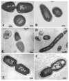
Transmission electron micrographs of pico-cyanobacteria isolated from the halocline of Lake Suigetsu. (A) Group I (CR2), (B) group II (CR3), (C) group III (CG3), (D) group IV (CG4), (E) group V (CG2), (F) group VI (CR5). Scale bars, 200 nm for (A), (B), (C), (E), (F) and 500 nm for (D).
Fig. 3
Fig. 3
Bayesian phylogenetic tree inferred from the 16S rRNA gene sequences. The corresponding posterior probabilities (left) and bootstrap values from ML (>50%; right) are given on each branch. Pigment information (PE, phycoerythrin rich; PC, phycocyanin rich) is based on Crosbie et al.(5). Accession numbers are shown in parentheses.
Fig. 4
Fig. 4
Specific growth rate of pico-cyanobacteria grown under different light intensities. Phycoerythrin-containing groups: (A) group I (CR2) grown under white light (black circles) or green light (gray circles); (B) group II (CR3) grown under white light (black triangles) or green light (gray triangles). Phycoerythrin-free groups; (C) group III (CG3) grown under white light (white circles) or green light (gray circles); (D) group IV (CG4) grown under white light (white triangles) or green light (gray triangles). Cells were cultured under white light of various intensities or under green light (460 to 600 nm) at 1.5 μmol m−2 s−1. Specific growth rate, μ (h−1), was determined from the increase in cell numbers during the exponential phase. Results are the mean±SD of three independent experiments with duplicate cultures. Error bar was omitted when±SD was too small to be shown.
Fig. 5
Fig. 5
Specific growth rate of pico-cyanobacteria grown in medium at different salinities. Group I (CR2, A), group II (CR3, B), group IV (CG4, C). Light intensity was 8 μmol m−2 s−1 (groups I and II) or 15 μmol m−2 s−1 (group IV). Specific growth rate, μ (h−1), was determined from the increase of cell numbers during the exponential growth phase. Results are the mean±SD of three independent experiments with duplicate cultures.
Fig. 6
Fig. 6
Effects of sulfide on the growth of pico-cyanobacteriaano-bacteria. Group I (CR2, black circles), group II (CR3, black triangles), group III (CG3, black triangles), group IV (CG4, white triangles, group VI, (CR5, crosses). Linear regression lines for Group I, group III and group IV are shown by dashed lines (r2=0.97), dotted lines (r2=0.96) and straight lines (r2=0.82). The sulfide concentration in the medium was determined after 7 d incubation. The relative inhibition of growth was estimated using the ratio of cell number increase in sulfide-containing medium compared to sulfide-free medium during 7 d incubation. Results are the mean±SD of three separate counts from triplicate cultures. Error bar was omitted when±SD was too small to be shown.

Similar articles

Cited by

References

    1. Bossshard PP, Santini Y, Grüter D, Stettler R, Bachofen R. Bacterial diversity and community composition in the chemocline of the meromictic alpine Lake Cadagno as revealed by 16S rDNA analysis. FEMS Microbiol Ecol. 2000;31:173–182. - PubMed
    1. Cohen Y, Padan E, Shilo M. Sulfide-dependent anoxygenic photosynthesis in the cyanobacteriuim Oscillatoria limnetica. Nature. 1975;257:489–491. - PMC - PubMed
    1. Cohen Y, Jørgensen BB, Revsbech NP, Poplawski R. Adaptation to hydrogen sulfide of oxygenic and anoxygenic photosynthesis among cyanobacteria. Appl Environ Microbiol. 1986;51:398–407. - PMC - PubMed
    1. Craig SR. The distribution and contribution of picoplankton to deep photosynthetic layers in some meromictic lakes. Acta Acad Aboensis. 1987;47:55–81.
    1. Crosbie ND, Pöckl M, Weisse T. Dispersal and phylogenetic diversity of nonmarine picocyanobacteria, inferred from 16S rRNA gene and cpcBA-intergenic spacer sequence analyses. Appl Environ Microbiol. 2003;69:5716–5721. - PMC - PubMed

Publication types

MeSH terms