. 2012 Oct 4;120(14):2925-9.
doi: 10.1182/blood-2012-04-422527. Epub 2012 Jul 12.

SOCS3 is an endogenous inhibitor of pathologic angiogenesis

Affiliations

SOCS3 is an endogenous inhibitor of pathologic angiogenesis

Andreas Stahl et al. Blood. .

Erratum in

Abstract

Inflammatory cytokines and growth factors drive angiogenesis independently; however, their integrated role in pathologic and physiologic angiogenesis is not fully understood. Suppressor of cytokine signaling-3 (SOCS3) is an inducible negative feedback regulator of inflammation and growth factor signaling. In the present study, we show that SOCS3 curbs pathologic angiogenesis. Using a Cre/Lox system, we deleted SOCS3 in vessels and studied developmental and pathologic angiogenesis in murine models of oxygen-induced retinopathy and cancer. Conditional loss of SOCS3 leads to increased pathologic neovascularization, resulting in pronounced retinopathy and increased tumor size. In contrast, physiologic vascularization is not regulated by SOCS3. In vitro, SOCS3 knockdown increases proliferation and sprouting of endothelial cells costimulated with IGF-1 and TNFα via reduced feedback inhibition of the STAT3 and mTOR pathways. These results identify SOCS3 as a pivotal endogenous feedback inhibitor of pathologic angiogenesis and a potential therapeutic target acting at the converging crossroads of growth factor- and cytokine-induced vessel growth.


Figures

Figure 1
Figure 1
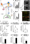
SOCS3 attenuates pathologic neovascularization. (A) Schematic representation of SOCS3 functioning as a negative feedback modulator of angiogenic activation. (B-C) Socs3 expression in wild-type mice exposed to OIR compared with normoxia both at the mRNA and protein level (n = 3-4). (D) Socs3 expression is significantly up-regulated in pathologic neovessels obtained by laser microdissection of retinal layers (outlined on the right; n = 4). GCL indicates ganglion cell layer; INL, inner nuclear layer; and ONL, outer nuclear layer. (E) Retinal vasculature of Cre reporter mice Rosa mTflox/mG. This strain universally expresses a red-fluorescent protein (mT) that is removed in tissues with Cre recombinase expression, revealing instead a green fluorescent protein (mGFP). In control mice, the retinal vasculature is red (top), whereas in Tie2-Cre mice, the retinal vasculature is green (bottom), demonstrating that Tie2-Cre recombinase is expressed in retinal vessels (n = 3). (F) Conditional Tie2-Socs3ko mice have more pathologic neovascularization in OIR compared with Socs3flox/flox control mice. The area of vaso-obliteration is unchanged (n = 13-34). (G,I) Increased growth of LLC (G; n = 5-7) and B16F10 melanoma tumors (I; n = 5-9) in Tie2-Socs3ko compared with Socs3flox/flox mice. (H,J) Increased tumor vessel density of LLC (H; n = 5-7) and B16F10 melanoma tumors (J; n = 5-9) in Tie2-Socs3ko compared with Socs3flox/flox mice. Scale bar in panel E indicates 200 μm; panel F, 1 mm; panels G and I, 1 cm; and in panels H and J, 100 μm. *P < .05, **P < .01, P < .001.
Figure 2
Figure 2
SOCS3 regulates vascular sprouting and endothelial activation. (A) Schematic representation of the signaling pathways regulated by SOCS3. (B) Increased sprouting of TNFα/IGF-1–stimulated aortic rings from Tie2-Socs3ko compared with Socs3flox/flox mice (n = 4-6). (C) Increased proliferation of human ECs treated with SOCS3 siRNA compared with controls (n = 9-10). (D) Expression of phospho-mTOR (P-mTOR) and P-STAT3 (green) in neovascular tufts (NV, red) of the OIR retina of Tie2-Socs3ko (not shown) and Socs3flox/flox control mice (n = 4). GCL indicates ganglion cell layer; INL, inner nuclear layer; and ONL, outer nuclear layer. (E) Increased mTOR and STAT3 phosphorylation, but not ERK, in IGF-1–stimulated ECs treated with Socs3 siRNA compared with controls (n = 3-5). (F) Prestimulation with TNFα (15 minutes) before IGF-1 further pronounces the increase of P-mTOR and P-STAT3 in IGF-1–stimulated ECs treated with Socs3 siRNA compared with controls (n = 3-6). Scale bar in panel B indicates 1 mm; and panel D, 50 μm. NS indicates not significant, *P < .05, **P < .01.

References

    1. Wang J, Campbell IL. Cytokine signaling in the brain: putting a SOCS in it? J Neurosci Res. 2002;67(4):423–427. - PubMed
    1. Starr R, Willson TA, Viney EM, et al. A family of cytokine-inducible inhibitors of signalling. Nature. 1997;387(6636):917–921. - PubMed
    1. Lebel E, Vallieres L, Rivest S. Selective involvement of interleukin-6 in the transcriptional activation of the suppressor of cytokine signaling-3 in the brain during systemic immune challenges. Endocrinology. 2000;141(10):3749–3763. - PubMed
    1. Jiang Y, Zhang Q, Soderland C, Steinle JJ. TNFalpha and SOCS3 regulate IRS-1 to increase retinal endothelial cell apoptosis. Cell Signal. 2012;24(5):1086–1092. - PMC - PubMed
    1. Dey BR, Furlanetto RW, Nissley P. Suppressor of cytokine signaling (SOCS)-3 protein interacts with the insulin-like growth factor-I receptor. Biochem Biophys Res Commun. 2000;278(1):38–43. - PubMed

Publication types

MeSH terms