. 2012 Aug 23;120(8):1742-51.
doi: 10.1182/blood-2012-03-417139. Epub 2012 Jul 12.

Inactivation of heparan sulfate 2-O-sulfotransferase accentuates neutrophil infiltration during acute inflammation in mice

Affiliations

Inactivation of heparan sulfate 2-O-sulfotransferase accentuates neutrophil infiltration during acute inflammation in mice

Jakob Axelsson et al. Blood. .

Abstract

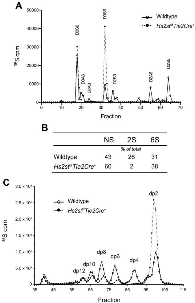
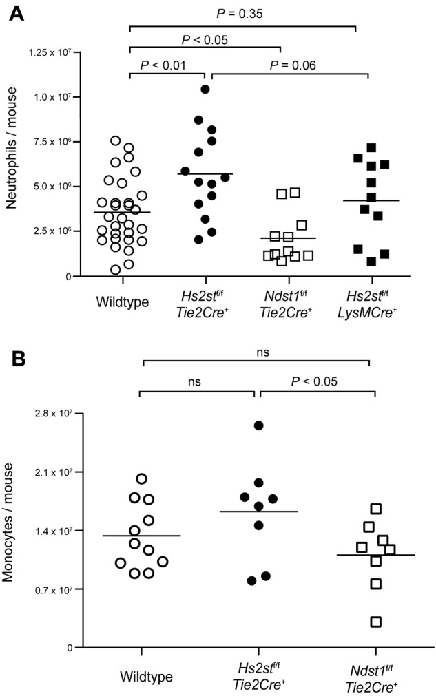
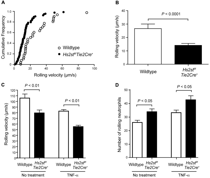
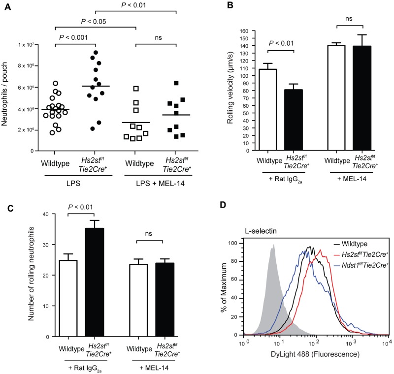
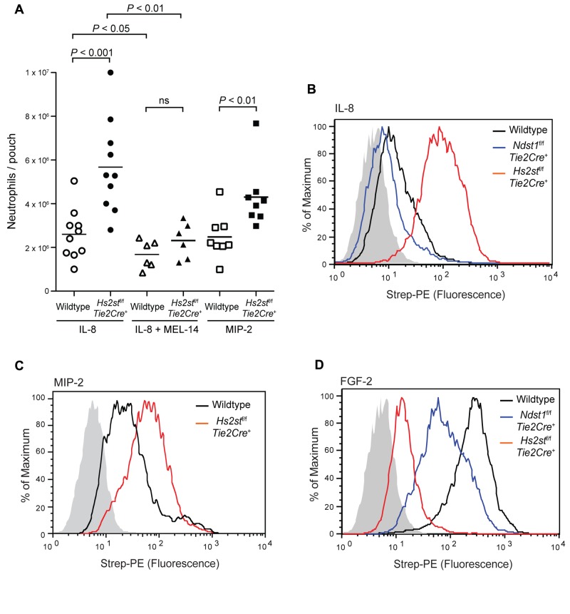
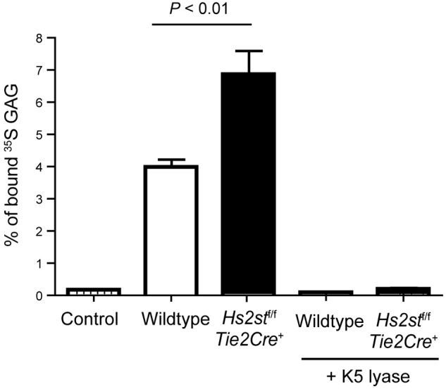
Neutrophil recruitment and extravasation at sites of inflammation provide a mechanism for host defense. We showed previously that heparan sulfate, a type of sulfated glycosaminoglycan, facilitates neutrophil recruitment based on the reduction of neutrophil infiltration in mice in which the overall sulfation of the chains was reduced by selective inactivation of N-acetylglucosamine N-deacetylase-N-sulfotransferase (Ndst1) in endothelial cells. Here we show that inactivation of uronyl 2-O-sulfotransferase in endothelial cells (Hs2st), an enzyme that acts downstream from Ndst1, results in enhanced neutrophil recruitment in several models of acute inflammation. Enhanced neutrophil infiltration resulted in part from reduced rolling velocity under flow both in vivo and in vitro, which correlated with stronger binding of neutrophil L-selectin to mutant endothelial cells. Hs2st-deficient endothelial cells also displayed a striking increase in binding of IL-8 and macrophage inflammatory protein-2. The enhanced binding of these mediators of neutrophil recruitment resulted from a change in heparan sulfate structure caused by increased N-sulfation and 6-O-sulfation of glucosamine units in response to the decrease in 2-O-sulfation of uronic acid residues. This gain-of-function phenotype provides formidable evidence demonstrating the importance of endothelial heparan sulfate in inflammation and suggests a novel enzyme target for enhancing the innate immune response.


Figures

Figure 1
Figure 1
Structural analysis of endothelial heparan sulfate isolated from Hs2stf/fTie2Cre+ mice. (A) Lung endothelial cells were metabolically labeled with 35SO4, and purified [35S]heparan sulfate was digested to completion with a combination of heparin lyases I, II, and III. The resulting disaccharides were analyzed by liquid chromatography. The 4-letter disaccharide structure codes (DSC) describe the composition of each recovered disaccharide. In DSC, the D designates a Δ4,5-unsaturated uronic acid and the absence or presence of a 2-O-sulfate group is designated by 0 or 2, respectively. The N-substituent of the glucosamine unit is designated A or S for acetate or sulfate, respectively. The absence or presence of 6-O-sulfate groups on the glucosamine units is designated by the numerals 0 or 6, respectively. (B) The number of [35S]sulfate groups at each position was determined and expressed as a percentage of the total. (C) Endothelial cells were metabolically labeled with 35SO4, and purified [35S]heparan sulfate was digested with heparin lyase III to completion. The resulting oligosaccharide fragments was resolved by gel filtration on a Bio-Gel P-10 column. dp indicates degree of polymerization (refers to the size of the oligosaccharide in the individual peaks).
Figure 2
Figure 2
Hs2stf/fTie2Cre+ mice show increased neutrophil and monocyte infiltration in thioglycolate-induced peritonitis. (A) Mice were injected intraperitoneally with sterile 3% thioglycolate broth. After 4 hours, the peritoneum was lavaged and infiltrated cells were collected. The cells were stained with a Gr-1 specific antibody followed by flow cytometry to quantify the total number of neutrophils. (B) In another set of animals, the peritoneum was lavaged after 18 hours and infiltrated cells were harvested. The cells were stained with antibodies specific for Gr-1 and F4/80 to quantify the total number of monocytes by flow cytometry. Each symbol represents a single mouse. ns indicates not significant.
Figure 3
Figure 3
Hs2stf/fTie2Cre+ mice show reduced neutrophil rolling velocity. (A) Rolling velocity of neutrophils in cremaster muscle venules was assessed by intravital microscopy. Up to 10 individual leucocytes were manually tracked over time using MATLAB software, and the velocity of the leucocytes was calculated. Each dot represents the cumulative frequency (summation of the velocity frequency and all frequencies below it). (B) Quantification of the rolling velocity of neutrophils in Hs2stf/fTie2Cre+ mutant mice and wild-type littermate control (n = 6-10). (C) Neutrophil rolling was assessed in vitro using flow chambers. Endothelial cells were grown to confluence on glass slides, and bone marrow-derived wild-type neutrophils were infused through the slides under a shear stress of 1 dyne/cm2. The rolling velocity of neutrophils was quantified with or without pretreatment with TNF-α (n = 3). (D) The number of rolling neutrophils was quantified. In a separate experiment, endothelial cells were pretreated with TNF-α (n = 3 for each experimental condition). Error bar represents SEM.
Figure 4
Figure 4
L-selectin contributes to the enhanced neutrophil infiltration in Hs2stf/fTie2Cre+ mice. (A) LPS (1 μg per mouse) was injected into dorsal air pouches; and after 4 hours, infiltrated neutrophils were collected and quantified. A set of wild-type and Hs2stf/fTie2Cre+ mice were injected intravenously with a blocking antibody to L-selectin (MEL-14) 1 hour before LPS injection. Each symbol represents a single mouse. (B) Effect of MEL-14 on neutrophil rolling velocity was assessed in vitro using flow chambers. Neutrophils were pretreated with either control IgG or MEL-14 and infused through the slides (n = 6). (C) The number of rolling neutrophils was quantified after pretreatment with either control IgG or MEL-14 (n = 6). Error bar represents SEM. (D) Binding of L-selectin-Fc fusion protein to wild-type or mutant lung endothelial cells was measured by flow cytometry. Black represents binding to wild-type cells; red, binding to Hs2stf/fTie2Cre+ cells; and blue, binding to Ndst1f/fTie2Cre+. The control sample was incubated only with anti–Fc-DyLight488 and is shown as filled gray histogram. ns indicates not significant.
Figure 5
Figure 5
Hs2stf/f Tie2Cre+ mice show increased neutrophil infiltration to IL-8 and MIP-2. (A) IL-8 or MIP-2 was injected into preformed dorsal air pouches. After 4 hours, pouches were lavaged and infiltrating neutrophils were quantified by flow cytometry. In a separate experiment, animals were treated with MEL-14 antibody by intravenous injection before injection of IL-8 into the pouch. Each symbol represents a single mouse. ns indicates not significant. (B) Binding of biotinylated IL-8 to wild-type or mutant lung endothelial cells was measured by flow cytometry. Black represents binding to wild-type cells; red, binding to Hs2stf/fTie2Cre+ cells; and blue, binding to Ndst1f/fTie2Cre+. The control sample was incubated only with streptavidin-PE and is shown as filled gray histogram. (C) Binding of MIP-2 to wild-type or mutant lung endothelial cells. (D) Binding of biotinylated FGF-2 to wild-type or mutant lung endothelial cells.
Figure 6
Figure 6
IL-8 shows enhanced binding to purified heparan sulfate derived from Hs2st-deficient endothelial cells. [35S]Heparan sulfate purified from mutant or wild-type endothelial cells was incubated with biotinylated IL-8 and collected by streptavidin-conjugated magnetic beads. One set of samples was treated with K5 lyases before incubation with IL-8. IL-8 was not added in the control sample.

References

    1. Ley K, Cerrito M, Arfors KE. Sulfated polysaccharides inhibit leukocyte rolling in rabbit mesentery venules. Am J Physiol. 1991;260:1667–1673. - PubMed
    1. Wang LC, Brown JR, Varki A, Esko JD. Heparin's anti-inflammatory effects require glucosamine 6-O-sulfation and are mediated by blockade of L- and P-selectins. J Clin Invest. 2002;110(1):127–136. - PMC - PubMed
    1. Young E. The anti-inflammatory effects of heparin and related compounds. Thromb Res. 2008;122(6):743–752. - PubMed
    1. Wang L, Fuster M, Sriramarao P, Esko JD. Endothelial heparan sulfate deficiency impairs L-selectin- and chemokine-mediated neutrophil trafficking during inflammatory responses. Nat Immunol. 2005;6(9):902–910. - PubMed
    1. Zuberi RI, Ge XN, Jiang S, et al. Deficiency of endothelial heparan sulfates attenuates allergic airway inflammation. J Immunol. 2009;183(6):3971–3979. - PMC - PubMed

Publication types

MeSH terms