. 2012 Sep;160(1):450-63.
doi: 10.1104/pp.112.198200. Epub 2012 Jul 12.

Toward stable genetic engineering of human O-glycosylation in plants

Affiliations

Toward stable genetic engineering of human O-glycosylation in plants

Zhang Yang et al. Plant Physiol. 2012 Sep.

Abstract

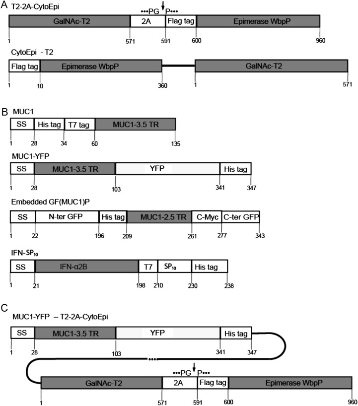
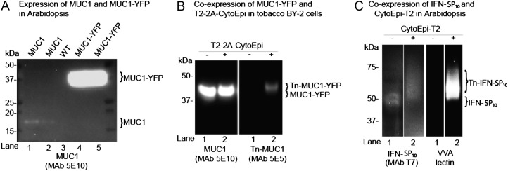
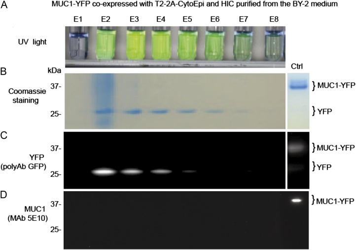
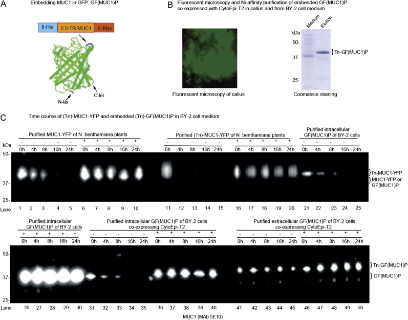
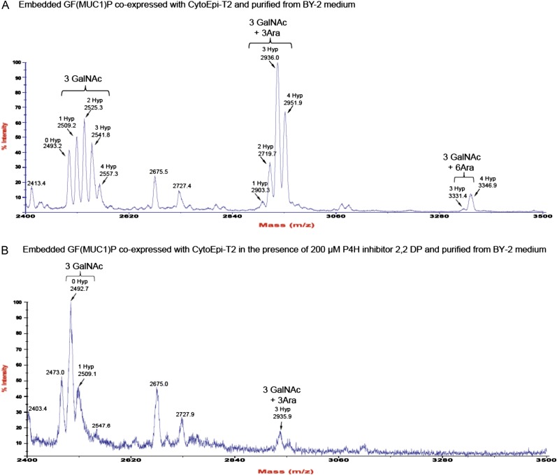
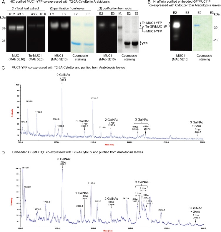
Glycosylation is the most abundant and complex posttranslational modification to be considered for recombinant production of therapeutic proteins. Mucin-type (N-acetylgalactosamine [GalNAc]-type) O-glycosylation is found in eumetazoan cells but absent in plants and yeast, making these cell types an obvious choice for de novo engineering of this O-glycosylation pathway. We previously showed that transient implementation of O-glycosylation capacity in plants requires introduction of the synthesis of the donor substrate UDP-GalNAc and one or more polypeptide GalNAc-transferases for incorporating GalNAc residues into proteins. Here, we have stably engineered O-glycosylation capacity in two plant cell systems, soil-grown Arabidopsis (Arabidopsis thaliana) and tobacco (Nicotiana tabacum) Bright Yellow-2 suspension culture cells. Efficient GalNAc O-glycosylation of two stably coexpressed substrate O-glycoproteins was obtained, but a high degree of proline hydroxylation and hydroxyproline-linked arabinosides, on a mucin (MUC1)-derived substrate, was also observed. Addition of the prolyl 4-hydroxylase inhibitor 2,2-dipyridyl, however, effectively suppressed proline hydroxylation and arabinosylation of MUC1 in Bright Yellow-2 cells. In summary, stably engineered mammalian type O-glycosylation was established in transgenic plants, demonstrating that plants may serve as host cells for the production of recombinant O-glycoproteins. However, the present stable implementation further strengthens the notion that elimination of endogenous posttranslational modifications may be needed for the production of protein therapeutics.


Figures

Figure 1.
Figure 1.
Construct designs for engineering O-glycosylation. A, Depiction of O-glycosylation machinery construct designs. Designs included Flag-tagged cytoplasm-targeted P. aeruginosa C4-epimerase WbpP (CytoEpi) and Golgi-targeted polypeptide GalNAc-T2 (T2) expressed as a single polycistronic protein interspaced by the 2A self-cleaving sequence (cleavage site indicated by the arrow; T2-2A-CytoEpi) or as separate transcriptional units (CytoEpi-T2). B, Depiction of the O-glycosylation reporter constructs used: MUC1-3.5TR (MUC1) with and without a C-terminal fusion to YFP (MUC1-YFP); MUC1 embedded into GFP [GF(MUC1)P]; Full-coding IFNα2B cytokine with a C-terminal T7 and SP10 glycomodule (IFN-SP10). All reporter constructs included N- or C-terminal His6 tags and N-terminal signal peptide sequences (SS). C, Depiction of a single combined construct with reporter MUC1-YFP and the 2A-linked O-glycosylation machinery under the control of two promoters (MUC1-YFP-T2-2A-CytoEpi).
Figure 2.
Figure 2.
Implementation of O-glycosylation in Arabidopsis and tobacco BY-2 suspension cells. A, SDS-PAGE western-blot analysis of four Arabidopsis lines expressing either MUC1 (lanes 1 and 2) or MUC1-YFP (lanes 4 and 5) alone. The contrast of the image was adjusted to visualize very weak bands for MUC1 (lanes 1 and 2). Lane 3 shows results for the wild type (WT). B, SDS-PAGE western-blot analysis of two tobacco BY-2 suspension cell lines expressing MUC1-YFP alone (lane 1) and MUC1-YFP together with the 2A-linked O-glycosylation machinery T2-2A-CytoEpi (lane 2). The absence (−) or presence (+) of O-glycosylation machinery is indicated above the lanes. C, An Arabidopsis line transgenic for T7-tagged full-coding secreted IFNα2B expressed alone (−) or coexpressed with the O-glycosylation machinery CytoEpi-T2 (+). The glycosylation of His tag-purified IFNα2B was detected by VVA lectin-blot analysis. Total protein extracts from transgenic Arabidopsis leaves or BY-2 cell callus were loaded and blots reacted with MUC1-specific MAbs 5E10 and 5E5, where 5E5 is specific for GalNAc-glycosylated MUC1 (Tn-MUC1) and does not react with nonglycosylated MUC1. Approximately 30 μg of total protein was loaded in each lane.
Figure 3.
Figure 3.
MUC1-YFP is expressed and glycosylated in BY-2 cells, but MUC1 is degraded in the medium. A, UV image of fractions (E1–E8) from HIC of MUC1-YFP secreted from BY-2 cells coexpressing T2-2A-CytoEpi. B, SDS-PAGE Coomassie blue-stained analysis of fractions. C, Western blot with anti-GFP antibody. D, Western blot with anti-MUC1 (5E10). Ten microliters of 2-mL eluate fractions was applied in each lane. [See online article for color version of this figure.]
Figure 4.
Figure 4.
O-Glycosylation and embedding in GFP stabilize MUC1 in BY-2 cell culture. A, Construct design for embedding MUC1-2.5TR into GFP [GF(MUC1)P], and corresponding barrel structure showing the loop into which MUC1-2.5 TR flanked with His8 and C-Myc tags was inserted after Asp-196 in the loop between the β-strands (blue) located opposite the N and C termini. This figure was adapted from Kobayashi et al. (2008). B, Fluorescence microscopy of a stable BY-2 transgenic line coexpressing GF(MUC1)P with the O-glycosylation machinery CytoEpi-T2 (left panel), and SDS-PAGE Coomassie blue staining of nickel chromatography-purified secreted GalNAc-glycosylated GF(MUC1)P [Tn-GF(MUC1)P] of this line (right panel). C, Analysis of the degradation of MUC1-YFP and GF(MUC1)P in BY-2 cell culture medium by SDS-PAGE western blotting: (1) MUC1-YFP (lanes 1–10) and GalNAc-glycosylated MUC1-YFP (Tn-MUC1-YFP; lanes 11–20) transiently produced in leaves of N. benthamiana plants; (2) intracellularly embedded GF(MUC1)P (lanes 21–30) purified from a stably transformed BY-2 cell line expressing GF(MUC1)P alone; (3) intracellularly (lanes 31–40) and extracellularly (lanes 41–50) embedded (Tn)-GF(MUC1)P purified from a transgenic BY-2 cell line additionally coexpressing CytoEpi-T2. The isolated proteins were added to 7-d-old unboiled (−) or boiled (+) wild-type BY-2 medium fractions, which were further incubated for up to 24 h under the same conditions. Approximately 5 μg of purified proteins was added to 1 mL of BY-2 medium fractions, of which approximately 15 μL was loaded and blots probed with anti-MUC1 MAb 5E10. The corresponding Tn-MUC1-specific western blot using MAb 5E5 is presented in Supplemental Figure S3. [See online article for color version of this figure.]
Figure 5.
Figure 5.
Inhibition of Pro hydroxylation of MUC1 in BY-2 cells. A, MALDI-TOF-MS analysis of endoproteinase Asp-released MUC1 tandem repeats (DTRPAPGSTAPPAHGVTSAP; indicated by arrows) from Tn-GF(MUC1)P expressed in BY-2 cells coexpressing CytoEpi-T2. The analysis revealed the presence of 3 mol of attached GalNAc (i.e. complete GalNAc-T2-mediated GalNAc occupancy of sites), with varying Pro hydroxylations of up to 4 mol per tandem repeat. Hydroxylated MUC1 peptides were further modified by the attachment of mainly three, and less frequently of six, Ara residues. B, MALDI-TOF-MS analysis of MUC1 tandem repeats (indicated by arrows) of GF(MUC1)P coexpressed with CytoEpi-T2 in BY-2 cells grown in the presence of a P4H inhibitor. Growth medium was exchanged at day 7 with fresh medium including 200 µm 2,2-DP, followed by incubation in similar growth conditions. [See online article for color version of this figure.]
Figure 6.
Figure 6.
MUC1-YFP and GF(MUC1)P expressed with or without O-glycosylation machinery in Arabidopsis. A, SDS-PAGE analysis of a transgenic Arabidopsis line expressing a combined construct (MUC1-YFP-T2-2A-CytoEpi) comprising both the reporter MUC1-YFP and the O-glycosylation machinery T2-2A-CytoEpi. Shown are MUC1-specific and Tn-MUC1-specific western-blot analysis of total leaf extracts (1) and MUC1-YFP purified from leaves (2) and roots (3) by HIC as visualized by Coomassie blue-stained SDS-PAGE gels and MUC1-specific MAb 5E10 western blots of the eluates. B, His tag purification of embedded GF(MUC1)P coexpressed with the O-glycosylation machinery CytoEpi-T2 as visualized by Coomassie blue-stained SDS-PAGE gels and MUC1-specific MAb 5E10 western blot. C and D, MALDI-TOF-MS analysis of MUC1 tandem repeats (DTRPAPGSTAPPAHGVTSAP; indicated by arrows) from His tag-purified Asp-N-digested MUC1-YFP (C) and GF(MUC1)P (D) coexpressed with the O-glycosylation machineries T2-2A-CytoEpi and CytoEpi-T2, respectively. [See online article for color version of this figure.]

Similar articles

Cited by

References

    1. Altmann F. (2007) The role of protein glycosylation in allergy. Int Arch Allergy Immunol 142: 99–115 - PubMed
    1. Amano K, Chiba Y, Kasahara Y, Kato Y, Kaneko MK, Kuno A, Ito H, Kobayashi K, Hirabayashi J, Jigami Y, et al. (2008) Engineering of mucin-type human glycoproteins in yeast cells. Proc Natl Acad Sci USA 105: 3232–3237 - PMC - PubMed
    1. Benchabane M, Goulet C, Rivard D, Faye L, Gomord V, Michaud D. (2008) Preventing unintended proteolysis in plant protein biofactories. Plant Biotechnol J 6: 633–648 - PMC - PubMed
    1. Bennett EP, Hassan H, Mandel U, Mirgorodskaya E, Roepstorff P, Burchell J, Taylor-Papadimitriou J, Hollingsworth MA, Merkx G, van Kessel AG, et al. (1998) Cloning of a human UDP-N-acetyl-alpha-D-galactosamine:polypeptide N-acetylgalactosaminyltransferase that complements other GalNAc-transferases in complete O-glycosylation of the MUC1 tandem repeat. J Biol Chem 273: 30472–30481 - PubMed
    1. Bennett EP, Mandel U, Clausen H, Gerken TA, Fritz TA, Tabak LA. (2012) Control of mucin-type O-glycosylation: a classification of the polypeptide GalNAc-transferase gene family. Glycobiology 22: 736–756 - PMC - PubMed

Publication types

MeSH terms