. 2012 Oct;93(Pt 10):2109-2117.
doi: 10.1099/vir.0.042713-0. Epub 2012 Jul 12.

Dynamin2 S-nitrosylation regulates adenovirus type 5 infection of epithelial cells

Affiliations

Dynamin2 S-nitrosylation regulates adenovirus type 5 infection of epithelial cells

Zhimin Wang et al. J Gen Virol. 2012 Oct.

Abstract

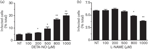
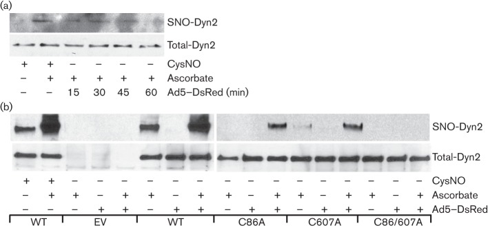
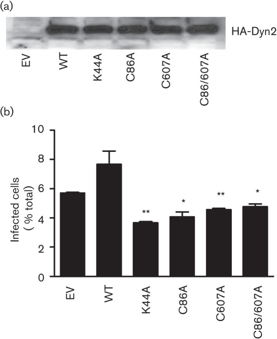
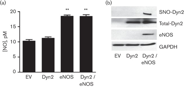
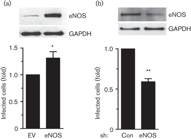
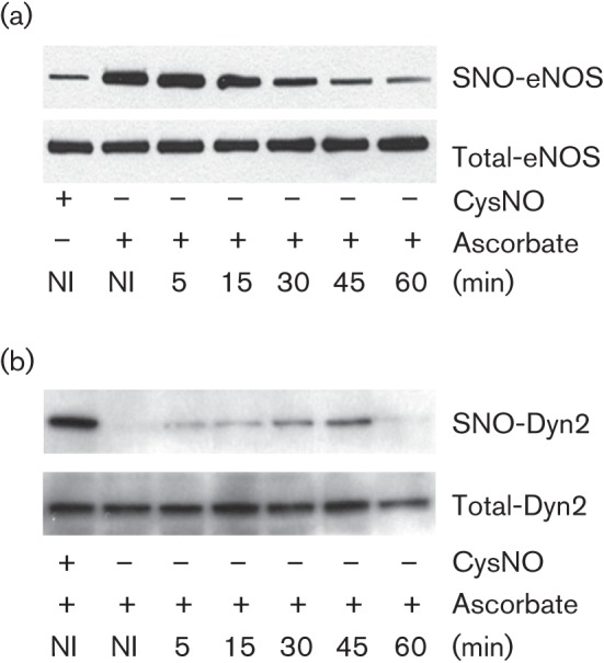
Dynamin2 is a large GTPase that regulates vesicle trafficking, and the GTPase activity of dynamin2 is required for the multistep process of adenovirus infection. Activity of dynamin2 may be regulated by post-translational phosphorylation and S-nitrosylation modifications. In this study, we demonstrate a role for dynamin2 S-nitrosylation in adenovirus infection of epithelial cells. We show that adenovirus serotype 5 (Ad5) infection augments production of nitric oxide (NO) in epithelial cells and causes the S-nitrosylation of dynamin2, mainly on cysteine 86 (C86) and 607 (C607) residues. Forced overexpression of dynamin2 bearing C86A and/or C607A mutations decreases Ad5 infection. Diminishing NO synthesis by RNAi-induced knockdown of endogenous endothelial NO synthase (eNOS) expression attenuates virus infection of target cells. Ad5 infection promotes the kinetically dynamic S-nitrosylation of dynamin2 and eNOS: there is a rapid decrease in eNOS S-nitrosylation and a concomitant increase in the dynamin2 S-nitrosylation. These results support the hypothesis that dynamin2 S-nitrosylation following eNOS activation facilitates adenovirus infection of host epithelial cells.


Figures

Fig. 1.
Fig. 1.
Effect of NO on Ad5–DsRed infection of epithelial cells. (a) BEC cells were treated with escalating doses of DETA-NO for 8 h before mixing with Ad5–DsRed. (b) BEC cells were treated with increasing concentrations of L-NAME for 8 h before mixing with Ad5–DsRed. In both cases, virus internalization was analysed 36 h after infection. Experiments were repeated three times and data are expressed as mean±sem. *, P<0.05 and **, P<0.01 versus non-treated (NT) samples.
Fig. 2.
Fig. 2.
Ad5–DsRed infection causes dynamin2 S-nitrosylation. (a) Ad5–DsRed infection promotes endogenous dynamin2 S-nitrosylation. BEC cells were infected with Ad5–DsRed for the indicated time and equal amounts of cell lysate were subjected to the biotin switch assay (top). (b) Ad5–DsRed infection promotes dynamin2 S-nitrosylation on C86 and C607 residues. BEC cells were transfected with cDNAs encoding HA-tagged dynamin2 [wild-type (WT), C86A, C607A, or C86/607A]. After 48 h, cells were mixed, or not, with Ad5–DsRed for 30 min. Cell lysates were harvested and subjected to the biotin switch method (top). For both panels, total cell lysates were immunoblotted with anti-dynamin2 antibody to show the equal protein loading (bottom), and lysate from cells treated with CysNO (100 µM) was used as a positive control. SNO-Dyn2, S-nitrosylated dynamin2.
Fig. 3.
Fig. 3.
Requirement of dynamin2 S-nitrosylation for Ad5–DsRed entry into cells. BEC cells were transfected with cDNAs encoding HA-tagged dynamin2 [wild-type (WT), K44A, C86A, C607A and C86/607A] for 48 h, and then infected with Ad5–DsRed for 24 h. (a) Cells were harvested and subjected to immunoblot analysis with an anti-HA antibody. (b) Virus infection efficiency was analysed by flow cytometry, exactly as described for Fig. 1. Experiments were repeated three times and data are expressed as mean±sem. *, P<0.05, **, P<0.01 versus empty vector (EV)-transfected cells.
Fig. 4.
Fig. 4.
Effect of Ad5–DsRed on NOS activation. (a) Ad5–DsRed infection promotes NO production. BEC cells were infected with Ad5–DsRed for the indicated time and then loaded with DAF-2DA for 2 min. The representative images were acquired using a Leica DM 6000 microscope at the indicated time (in min). (b) eNOS is phosphorylated at S1177 upon Ad5–DsRed infection. BEC cells were mixed with Ad5–DsRed for the indicated time and cell lysates were analysed by immunoblotting with an antibody against phospho-S1177-eNOS. The nitrocellulose membrane was stripped and immunoblotted with an antibody against eNOS to establish equal protein loading.
Fig. 5.
Fig. 5.
eNOS promotes dynamin2 S-nitrosylation. (a) COS-7 cells were transfected with cDNAs encoding HA-tagged dynamin2, WT bovine eNOS or both. After 24 h, the culture medium from the different transfected groups was mixed with an equal volume of ethanol for 20 min, followed by centrifugation. NO levels in the supernatant were quantified by a chemiluminescence detector after reaction with ozone using an NO analyser (Sievers). Experiments were repeated three times and data are expressed as mean±sem. **, P<0.01 versus empty vector (EV) pcDNA3.1-transfected cells. (b) Transfected COS-7 cells were harvested and analysed for dynamin2 S-nitrosylation (SNO-Dyn2) using the biotin switch assay. The cell lysates were immunoblotted with antibodies against dynamin2 (Total-Dyn2), eNOS or GAPDH.
Fig. 6.
Fig. 6.
eNOS regulates Ad5–DsRed infection. (a) Ad5–DsRed infection is increased as a result of forced overexpression of eNOS. BEC cells were transfected with pcDNA3.1 (EV) or pcDNA3.1-eNOS (eNOS) for 24 h. Cells were then infected with Ad5–DsRed for an additional 24 h and cell lysates were harvested and immunoblotted with antibodies against eNOS or GAPDH. (b) Ad5–DsRed infection is decreased in response to the knockdown of endogenous eNOS expression. Cells were infected with adenovirus encoding control shRNA that targets green fluorescent protein (GFP) (Con) or shRNA that targets eNOS (eNOS) for 36 h. Next, cells were infected with Ad5–DsRed for 24 h and cell lysates were immunoblotted with antibodies against eNOS or GAPDH. For both panels, the cells were also analysed for Ad5–DsRed internalization using flow cytometry, as described. Infection ratio of the control group was arbitrarily assigned a value of 1, to which the infection ratio of cells with overexpression of eNOS or knockdown of endogenous eNOS was normalized. Experiments were repeated three times and data are expressed as mean±sem. *, P<0.05, **, P<0.01 versus empty vector (EV)-transfected, or shRNA-GFP (Con)-infected cells.
Fig. 7.
Fig. 7.
Impact of Akt on Ad5–DsRed-induced eNOS phosphorylation. BEC cells were treated, or not, with PI3K inhibitor LY294002 (50 µM) for 2 h and then infected with Ad5–DsRed. Cells were harvested and lysates were fractionated on SDS-PAGE. Antibodies against phospho-S1177-eNOS and phosho-S473-Akt were used to detect phosphorylation of eNOS and Akt, respectively. The nitrocellulose membrane was stripped and immunoblotted with antibodies against total eNOS and Akt to show the equal protein loading. NI, Not-infected.
Fig. 8.
Fig. 8.
eNOS and dynamin2 are dynamically S-nitrosylated in response to Ad5–DsRed infection. BEC cells were transfected with cDNAs encoding HA-dynamin2 and bovine eNOS for 48 h and then infected with Ad5–DsRed for the indicated time. BEC cell lysates were subjected to a biotin switch assay to detect S-nitrosylated (a) eNOS (SNO-eNOS) or (b) dynamin2 (SNO-Dyn2). Treatment with CysNO served as a positive control and total dynamin2 or eNOS levels are shown to evidence equal protein loading among the different samples.

Similar articles

Cited by

References

    1. Ahn S., Maudsley S., Luttrell L. M., Lefkowitz R. J., Daaka Y. (1999). Src-mediated tyrosine phosphorylation of dynamin is required for β2-adrenergic receptor internalization and mitogen-activated protein kinase signaling. J Biol Chem 274, 1185–1188 10.1074/jbc.274.3.1185 - DOI - PubMed
    1. Ahn S., Kim J., Lucaveche C. L., Reedy M. C., Luttrell L. M., Lefkowitz R. J., Daaka Y. (2002). Src-dependent tyrosine phosphorylation regulates dynamin self-assembly and ligand-induced endocytosis of the epidermal growth factor receptor. J Biol Chem 277, 26642–26651 10.1074/jbc.M201499200 - DOI - PubMed
    1. Bauer P. M., Fulton D., Boo Y. C., Sorescu G. P., Kemp B. E., Jo H., Sessa W. C. (2003). Compensatory phosphorylation and protein-protein interactions revealed by loss of function and gain of function mutants of multiple serine phosphorylation sites in endothelial nitric-oxide synthase. J Biol Chem 278, 14841–14849 10.1074/jbc.M211926200 - DOI - PubMed
    1. Ben-Israel H., Kleinberger T. (2002). Adenovirus and cell cycle control. Front Biosci 7, d1369–d1395 10.2741/ben - DOI - PubMed
    1. Bergelson J. M., Cunningham J. A., Droguett G., Kurt-Jones E. A., Krithivas A., Hong J. S., Horwitz M. S., Crowell R. L., Finberg R. W. (1997). Isolation of a common receptor for coxsackie B viruses and adenoviruses 2 and 5. Science 275, 1320–1323 10.1126/science.275.5304.1320 - DOI - PubMed

Publication types

MeSH terms

LinkOut - more resources