. 2013 Feb 1;521(2):299-311.
doi: 10.1002/cne.23183.

Development of the principal nucleus trigeminal lemniscal projections in the mouse

Affiliations

Development of the principal nucleus trigeminal lemniscal projections in the mouse

Beril G Kivrak et al. J Comp Neurol. .

Abstract

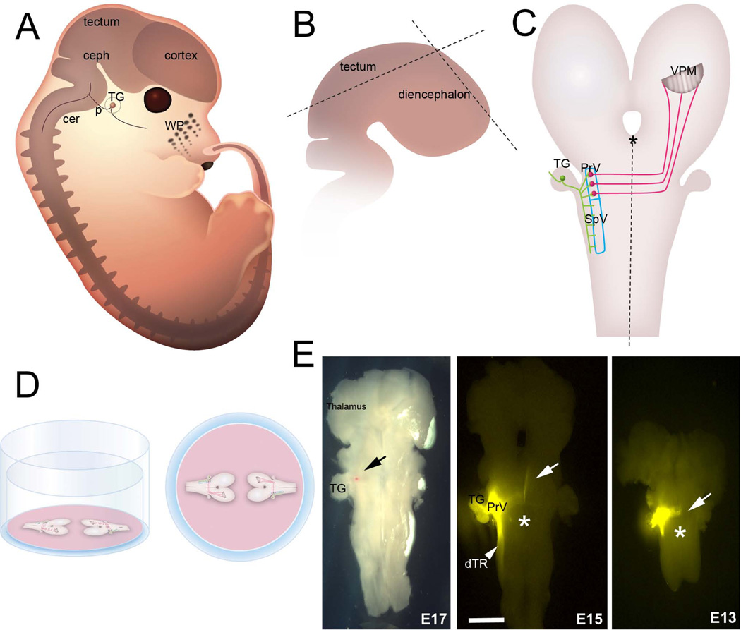
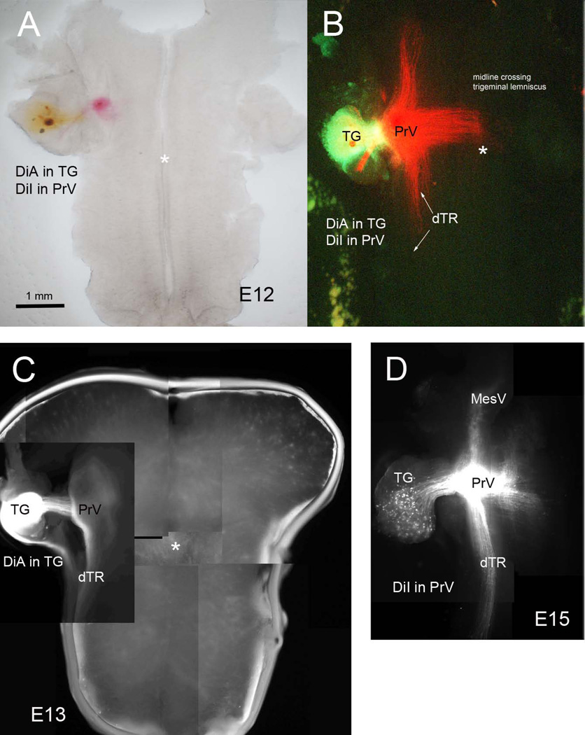
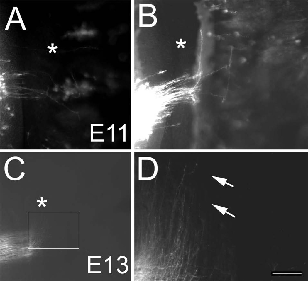
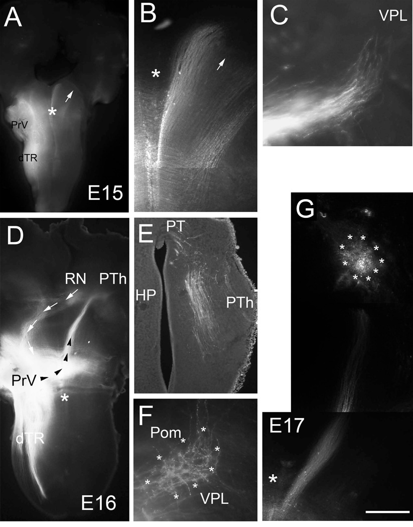
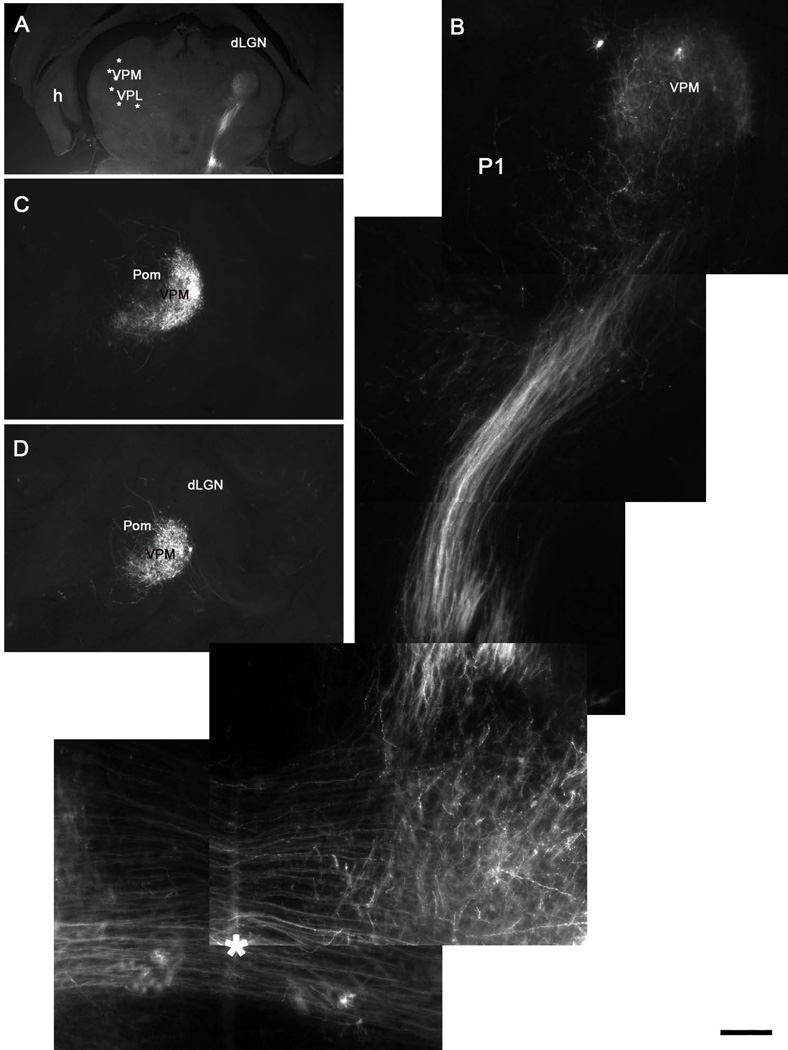
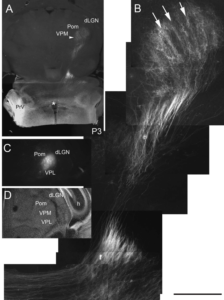
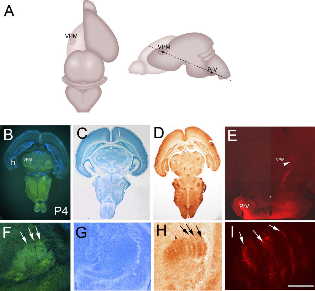
The principal sensory (PrV) nucleus-based trigeminal lemniscus conveys whisker-specific neural patterns to the ventroposteromedial (VPM) nucleus of the thalamus and subsequently to the primary somatosensory cortex. Here we examined the perinatal development of this pathway with carbocyanine dye labeling in embryonic and early postnatal mouse brains. We developed a novel preparation in which the embryonic hindbrain and the diencephalon are flattened out, allowing a birds-eye view of the PrV lemniscus in its entirety. For postnatal brains we used another novel approach by sectioning the brain along an empirically determined oblique horizontal angle, again preserving the trigeminothalamic pathway. PrV neurons are born along the hindbrain ventricular zone and migrate radially for a short distance to coalesce into a nucleus adjacent to the ascending trigeminal tract. During migration of the spindle-shaped cell bodies, slender axonal processes grow along the opposite direction towards the floor plate. As early as embryonic day (E) 11, pioneering axons tipped with large growth cones cross the ventral midline and immediately make a right angle turn. By E13 many PrV axons form fascicles crossing the midline and follow a rostral course. PrV axons reach the midbrain by E15 and the thalamus by E17. While the target recognition and invasion occurs prenatally, organization of PrV axon terminals into whisker-specific rows and patches takes place during the first 4 postnatal (P) days. Initially diffuse and exuberant projections in the VPM at P1 coalesce into row and whisker specific terminal zones by P4.


Conflict of interest statement

Conflict of interest statement.

The authors state that there is no conflict of interest.

Figures

Figure 1
Figure 1
Preparation of the embryonic trigeminal lemniscal pathway wholemounts. A: Schematic drawing of an E15 mouse embryo from a parasagittal view. B: For embryonic brainstem wholemounts, the cerebral cortex and tectum are removed, a cut is made along the dorsal diencephalic midline (dashed lines) and the entire specimen is splayed out (C) on a Millicell membrane as described in the Methods. D: Side view and birds-eye view of two brainstem wholemounts on a Millicell membrane in a six-well plate. Up to 4 explants can be flattened and kept on the same membrane. E: DiI labeling in flattened brainstem specimens at 3 different embryonic ages. The red dot (black arrow) in the E17 specimen is an exemplary DiI crystal. In other photomicrographs, white arrows point to the axons arriving at and crossing the midline. Asterisks indicate the midline. WP: whisker pad, ceph: cephalic flexure, p: pontine flexure, cer: cervical flexure, TG: trigeminal ganglion, PrV: principal sensory nucleus of the trigeminal nerve, SpV: spinal trigeminal nucleus; VPM: ventroposteromedial nucleus of the thalamus. Scale bar = 300 µm.
Figure 2
Figure 2
Double carbocyanine dye labeling to localize the position of the PrV in embryonic wholemounts. Placement of DiA in the ganglion and DiI in the PrV (A and B). DiA in the ganglion (C) allowed initial localization of the PrV. Backlabeling of the trigeminal ganglion (TG) cells and the descending trigeminal tract (dTR) also ensured that DiI placement was in the PrV (D).
Figure 3
Figure 3
Onset of midline crossing by PrV axons. A, B: Examples of pioneering PrV axons crossing the midline on E11. Note that the PrV axons do not cross the midline en masse, rather a few pioneers lead the way, stall on the other side of the midline and make an abrupt rostral turn. Asterisks indicate the midline. C: A large group of PrV axons have crossed the midline by E13, made a sharp rostral turn and waves of growth cone-tipped axons are advancing towards the midbrain. D: higher magnification view of the boxed area in C. Pioneering front of the PrV lemniscal pathway (arrows). Scale bar = 50 µm for B and 100 µm for D.
Figure 4
Figure 4
Developmental progression of the trigeminothalamic tract between E15-E17. Low (A,) and high (B) power views of the PrV lemniscal pathway on E15. At this age, trigeminal axons have reached the midbrain tegmental levels and spread out to form a ribbon. Arrows point to the same frontier edge of the lemniscal fibers. C: In a few late E15 cases, trigeminal axons are seen approaching the VPL (coronal section). D: Low power view of a brainstem-diencephalon flat mount from an E16 case. The trigeminal axons (black arrowheads) have reached the posterior thalamus (PTh) slightly rostral to the red nucleus (RN). In this case rubrotrigeminal (to trigeminal motor nucleus) axons are also labeled as a contralaterally projecting pathway (white arrows). E: A coronal section through the pretectal (PT)- posterior thalamus (PTh) area from another E16 case showing the progression of trigeminothalamic axons in this region; some axons are destined towards the tectum. E17 marks the time when PrV axons arrive at ventroposteromedial thalamic nucleus (VPM). F: Initial termination pattern in the VPM (boundaries marked by asterisks; E17-coronal section) at E17 is diffuse but mostly restricted to the VPM nucleus. G: Photomontage of low power views of the PrV lemniscal pathway in a wholemount preparation. Scale bar = 1mm for A, 500 µm for D and 200 µm for G. Large asterisks mark the midline; HP: habenulopeduncular tract; Pom: posteromedial thalamic nucleus; dTR: descending trigeminal tract.
Figure 5
Figure 5
Postnatal development of the trigeminal lemniscal pathway. A, B: Oblique horizontal sections show that the trigeminal lemniscus has invaded the VPM and axons branch diffusely across the entire nucleus at P1. C, D: Consecutive coronal sections showing the confinement of PrV axons to the VPM, with only a few axons spilling into the neighboring posteromedial nucleus (Pom). Note that no lemniscal axons enter the dorsal lateral geniculate nucleus (dLGN). Asterisk in B indicates the midline crossing of the PrV axons. Scale bar = 1200µm for A, 200µm for B and 500µm for C, D.
Figure 6
Figure 6
Emergence of barreloid rows on P3. Low power (A) and higher magnification (B) views of the PrV lemniscal pathway in oblique horizontal sections. Note that rows (arrows) of axon terminal bands are emerging from an initially diffuse terminal projection field in the VPM. C, D: DiI labeling and Nissl staining in coronal sections through the VPM at P3. H: hippocampus, dLGN: dorsal lateral geniculate nucleus, VPL: ventroposterolateral nucleus, Pom: posteromedial nucleus, asterisk indicates the midline crossing of the PrV axons. Scale bar = 400µm for B.
Figure 7
Figure 7
A: Schematic illustration of the oblique horizontal plane of sectioning to preserve the PrV-thalamic pathway. B: VGLUT2 immunostained oblique horizontal section with DAPI counterstain from a P4 brain. C: Nissl stain. D: Cytochrome oxidase histochemistry. E: DiI labeling at P4. H: hippocampus; VPM: ventroposteromedial nucleus. F–I show higher magnification views of the VPM with VGLUT2 immunohistochemistry (F), Nissl (G), CO (H) staining and DiI (I) labeling. Barreloid rows are clearly visible with VGLUT2 immuno- and CO histochemistry (arrows). Note also alignment of axon terminals along bands (arrows in I), reflecting the barreloid organization in the VPM. Scale bar in F = 200µm, I = 200µm.

Similar articles

Cited by

References

    1. Agmon A, Yang LT, O'Dowd DK, Jones EG. Organized growth of thalamocortical axons from the deep tier of terminations into layer IV of developing mouse barrel cortex. J Neurosci. 1993;13:5365–5382. - PMC - PubMed
    1. Al-Ghoul WM, Miller WM. Orderly migration of neurons to the principal sensory nucleus of the trigeminal nerve of the rat. J Comp Neurol. 1993;330:464–475. - PubMed
    1. Altman J, Bayer SA. Development of the brain stem in the rat IV. Thymidine-autoradiographic study of the time of origin of neurons in the pontine region. J Comp Neurol. 1980;194:905–929. - PubMed
    1. Bates CA, Killackey HP. The organization of the neonatal rat's brainstem trigeminal complex and its role in the formation of central trigeminal patterns. J Comp Neurol. 1985;240:265–287. - PubMed
    1. Belford GR, Killackey HP. The development of vibrissae representation in subcortical trigeminal centers of the neonatal rat. J Comp Neurol. 1979;188:63–74. - PubMed

Publication types

Substances