Development of the principal nucleus trigeminal lemniscal projections in the mouse
- PMID: 22791623
- PMCID: PMC3492550
- DOI: 10.1002/cne.23183
Development of the principal nucleus trigeminal lemniscal projections in the mouse
Abstract
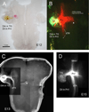
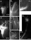
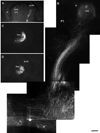
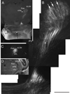
The principal sensory (PrV) nucleus-based trigeminal lemniscus conveys whisker-specific neural patterns to the ventroposteromedial (VPM) nucleus of the thalamus and subsequently to the primary somatosensory cortex. Here we examined the perinatal development of this pathway with carbocyanine dye labeling in embryonic and early postnatal mouse brains. We developed a novel preparation in which the embryonic hindbrain and the diencephalon are flattened out, allowing a birds-eye view of the PrV lemniscus in its entirety. For postnatal brains we used another novel approach by sectioning the brain along an empirically determined oblique horizontal angle, again preserving the trigeminothalamic pathway. PrV neurons are born along the hindbrain ventricular zone and migrate radially for a short distance to coalesce into a nucleus adjacent to the ascending trigeminal tract. During migration of the spindle-shaped cell bodies, slender axonal processes grow along the opposite direction towards the floor plate. As early as embryonic day (E) 11, pioneering axons tipped with large growth cones cross the ventral midline and immediately make a right angle turn. By E13 many PrV axons form fascicles crossing the midline and follow a rostral course. PrV axons reach the midbrain by E15 and the thalamus by E17. While the target recognition and invasion occurs prenatally, organization of PrV axon terminals into whisker-specific rows and patches takes place during the first 4 postnatal (P) days. Initially diffuse and exuberant projections in the VPM at P1 coalesce into row and whisker specific terminal zones by P4.
Copyright © 2012 Wiley Periodicals, Inc.
Conflict of interest statement
The authors state that there is no conflict of interest.
Figures


References
-
- Al-Ghoul WM, Miller WM. Orderly migration of neurons to the principal sensory nucleus of the trigeminal nerve of the rat. J Comp Neurol. 1993;330:464–475. - PubMed
-
- Altman J, Bayer SA. Development of the brain stem in the rat IV. Thymidine-autoradiographic study of the time of origin of neurons in the pontine region. J Comp Neurol. 1980;194:905–929. - PubMed
-
- Bates CA, Killackey HP. The organization of the neonatal rat's brainstem trigeminal complex and its role in the formation of central trigeminal patterns. J Comp Neurol. 1985;240:265–287. - PubMed
-
- Belford GR, Killackey HP. The development of vibrissae representation in subcortical trigeminal centers of the neonatal rat. J Comp Neurol. 1979;188:63–74. - PubMed
Publication types
MeSH terms
Substances
Grants and funding
LinkOut - more resources
Full Text Sources
Research Materials

