. 2012 Sep 7;287(37):31085-94.
doi: 10.1074/jbc.M112.383869. Epub 2012 Jul 12.

Design and characterization of modular scaffolds for tubulin assembly

Affiliations

Design and characterization of modular scaffolds for tubulin assembly

Ingrid Mignot et al. J Biol Chem. .

Abstract

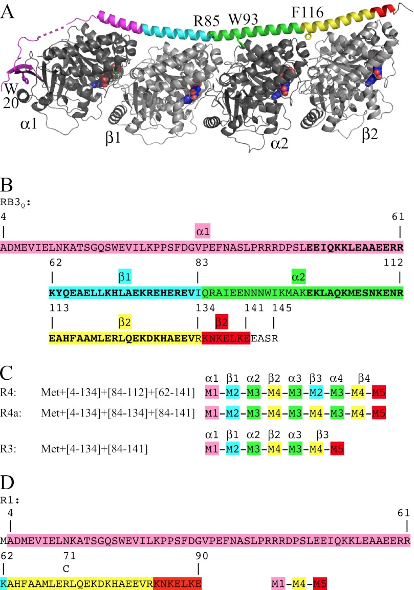
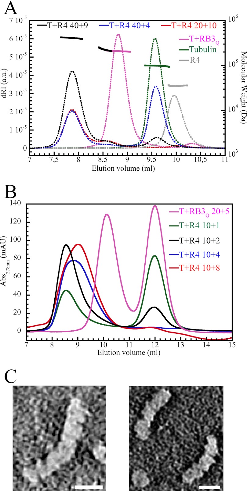
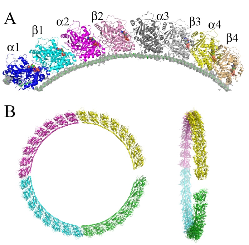
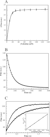
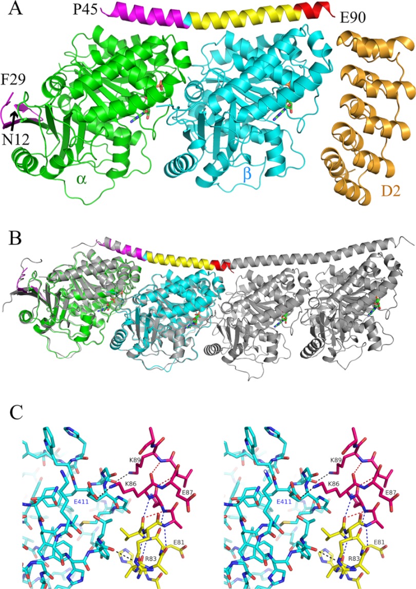
In cells, microtubule dynamics is regulated by stabilizing and destabilizing factors. Whereas proteins in both categories have been identified, their mechanism of action is rarely understood at the molecular level. This is due in part to the difficulties faced in structural approaches to obtain atomic models when tubulin is involved. Here, we design and characterize new stathmin-like domain (SLD) proteins that sequester tubulins in numbers different from two, the number of tubulins bound by stathmin or by the SLD of RB3, two stathmin family members that have been extensively studied. We established rules for the design of tight tubulin-SLD assemblies and applied them to complexes containing one to four tubulin heterodimers. Biochemical and structural experiments showed that the engineered SLDs behaved as expected. The new SLDs will be tools for structural studies of microtubule regulation. The larger complexes will be useful for cryo-electron microscopy, whereas crystallography or nuclear magnetic resonance will benefit from the 1:1 tubulin-SLD assembly. Finally, our results provide new insight into SLD function, suggesting that a major effect of these phosphorylatable proteins is the programmed release of sequestered tubulin for microtubule assembly at the specific cellular locations of members of the stathmin family.


Figures

FIGURE 1.
FIGURE 1.
The design of RB3SLD-based constructs for binding tubulin with a predefined stoichiometry. A, the T2R structure in which RB3SLD is colored according to the modules used in the design of the new SLDs. The RB3SLD according to which the modules are defined (RB3Q) contains four mutations that are modeled in the structure, F20W, K85R, F93W, and L116F (numbering is in reference to stathmin), plus the additional C14A mutation. The linker between the N-terminal β hairpin and the C-terminal helix, starting at residue Leu47, contains the least ordered region of RB3SLD; part of it is shown as a dashed line. Figs. 1A, 3, and 5 were generated using PyMOL (44). B, the RB3Q sequence colored according to the same modules as in A. The residues of the two stretches of the internal repeat (Glu48–Val82 and Glu99–Val133) are highlighted in boldface type. C, the design in terms of the modules defined in panel B of SLDs engineered to bind three (R3) or four tubulin heterodimers (R4 and R4a). In A–C, the tubulin subunits interacting with the modules we defined are indicated. D, sequence of R1 designed to bind one tubulin molecule. The position of the residue mutated to cysteine (R71C) and used to label R1 for affinity measurements is indicated.
FIGURE 2.
FIGURE 2.
R4 forms mainly a T4R4 complex with tubulin. A, SEC-MALLS analysis. The differential refractive index (dRI, arbitrary unit (a.u.) on the left axis, dotted lines) and molecular mass (displayed as solid lines for the regions of interest, i.e. for the chromatographic peaks, with the scale on the right axis) are plotted as a function of the column elution volume. The samples analyzed were as follows: tubulin (40 μm, green), R4 (60 μm, gray), T2R (40 μm tubulin and 30 μm RB3Q, magenta), and tubulin-R4 (20:10 μm, red; 40:4 μm, blue; 40:9 μm, black). The molecular masses of tubulin-R4 complexes are only displayed in the case of the 40:9 μm sample. B, gel filtration profiles obtained with a low salt buffer. Samples (100 μl) containing 10 μm tubulin and increasing concentrations of R4 (1 μm, green curve; 2 μm, black; 4 μm, blue; and 8 μm, red) were injected on the column. As a control, a sample containing 20 μm tubulin and 5 μm RB3Q was also analyzed (magenta). mAU, milliabsorbance units. C, electron micrographs of negatively stained tubulin-R4 complexes. Species comprising four tubulin heterodimers (left) predominate, whereas complexes with three tubulins are also identified (right). Their dimensions (∼55 Å × 355 Å and 55 Å × 265 Å, respectively) are consistent with those of a smaller SLD complex comprising two tubulins (15). Scale bar, 100 Å.
FIGURE 3.
FIGURE 3.
The T4R4 structure. A, overview of the complex in which each tubulin is colored differently. The α (β) subunits are in brighter (lighter) colors. The 4.2 Å resolution 2FobsFcalc electron density map of the R4 molecule, contoured at the 1σ level, is displayed. B, the relative orientations of the tubulin subunits in T4R4 are close to those in a ring. The model resulting from the repetition of T4R4 was obtained by superimposing the β1 moiety of the (m+1)th complex onto the β3 moiety of the mth complex and by keeping in the final model the 1st, 3rd, 5th, and 7th complexes. Each T4R4 is colored differently. The resulting flat helix is viewed along its axis (left) and nearly perpendicularly to it (right).
FIGURE 4.
FIGURE 4.
The tubulin-R1 interaction monitored by fluorescence spectroscopy. A, fluorescence variation of 13 nm R1* upon addition of tubulin. The curve is the fit of the experimental points with Equation 1, from which the Kd (1 nm) is extracted. Error bars correspond to the S.D. of the variation of fluorescence signal upon tubulin addition. B, dissociation of R1* from tubulin. The fluorescent TR1* complex was formed by mixing 30 nm R1* with 50 nm tubulin. The fluorescence decrease following addition of 4.2 μm R1 to this sample was monitored in a stopped-flow apparatus. The curve is the fit of the experimental points (5% of which are shown) with Equation 2. The same rate constant was obtained with two R1 concentrations (see “Experimental Procedures”) and is interpreted to be the dissociation rate constant of the complex (koff = 0.016 ± 0.003 s−1). C, determination of the association rate constant. Tubulin, at concentrations ranging from 200 nm to 1 μm, was added to a fixed concentration of R1* (30 nm). Fluorescence variations upon addition of 200 nm (square symbols) and 400 nm (dots) tubulin are shown (5% of the experimental points are displayed). The data were fitted according to Equation 3. The variation of kobs as a function of tubulin concentration is linear. kon is the slope (8 × 10⁶ m−1 s−1) of that curve (inset). a.u., arbitrary units.
FIGURE 5.
FIGURE 5.
The TR1 structure. A, overview of the TR1-D2 structure. The tubulin α and β subunits are in green and cyan, respectively. R1 is colored by modules as in Fig. 1. D2, the tubulin-binding DARPin with which crystals were obtained, is in orange. The nucleotides (GTP on α, GDP on β) are in green. The disordered eight N-terminal residues of R1 as well as the disordered linker between its N-terminal β hairpin and the C-terminal α helix (residues 30 to 44) are not displayed. B, comparison of TR1 with T2R. TR1 is colored as in A and superimposed on T2R (gray). The root mean square deviation after superposition (45) of Cαs of α, β, and R1 from TR1 and of α1, β1, and RB3SLD from T2R is 0.691 Å (872 atoms compared). C, stereo view of the interaction of the C-terminal end of R1 with tubulin in TR1. R1 and the tubulin β subunit are colored as in A. The hydrogen bonds between R1 and tubulin, including the Lys86–Gluβ411 salt bridge, are displayed as black dotted lines. The main chain hydrogen bonds in the R1 helix are displayed as red (310-helix) or blue (α-helix) dotted lines. R1 residues Glu80, Lys84, and Glu90, whose side chains are not defined, have been modeled as alanines. See supplemental Fig. S2 for the same view with R1 in its electron density map.

Similar articles

Cited by

References

    1. Desai A., Mitchison T. J. (1997) Microtubule polymerization dynamics. Annu. Rev. Cell Dev. Biol. 13, 83–117 - PubMed
    1. Amos L. A., Schlieper D. (2005) Microtubules and maps. Adv. Protein Chem. 71, 257–298 - PubMed
    1. Brouhard G. J., Stear J. H., Noetzel T. L., Al-Bassam J., Kinoshita K., Harrison S. C., Howard J., Hyman A. A. (2008) XMAP215 is a processive microtubule polymerase. Cell 132, 79–88 - PMC - PubMed
    1. Howard J., Hyman A. A. (2007) Microtubule polymerases and depolymerases. Curr. Opin. Cell Biol. 19, 31–35 - PubMed
    1. Jourdain L., Curmi P., Sobel A., Pantaloni D., Carlier M. F. (1997) Stathmin: A tubulin-sequestering protein, which forms a ternary T2S complex with two tubulin molecules. Biochemistry 36, 10817–10821 - PubMed

Publication types

LinkOut - more resources