. 2012 Oct 15;21(20):4394-405.
doi: 10.1093/hmg/dds272. Epub 2012 Jul 12.

Altered LKB1/AMPK/TSC1/TSC2/mTOR signaling causes disruption of Sertoli cell polarity and spermatogenesis

Affiliations

Altered LKB1/AMPK/TSC1/TSC2/mTOR signaling causes disruption of Sertoli cell polarity and spermatogenesis

Pradeep S Tanwar et al. Hum Mol Genet. .

Abstract

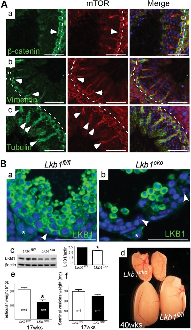
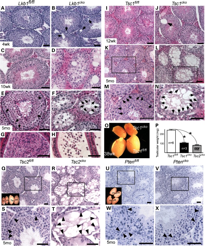
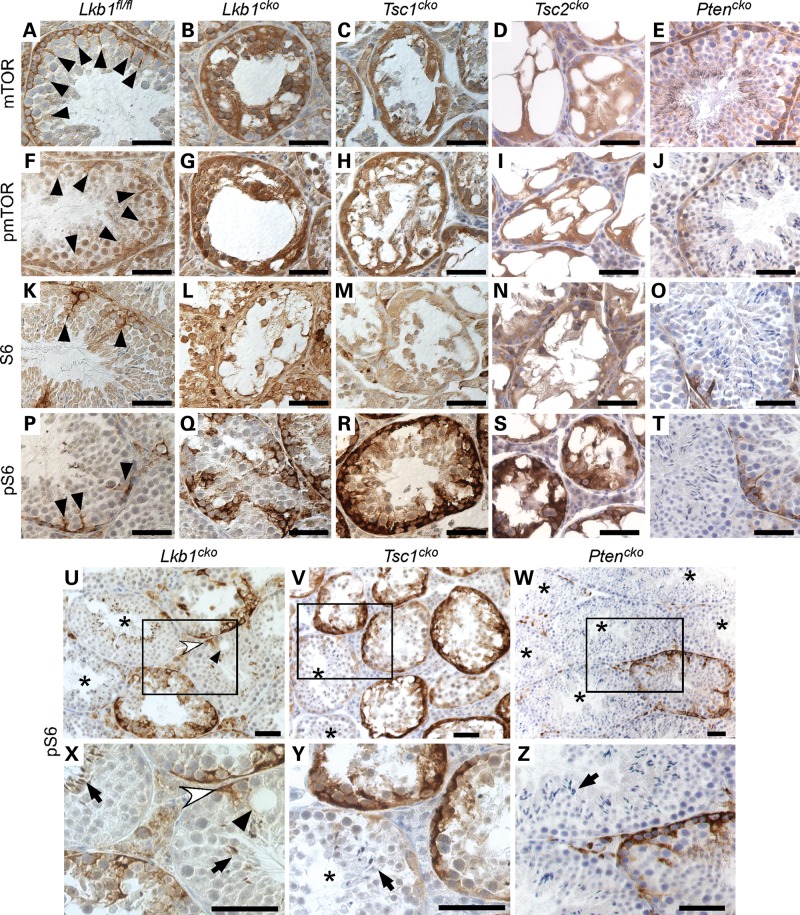
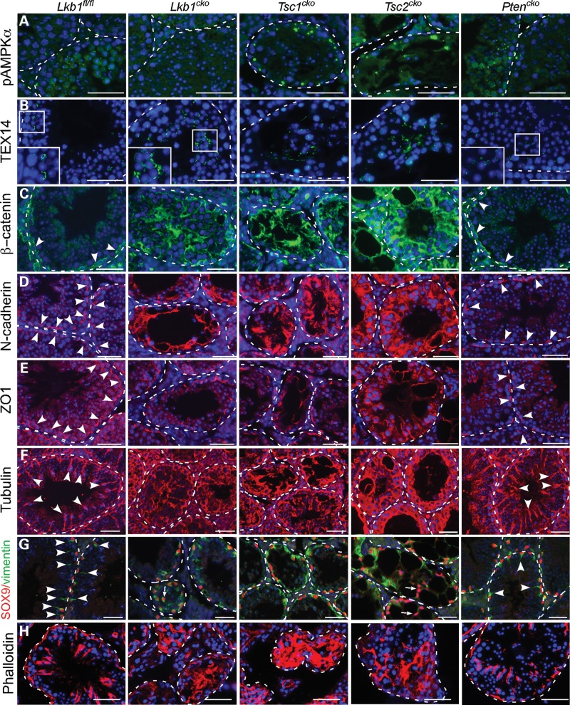
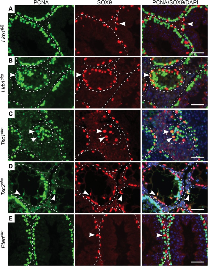
Male patients with Peutz-Jeghers syndrome (PJS) have defective spermatogenesis and are at increased risk of developing Sertoli cell tumors. Mutations in the Liver Kinase B1 (LKB1/STK11) gene are associated with the pathogenesis of PJS and have been identified in non-PJS patients with sporadic testicular cancers. The mechanisms controlled by LKB1 signaling in Sertoli cell functions and testicular biology have not been described. We have conditionally deleted the Lkb1 gene (Lkb1(cko)) in somatic testicular cells to define the molecular mechanisms involved in the development of the testicular phenotype observed in PJS patients. Focal vacuolization in some of the seminiferous tubules was observed in 4-week-old mutant testes but germ cell development appeared to be normal. However, similar to PJS patients, we observed progressive germ cell loss and Sertoli cell only tubules in Lkb1(cko) testes from mice older than 10 weeks, accompanied by defects in Sertoli cell polarity and testicular junctional complexes and decreased activation of the MAP/microtubule affinity regulating and focal adhesion kinases. Suppression of AMP kinase and activation of mammalian target of rapamycin (mTOR) signaling were also observed in Lkb1(cko) testes. Loss of Tsc1 or Tsc2 copies the progressive Lkb1(cko) phenotype, suggesting that dysregulated activation of mTOR contributes to the pathogenesis of the Lkb1(cko) testicular phenotype. Pten(cko) mice had a normal testicular phenotype, which could be explained by the comparative lack of mTOR activation detected. These studies describe the importance of LKB1 signaling in testicular biology and the possible molecular mechanisms driving the pathogenesis of the testicular defects observed in PJS patients.


Figures

Figure 1.
Figure 1.
Conditional deletion of LKB1 in mouse testes. (A) Colocalization of mTOR with β-catenin (a; a marker for adherens junctions; arrowheads), vimentin (b; a marker for Sertoli cells apical extensions; arrowheads) and tyrosinated α-tubulin (c; a marker for Sertoli cell microtubules; arrowheads) by IF. (B) (a and b) Localization of LKB1 in control and Lkb1cko mutant testes (Bc) by IF. Arrowheads indicate Sertoli cell nuclei and asterisks indicate interstitial Leydig cells. Western blot and semiquantitative analyses of LKB1 in control and mutant testes (Bc). Gross testes from Lkb1 control and mutant mice (Bd). Weight of Lkb1cko mutant and control testes (Be) and seminal vesicles (Bf). Columns represent the mean in n= 3 testes, bars equal the SEM and asterisks indicate P< 0.05. Bars: 50 μm. Nuclei are stained with DAPI.
Figure 2.
Figure 2.
Premature germ cell loss and Sertoli cell only tubules of Lkb1cko, Tsc1cko and Tsc2cko but not in Ptencko mice testes. (A–F) Histology of control and Lkb1cko mutant testes by H&E. (G and H) Epididymides from control and mutant mice. Histology of control and Tsc1cko (I–N) testes. (O) Gross picture of control and Tsc1cko mutant testes. Arrows in (B and J) indicate vacuolated seminiferous epithelium. Arrows in (E), (F), (M), and (N) are pointing towards Sertoli cells. (P) Weight of adult Tsc1cko, Tsc2cko and control testes. Columns represent the mean in n= 3 testes, bars equal the SEM and asterisks indicate P< 0.05. Histology of control, Tsc2cko (Q–T), and Ptencko (U–X) mutant testes. (M), (N), (S), (T), (W) and (X) are higher magnification images of the boxed areas in K, L, Q, R, U and V, respectively. Arrowheads in (S), (T), (W) and (X) indicate Sertoli cells. Insets in (Q and U) show adult testes from control and mutant mice. Bars: 50 μm.
Figure 3.
Figure 3.
Overactivation of mTOR signaling in Lkb1cko, Tsc1cko and Tsc2cko testes. IHC for mTOR (A–E), pmTOR (F–J), S6 (K–O) and pS6 (P–T) in Lkb1fl/fl, Lkb1cko, Tsc1cko, Tsc2cko and Ptencko testes. Black arrowheads in (A), (F), (K), (P) are pointing towards Sertoli cells. (U and X) Vacuolation (black arrowheads) of seminiferous epithelium next to Sertoli cell positive (white arrowhead) for pS6 immunostaining. (X), (Y), and (Z) are higher magnification images of the boxed areas in U, V and W, respectively. Seminiferous tubules in (U–Z) with elongated spermatids (black arrow) are marked by asterisks. Bars: 50 μm.
Figure 4.
Figure 4.
Disruption of the Sertoli cell polarity and testicular junctions in Lkb1cko, Tsc1cko and Tsc2cko mice. Expression of pAMPKα (A), TEX14 (B; a marker of testicular intercellular junctions), β-catenin (C), N-cadherin (D; markers of adherens junctions), ZO-1 (E; a marker for tight junctions), tyrosinated α-tubulin (F; a marker for Sertoli cell microtubules), SOX9 (a Sertoli cell nuclear marker)/vimentin (G; a marker for Sertoli apical extensions) and Phalloidin (H) in Lkb1fl/fl, Lkb1cko, Tsc1cko, Tsc2cko and Ptencko testes. Arrowheads in (C), (D), (E) indicate positive staining for β-catenin, N-Cadherin and ZO-1 at the site of the blood–testis barrier. Arrowheads in (F) indicate Sertoli cell microtubules. White-dotted lines mark the basement membrane of the seminiferous tubules. Arrowheads in (G) indicate the Sertoli cells apical extensions. Bars: 50 μm. Nuclei are stained with DAPI.
Figure 5.
Figure 5.
Proliferation of Sertoli cells in Lkb1cko, Tsc1cko and Tsc2cko adult murine testes. Colocalization of PCNA (a marker for proliferating cells) and SOX9 (a nuclear marker for Sertoli cells) in Lkb1fl/fl (A) Lkb1cko, (B) Tsc1cko, (C) Tsc2cko, (D) and Ptencko (E) testes. Arrowheads in (A–E) represent Sertoli cells. White-dotted lines indicate the basement membranes of the seminiferous tubules. Bars: 50 μm.

Similar articles

Cited by

References

    1. Hezel A.F., Bardeesy N. LKB1; linking cell structure and tumor suppression. Oncogene. 2008;27:6908–6919. doi:10.1038/onc.2008.342. - DOI - PubMed
    1. Jeghers H., Mc K.V., Katz K.H. Generalized intestinal polyposis and melanin spots of the oral mucosa, lips and digits; a syndrome of diagnostic significance. N. Engl. J. Med. 1949;241:1031–1036. doi:10.1056/NEJM194912292412601. - DOI - PubMed
    1. Hemminki A., Markie D., Tomlinson I., Avizienyte E., Roth S., Loukola A., Bignell G., Warren W., Aminoff M., Hoglund P., et al. A serine/threonine kinase gene defective in Peutz–Jeghers syndrome. Nature. 1998;391:184–187. doi:10.1038/34432. - DOI - PubMed
    1. Jenne D.E., Reimann H., Nezu J., Friedel W., Loff S., Jeschke R., Muller O., Back W., Zimmer M. Peutz–Jeghers syndrome is caused by mutations in a novel serine threonine kinase. Nat. Genet. 1998;18:38–43. doi:10.1038/ng0198-38. - DOI - PubMed
    1. Shackelford D.B., Shaw R.J. The LKB1–AMPK pathway: metabolism and growth control in tumour suppression. Nat. Rev. Cancer. 2009;9:563–575. doi:10.1038/nrc2676. - DOI - PMC - PubMed

Publication types

MeSH terms

Substances