Recurrent attacks of headache and neck pain caused by congenital aplasia of the posterior arch of atlas in an adult
- PMID: 22791780
- PMCID: PMC3029836
- DOI: 10.1136/bcr.05.2010.3053
Recurrent attacks of headache and neck pain caused by congenital aplasia of the posterior arch of atlas in an adult
Abstract
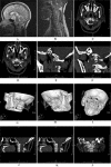
A 47-year-old Chinese woman, with a history of recurrent attacks of vertigo and vomiting for the past 5 years, presented with intermittent radicular pain in the left upper limb for the past 2 years. She also reported recurrent attacks of severe headache and neck pain for more than 10 years. The pain might be aggravated by coughing or sneezing and relieved after sleeping in the decubitus position. The MRI depicted Chiari malformation. A multidetector CT scan and three-dimensional CT reconstruction revealed partial aplasia of the left posterior arch of atlas of a small gap. The patient underwent plastic surgeries in Beijing. The disappearance of the recurrent pain syndrome was confirmed by follow-up after surgery.
Conflict of interest statement
Figures


References
-
- Menezes AH, Vogel TW. Specific entities affecting the craniocervical region: syndromes affecting the craniocervical junction. Childs Nerv Syst 2008;24:1155–63 - PubMed
-
- Martich V, Ben-Ami T, Yousefzadeh DK, et al. Hypoplastic posterior arch of C-1 in children with Down syndrome: a double jeopardy. Radiology 1992;183:125–8 - PubMed
-
- Klimo P, Jr, Blumenthal DT, Couldwell WT. Congenital partial aplasia of the posterior arch of the atlas causing myelopathy: case report and review of the literature. Spine 2003;28:E224–8 - PubMed
-
- Speer MC, Enterline DS, Mehltretter L, et al. Chiari Type I malformation with or without syringomyelia: prevalence and genetics . J Genet Couns 2003;12:297–311 - PubMed
-
- Tubbs RS, Lyerly MJ, Loukas M, et al. The pediatric Chiari I malformation: a review. Childs Nerv Syst 2007;23:1239–50 - PubMed
Publication types
MeSH terms
LinkOut - more resources
Full Text Sources
Medical
Miscellaneous
