

FoxP2 is a parvocellular-specific transcription factor in the visual thalamus of monkeys and ferrets
- PMID: 22791804
- PMCID: PMC3729200
- DOI: 10.1093/cercor/bhs207
FoxP2 is a parvocellular-specific transcription factor in the visual thalamus of monkeys and ferrets
Abstract
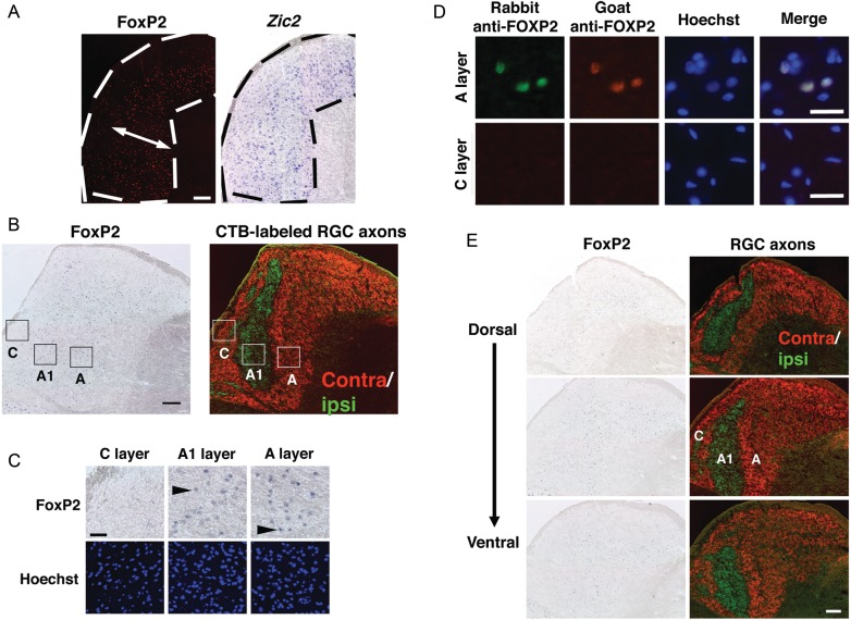
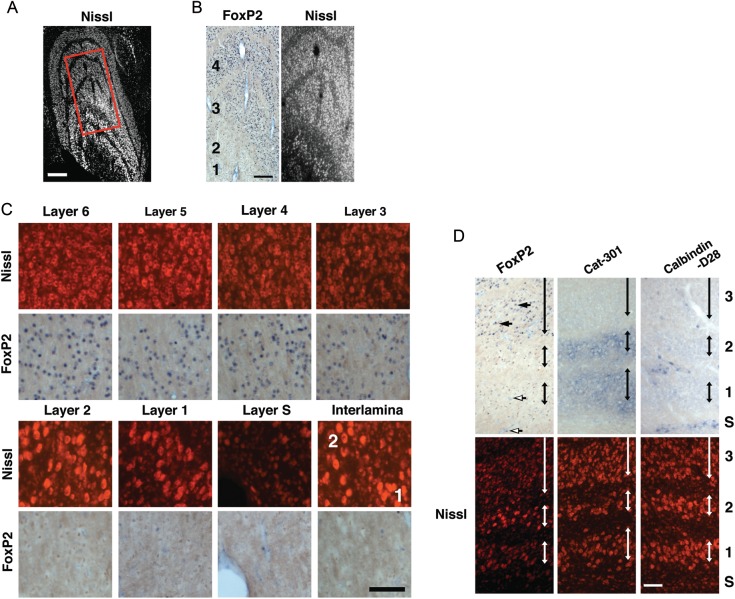
Although the parallel visual pathways are a fundamental basis of visual processing, our knowledge of their molecular properties is still limited. Here, we uncovered a parvocellular-specific molecule in the dorsal lateral geniculate nucleus (dLGN) of higher mammals. We found that FoxP2 transcription factor was specifically expressed in X cells of the adult ferret dLGN. Interestingly, FoxP2 was also specifically expressed in parvocellular layers 3-6 of the dLGN of adult old world monkeys, providing new evidence for a homology between X cells in the ferret dLGN and parvocellular cells in the monkey dLGN. Furthermore, this expression pattern was established as early as gestation day 140 in the embryonic monkey dLGN, suggesting that parvocellular specification has already occurred when the cytoarchitectonic dLGN layers are formed. Our results should help in gaining a fundamental understanding of the development, evolution, and function of the parallel visual pathways, which are especially prominent in higher mammals.
Keywords: FoxP2; X cells; ferret; monkey; parvocellular.
Figures


Similar articles
-
Cell type-specific expression of FoxP2 in the ferret and mouse retina.Neurosci Res. 2017 Apr;117:1-13. doi: 10.1016/j.neures.2016.11.008. Epub 2016 Nov 22. Neurosci Res. 2017. PMID: 27888071
-
Shrinkage of X cells in the lateral geniculate nucleus after monocular deprivation revealed by FoxP2 labeling.Vis Neurosci. 2014 May;31(3):253-61. doi: 10.1017/S0952523813000643. Epub 2014 Jan 30. Vis Neurosci. 2014. PMID: 24480423
-
Molecular correlates of laminar differences in the macaque dorsal lateral geniculate nucleus.J Neurosci. 2008 Nov 12;28(46):12010-22. doi: 10.1523/JNEUROSCI.3800-08.2008. J Neurosci. 2008. PMID: 19005066 Free PMC article.
-
Tracking blue cone signals in the primate brain.Clin Exp Optom. 2013 May;96(3):259-66. doi: 10.1111/j.1444-0938.2012.00819.x. Epub 2012 Nov 27. Clin Exp Optom. 2013. PMID: 23186138 Review.
-
Molecular guidance cues in the development of visual pathway.Protein Cell. 2018 Nov;9(11):909-929. doi: 10.1007/s13238-017-0490-7. Epub 2017 Nov 27. Protein Cell. 2018. PMID: 29181831 Free PMC article. Review.
Cited by
-
Glial cell type-specific gene expression in the mouse cerebrum using the piggyBac system and in utero electroporation.Sci Rep. 2021 Mar 1;11(1):4864. doi: 10.1038/s41598-021-84210-z. Sci Rep. 2021. PMID: 33649472 Free PMC article.
-
Role of Feedback Connections in Central Visual Processing.Annu Rev Vis Sci. 2020 Sep 15;6:313-334. doi: 10.1146/annurev-vision-121219-081716. Epub 2020 Jun 17. Annu Rev Vis Sci. 2020. PMID: 32552571 Free PMC article. Review.
-
Diverse visual features encoded in mouse lateral geniculate nucleus.J Neurosci. 2013 Mar 13;33(11):4642-56. doi: 10.1523/JNEUROSCI.5187-12.2013. J Neurosci. 2013. PMID: 23486939 Free PMC article.
-
Single-cell and single-nucleus RNA-seq uncovers shared and distinct axes of variation in dorsal LGN neurons in mice, non-human primates, and humans.Elife. 2021 Sep 2;10:e64875. doi: 10.7554/eLife.64875. Elife. 2021. PMID: 34473054 Free PMC article.
-
Molecular investigations of development and diseases of the brain of higher mammals using the ferret.Proc Jpn Acad Ser B Phys Biol Sci. 2017;93(5):259-269. doi: 10.2183/pjab.93.017. Proc Jpn Acad Ser B Phys Biol Sci. 2017. PMID: 28496051 Free PMC article. Review.
References
-
- Ako R, Wakimoto M, Ebisu H, Tanno K, Hira R, Kasai H, Matsuzaki M, Kawasaki H. Simultaneous visualization of multiple neuronal properties with single-cell resolution in the living rodent brain. Mol Cell Neurosci. 2011;48:246–257. - PubMed
-
- Boyden ES, Zhang F, Bamberg E, Nagel G, Deisseroth K. Millisecond-timescale, genetically targeted optical control of neural activity. Nat Neurosci. 2005;8:1263–1268. - PubMed
-
- Chan AW, Chong KY, Martinovich C, Simerly C, Schatten G. Transgenic monkeys produced by retroviral gene transfer into mature oocytes. Science. 2001;291:309–312. - PubMed
