. 2012 Aug;139(16):3032-9.
doi: 10.1242/dev.075812. Epub 2012 Jul 12.

RTK signaling modulates the Dorsal gradient

Affiliations

RTK signaling modulates the Dorsal gradient

Aharon Helman et al. Development. 2012 Aug.

Abstract

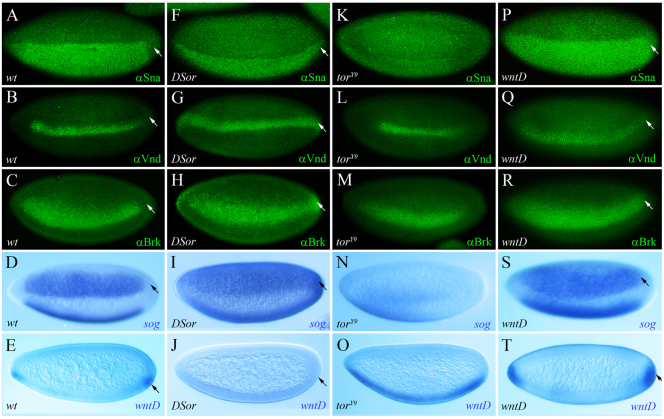
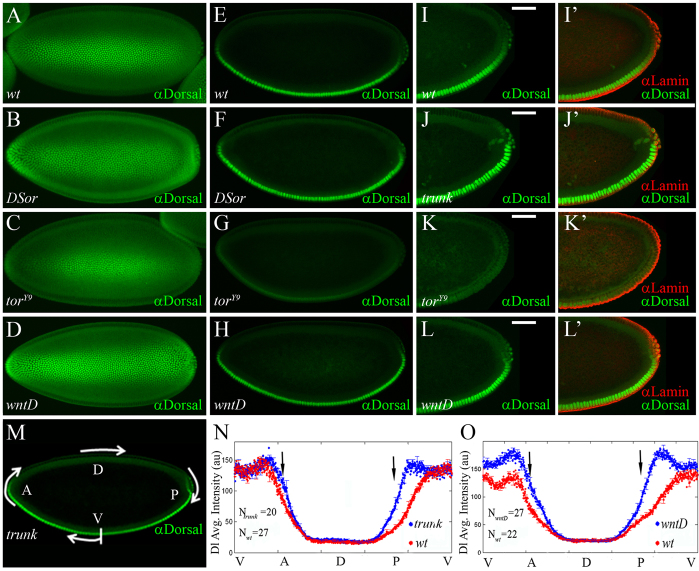
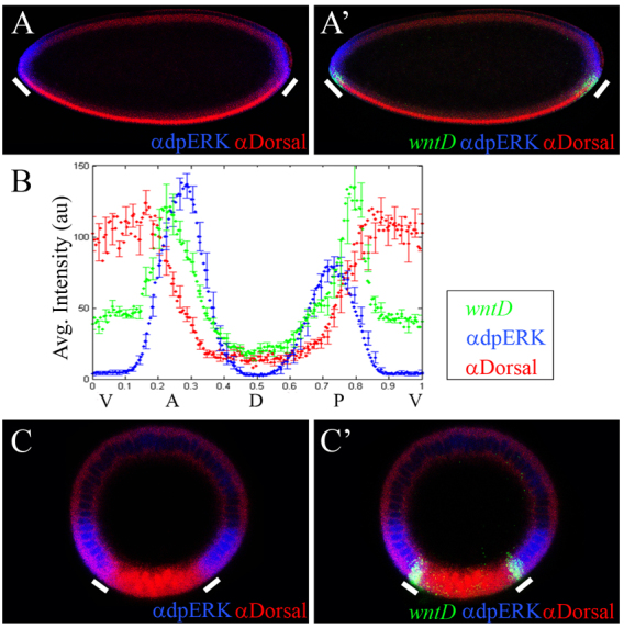
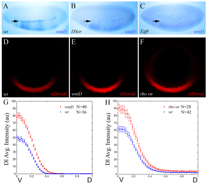
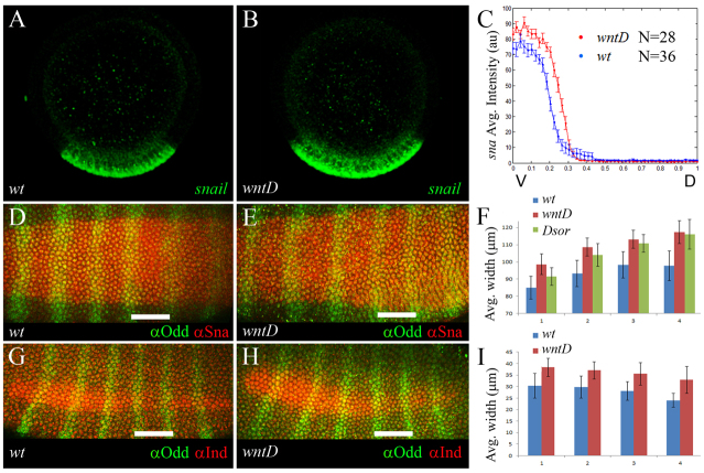
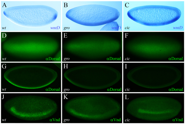
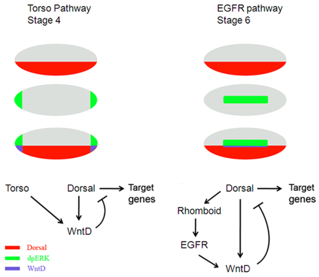
The dorsoventral (DV) axis of the Drosophila embryo is patterned by a nuclear gradient of the Rel family transcription factor, Dorsal (Dl), that activates or represses numerous target genes in a region-specific manner. Here, we demonstrate that signaling by receptor tyrosine kinases (RTK) reduces nuclear levels and transcriptional activity of Dl, both at the poles and in the mid-body of the embryo. These effects depend on wntD, which encodes a Dl antagonist belonging to the Wingless/Wnt family of secreted factors. Specifically, we show that, via relief of Groucho- and Capicua-mediated repression, the Torso and EGFR RTK pathways induce expression of WntD, which in turn limits Dl nuclear localization at the poles and along the DV axis. Furthermore, this RTK-dependent control of Dl is important for restricting expression of its targets in both contexts. Thus, our results reveal a new mechanism of crosstalk, whereby RTK signals modulate the spatial distribution and activity of a developmental morphogen in vivo.


Figures

Fig. 1.
Fig. 1.
Torso RTK signaling restricts expression of multiple Dorsal targets at the embryonic termini. (A-T) Lateral view of stage 5 wild-type (A-E), DSor (F-J), torY9 (K-O) and wntD (P-T) mutant embryos, immunostained for Sna (A,F,K,P), Vnd (B,G,L,Q) and Brk (C,H,M,R), or hybridized using digoxigenin-labeled RNA probes for sog (D,I,N,S) and wntD (E,J,O,T). Torso-dependent activity limits the expression of Dl targets at the poles. (A-D) Expression of four Dl targets, Sna, Vnd, Brk and sog, is confined to the trunk region and is excluded from the termini. (F-I) In DSor mutant embryos, where Torso signaling is blocked, expression of these Dl targets expands into terminal regions. (K-N) In torY9 embryos, where Torso is overactive, expression of these Dl targets retracts towards more central locations. (E,J,O) Torso signaling regulates wntD expression. Expression of wntD, normally observed in ventro-terminal positions (E), is lost in DSor mutants (J) and expands in torY9 embryos throughout the ventral region (O). (P-T) The Dl targets Sna, Vnd, Brk, sog and wntD are ectopically expressed at the poles of wntD mutants, where Dl is nuclear owing to the lack of functional WntD (see Fig. 2). Embryos are oriented with anterior to the left and dorsal side upwards. Arrows point to the posterior pole.
Fig. 2.
Fig. 2.
The Torso pathway antagonizes nuclear localization of Dorsal. (A-M) Stage 5 embryos stained for Dl (green). (A-D) Confocal z-stack images of ventral views, with anterior towards the left. (I-L) High magnification views of posterior poles of embryos stained for Dl (green). (I′-L′) Embryos were also co-stained for Lamin (red), showing that images truly correspond to sagittal cross-sections. Scale bars: 50 μm. (A,E,I,I′) Wild-type embryos. Dl is nuclear on the ventral side and cytoplasmic dorsally. Note the declining nuclear Dl accumulation towards the termini. (B,F) DSor mutants. (J,J′) trunk mutants. Dl is nuclear at the termini. (C,G,K,K′) torY9 embryos. The domain of nuclear Dl retracts towards the center of the embryo. (D,H,L,L′) wntD mutants. Dl is nuclear at the poles, as in DSor and trunk embryos. (M-O) Quantification of nuclear Dl levels. (M) trunk mutant. The arrows indicate the clockwise, left-to-right direction of quantitative measurements of levels of nuclear Dl, presented in the graphs. (E-M) Embryos are oriented with anterior to the left and dorsal side upwards. (N,O) Quantifying nuclear Dl gradients in wild-type and mutant embryos. Solid line designates the average gradient; error bars indicate s.e.m. Levels of nuclear Dl are significantly higher at the anterior and the posterior poles (black arrows) of trunk (N; blue line; n=20 embryos) and wntD (O; blue line; n=27 embryos) mutants, compared with wild-type embryos (red lines; n=27 and 22 embryos, respectively), suggesting that Torso-induced WntD antagonizes nuclear accumulation of Dl at the termini.
Fig. 3.
Fig. 3.
wntD is expressed at the intersection of RTK signaling and Dorsal activity. (A-C′) Wild-type embryos, triple stained for wntD transcripts (green), Dl (red) and dpERK (blue). (A,A′) Sagittal cross-sections of a stage 4 embryo. At this stage, dpERK staining reflects the activity of the Torso pathway. White bars indicate domains of wntD expression. (B) Quantification of wntD expression (green), nuclear Dl (red) and dpERK (blue) in sagittal cross-sections of stage 4 wild-type embryos (n=14 embryos). Error bars indicate s.e.m. wntD is expressed at the junction of the two inputs and there is inverse correlation between the amounts of wntD and nuclear Dorsal. (C,C′) Cross-section views of a stage 6 wild-type embryo. At this stage, dpERK staining reflects MAPK/Erk activation that is dependent on EGFR signaling. wntD is expressed at the point of intersection of the domains of nuclear Dl and activated MAPK/Erk. (A,B) Embryos are oriented with anterior to the left and dorsal side upwards.
Fig. 4.
Fig. 4.
The EGFR pathway induces wntD expression and limits the dorsoventral concentration gradient of nuclear Dorsal. (A-C) Stage 6 wild-type (A) and mutant DSor (B) and Egfr (C) embryos were hybridized using a digoxigenin-labeled wntD RNA probe. RTK signaling is impeded in DSor (B) and Egfr (C) mutants, and the expression of wntD is blocked. The weak ventral wntD expression in the mutant embryos (B,C) probably results from ineffective induction by Dl alone. Arrows point to the domain of wntD expression. (D-F) Representative cross-section images of stage 6 wild-type (D), wntD (E) and rho vn (F) embryos, stained for Dl (red). (A-C) Embryos are oriented with anterior to the left and dorsal side upwards. (G,H) Quantification of nuclear Dl gradients along the DV axis in wild-type and mutant embryos. Data are the average gradients±s.e.m. Ventral levels of nuclear Dl are significantly higher in wntD (G; red; n=40 measurements) and rho vn mutants (H; red; n=28 measurements), relative to wild-type controls (blue; n=36 and 42 measurements, respectively), suggesting that EGFR-induced WntD antagonizes nuclear accumulation of Dl along the DV axis. For the nuclear Dl gradient, two values were extracted from each embryo (see Materials and methods).
Fig. 5.
Fig. 5.
WntD restricts the dorsoventral extent of Dorsal target expression. (A,B) Cross-sections of stage 6 wild-type (A) and wntD mutant (B) embryos, hybridized using a fluorescent sna RNA probe (green). (C) Expression levels of sna were quantified along the DV axis (wild-type in blue and wntD in red; n=28 and 36 measurements, respectively). Data are the average gradients±s.e.m. Two values were extracted from each embryo (see Materials and methods). (D,E,G,H) Stage 6 wild-type (D,G) and wntD mutant (E,H) embryos, stained for the Dl targets Sna (red; D,E) or Ind (red; G,H), together with Odd-skipped (Odd; green; D,E,G,H). The DV extent of the Sna and Ind expression domains was measured along Odd stripes 1-4. Scale bars: 50 μm. (D) Embryos are oriented with anterior to the left. (F) The average width (μm) of Sna expression in wild-type, wntD and DSor embryos, along Odd stripes 1-4 (blue, red and green bars, respectively; n=12, 11 and 6 embryos, respectively). Error bars indicate s.d. (I) The average width (μm) of Ind expression in wild-type and wntD embryos, along Odd stripes 1-4 (blue and red, respectively; n=10 embryos for each genotype). Ind is not expressed in DSor mutants. Error bars indicate s.d.
Fig. 6.
Fig. 6.
RTK signaling promotes wntD expression via relief of Groucho- and Capicua-dependent repression. (A-C) Stage 4 wild-type (A), gro (B) and cic maternal mutant (C) embryos, hybridized using a digoxigenin-labeled wntD RNA probe. There is ventral expansion of wntD expression in the mutants. (D-I) Stage 5 wild-type (D,G), gro (E,H) and cic mutant (F,I) embryos stained for Dl (green). The reduced accumulation of nuclear Dl in the two mutant backgrounds is evident both in ventral views (E,F; compare with D) and in sagittal cross-sections (H,I; compare with G). (J-L) Stage 5 embryos stained for Vnd (green). Note the weaker expression in gro (K) and cic (L) mutant embryos, compared with wild-type control (J). Embryos are oriented with anterior to the left.
Fig. 7.
Fig. 7.
Combinatorial induction of wntD expression by Dorsal and by RTK-mediated signaling. Nuclear Dl (red) and RTK signaling (green) are both required for induction of wntD expression (blue). Accordingly, wntD is transcribed only where the domains of nuclear Dl and activated MAPK/Erk converge, at the termini (stage 4; left) and in the neuroectoderm (stage 6; right). WntD antagonizes nuclear localization of Dl, attenuating Dl function as a transcriptional regulator. Early, at stage 4, the Torso pathway is activated independently of Dl; hence, it acts, via wntD, as a gating mechanism that blocks Dl-mediated activation and repression at the embryonic poles. Later on in development, at stage 6, Dl-dependent EGFR pathway activity provides a negative-feedback regulatory mechanism that restricts Dl target gene expression along the DV axis. Embryos are oriented with anterior to the left and dorsal side upwards.

Similar articles

Cited by

References

    1. Astigarraga S., Grossman R., Díaz-Delfín J., Caelles C., Paroush Z., Jiménez G. (2007). A MAPK docking site is critical for downregulation of Capicua by Torso and EGFR RTK signaling. EMBO J. 26, 668–677 - PMC - PubMed
    1. Bang A. G., Kintner C. (2000). Rhomboid and Star facilitate presentation and processing of the Drosophila TGF-alpha homolog Spitz. Genes Dev. 14, 177–186 - PMC - PubMed
    1. Brönner G., Jäckle H. (1991). Control and function of terminal gap gene activity in the posterior pole region of the Drosophila embryo. Mech. Dev. 35, 205–211 - PubMed
    1. Casanova J. (1991). Interaction between torso and dorsal, two elements of different transduction pathways in the Drosophila embryo. Mech. Dev. 36, 41–45 - PubMed
    1. Chopra V. S., Levine M. (2009). Combinatorial patterning mechanisms in the Drosophila embryo. Brief. Funct. Genomics Proteomics 8, 243–249 - PubMed

Publication types

MeSH terms