. 2012 Aug;139(16):2866-77.
doi: 10.1242/dev.078519. Epub 2012 Jul 12.

Conversion from mouse embryonic to extra-embryonic endoderm stem cells reveals distinct differentiation capacities of pluripotent stem cell states

Affiliations

Conversion from mouse embryonic to extra-embryonic endoderm stem cells reveals distinct differentiation capacities of pluripotent stem cell states

Lily T Y Cho et al. Development. 2012 Aug.

Abstract

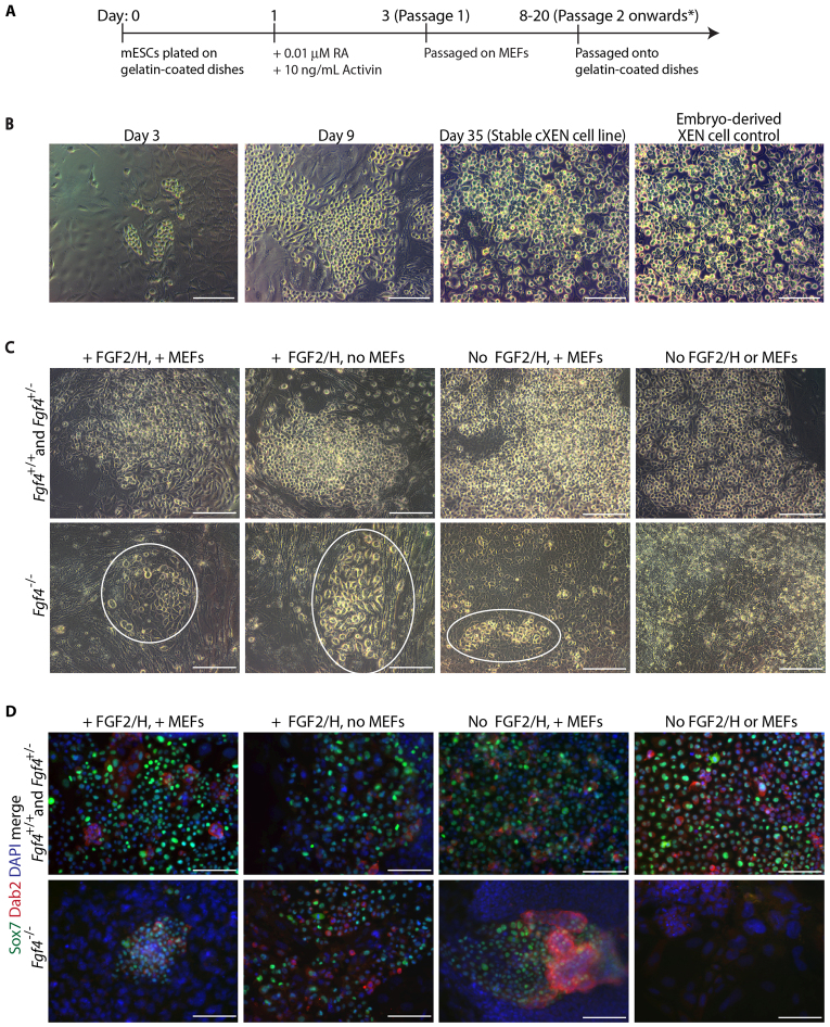
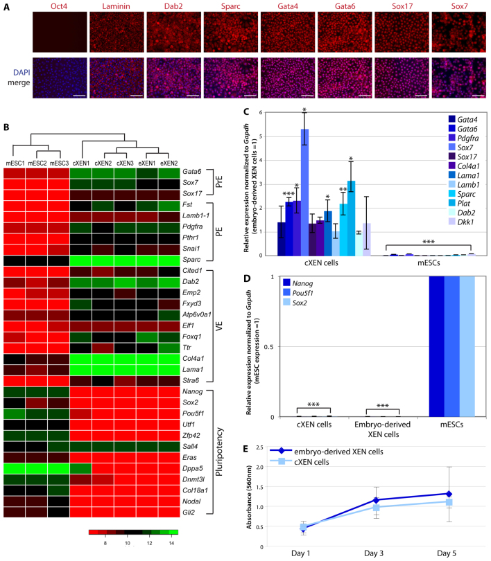
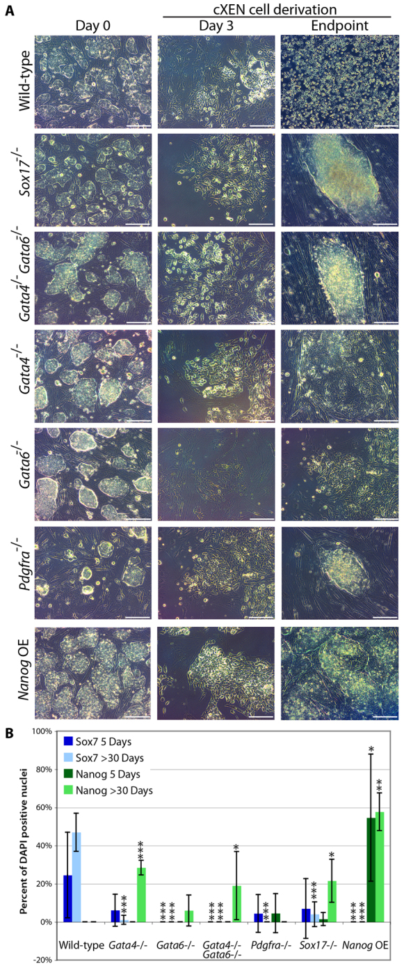
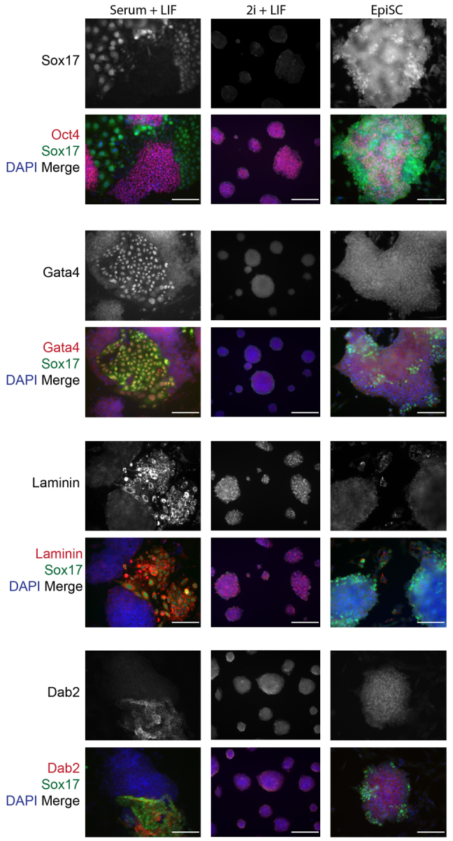
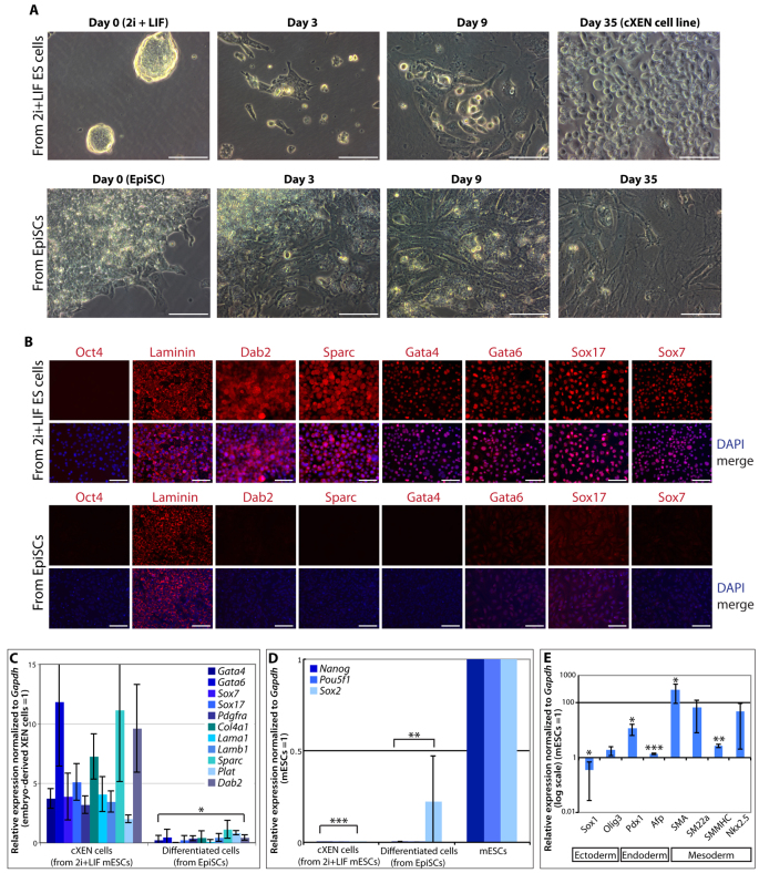
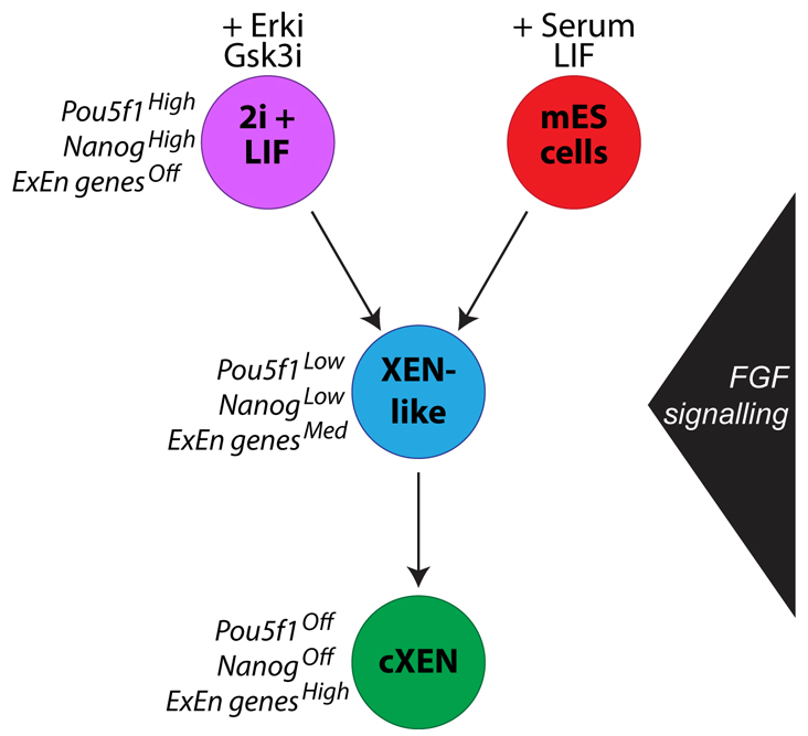
The inner cell mass of the mouse pre-implantation blastocyst comprises epiblast progenitor and primitive endoderm cells of which cognate embryonic (mESCs) or extra-embryonic (XEN) stem cell lines can be derived. Importantly, each stem cell type retains the defining properties and lineage restriction of their in vivo tissue of origin. Recently, we demonstrated that XEN-like cells arise within mESC cultures. This raises the possibility that mESCs can generate self-renewing XEN cells without the requirement for gene manipulation. We have developed a novel approach to convert mESCs to XEN cells (cXEN) using growth factors. We confirm that the downregulation of the pluripotency transcription factor Nanog and the expression of primitive endoderm-associated genes Gata6, Gata4, Sox17 and Pdgfra are necessary for cXEN cell derivation. This approach highlights an important function for Fgf4 in cXEN cell derivation. Paracrine FGF signalling compensates for the loss of endogenous Fgf4, which is necessary to exit mESC self-renewal, but not for XEN cell maintenance. Our cXEN protocol also reveals that distinct pluripotent stem cells respond uniquely to differentiation promoting signals. cXEN cells can be derived from mESCs cultured with Erk and Gsk3 inhibitors (2i), and LIF, similar to conventional mESCs. However, we find that epiblast stem cells (EpiSCs) derived from the post-implantation embryo are refractory to cXEN cell establishment, consistent with the hypothesis that EpiSCs represent a pluripotent state distinct from mESCs. In all, these findings suggest that the potential of mESCs includes the capacity to give rise to both extra-embryonic and embryonic lineages.


Figures

Fig. 1.
Fig. 1.
A growth factor-based method to convert mESCs into stable cXEN cell lines. (A) cXEN derivation protocol in standard XEN media. Cells were passaged onto MEFs at passage 1 and maintained on gelatin thereafter. *cXEN cells were passaged when they reached 70-80% confluency. (B) Representative phase-contrast images of wild-type cells at the defined time points during cXEN derivation (day 3 prior to the first passage, day 8 after the second passage and day 35 stable cXEN cell lines) compared with embryo derived XEN cell control. Scale bars: 100 μm. (C) Representative phase-contrast images of Fgf4+/+, Fgf4+/– and Fgf4–/– mESCs at day 13 following initiation of the cXEN protocol in the conditions indicated. XEN-like cells are circled in the Fgf4–/– conditions. Scale bars: 100 μm. (D) Immunofluorescence analysis for Sox7 (green) and Dab2 (red) expression with DAPI merge (blue) of Fgf4+/+, Fgf4+/– and Fgf4–/– in cXEN cell conditions at day 18. Scale bars: 100 μm.
Fig. 2.
Fig. 2.
cXEN cells are identical to embryo-derived XEN cells. (A) Immunofluorescence analysis of stable cXEN cells (day 35) for Oct4, laminin, Dab2, Sparc, Gata4, Gata6, Sox17 and Sox7 expression (red) and DAPI (blue) merge (representative of three cXEN cell biological replicates). Scale bars: 100 μm. (B) Heat map of microarray data for pluripotency, primitive endoderm (PrE), visceral endoderm (VE), parietal endoderm (PE) or additional XEN cell-associated genes. Normalized gene expression values are represented by a colour heat map spectrum from high expression (green) to low expression (red). Data shown are biological replicates of cXEN cells, embryo-derived XEN cells (eXEN) and mESCs. (C,D) qRT-PCR analysis of cXEN cells (day 35) and mESCs for the expression of ExEn lineage-associated genes (C) (Gata4, Gata6, Pdgfra, Sox7, Sox17, Col4a1, Lama1, Lamb1, Sparc, Plat, Dab2 and Dkk1) or pluripotency associated genes (D) (Nanog, Pou5f1 and Sox2). Relative expression reflected as a fold difference compared with embryo-derived XEN cells or mESCs (=1), respectively. Data are mean±s.e.m. Three biological replicates. *P<0.05; **P<0.01; ***P<0.001. (E) MTT assay of the rate of proliferation of cXEN cells (day 35) compared with embryo-derived XEN cells. Cells were plated at the same cell density and analyzed after 1, 3 or 5 days of culture in standard XEN media. Data are the mean±s.e.m. Three biological and three technical replicates.
Fig. 3.
Fig. 3.
Gata6, Gata4, Sox17 and Pdgfra are required for cXEN establishment. (A) mESCs mutant for Sox17, Gata4, Gata6, Pdgfra or Gata4 and Gata6 double-mutant cells were subjected to the cXEN protocol along with wild-type or Nanog overexpressing (OE) mESCs. Representative phase-contrast images were taken at defined time points during the course of derivation [day 0 in pluripotency conditions, day 3 prior to the first passage and the endpoint: after prolonged culture (day 30)]. Scale bars: 100 μm. (B) Quantification of Sox7 and Nanog expression following immunofluorescence analysis at 5 days and 30 days after cXEN derivation. Data are mean±s.e.m. of five separate counts. *P<0.05; **P<0.01; ***P<0.001.
Fig. 4.
Fig. 4.
XEN-like cells are present in mESC cultures in serum and LIF. Immunofluorescence analysis of Sox17 expression: in mESCs maintained in serum and LIF; in mESC maintained in Erk and Gsk3 inhibitors, and LIF (2i+LIF); and in EpiSC maintained in the presence of Fgf2 and activin. Sox17-high-expressing cells were analyzed for colocalization with Oct4, Gata4, laminin or Dab2 (red) expression along with DAPI (blue) merge. Scale bars: 100 μm.
Fig. 5.
Fig. 5.
mESCs from 2i and LIF conditions can give rise to cXEN cells, unlike EpiSCs. (A) Representative phase-contrast images of mESCs from 2i and LIF conditions or EpiSCs at defined time-points during the cXEN protocol (day 0 in pluripotency conditions, day 3 prior to the first passage, day 9 after the second passage and day 35) (representative image of three biological replicates of stem cell lines from 2i and LIF conditions or two biological and six technical replicates of EpiSCs). Scale bars: 50 μm. (B) Immunofluorescence analysis of cXEN cells from 2i and LIF or differentiated cells from EpiSCs at day 35 of the protocol. Cells were immunostained and analyzed for the expression of Oct4, laminin, Dab2, Sparc, Gata4, Gata6, Sox17 and Sox7 expression (red), and DAPI (blue) merge. Scale bars: 100 μm. (C,D) qRT-PCR analysis of cXEN cells from 2i and LIF conditions or differentiated cells from EpiSCs for the expression of XEN-associated genes (C) or pluripotency-associated genes (D). Relative expression reflected as a fold difference compared with embryo-derived XEN or mESCs (=1), respectively. Data are the mean±s.e.m. Three biological replicates (from 2i and LIF) or two biological and three technical replicates (from EpiSCs). (E) qRT-PCR analysis of differentiated cells from EpiSCs. Relative expression reflected as a fold difference (log scale) compared with mESCs (=1) for germ cell lineage-associated genes: Sox1 and Olig3 (ectoderm); Pdx1 and Afp (endoderm); Sma, Sm22a, Smmhc and Nkx2.5 (mesoderm). Data are the mean±s.e.m. Two biological and three technical replicates. *P<0.05; **P<0.01; ***P<0.001.
Fig. 6.
Fig. 6.
A schematic model of cXEN cell derivation from mESCs. mESCs maintained in LIF and either 2i (purple) or serum (red) can give rise to cXEN cells. Our data suggest that, during cXEN derivation, cells transit through a XEN-like cell state (blue), whereby pluripotency genes are downregulated and extra-embryonic endoderm (ExEn) genes are upregulated. FGF signalling is required to exit mESC self-renewal but not for cXEN maintenance (green).

Similar articles

Cited by

References

    1. Arman E., Haffner-Krausz R., Chen Y., Heath J. K., Lonai P. (1998). Targeted disruption of fibroblast growth factor (FGF) receptor 2 suggests a role for FGF signaling in pregastrulation mammalian development. Proc. Natl. Acad. Sci. USA 95, 5082–5087 - PMC - PubMed
    1. Artus J., Panthier J. J., Hadjantonakis A. K. (2010). A role for PDGF signaling in expansion of the extra-embryonic endoderm lineage of the mouse blastocyst. Development 137, 3361–3372 - PMC - PubMed
    1. Artus J., Piliszek A., Hadjantonakis A. K. (2011). The primitive endoderm lineage of the mouse blastocyst: sequential transcription factor activation and regulation of differentiation by Sox17. Dev. Biol. 350, 393–404 - PMC - PubMed
    1. Artus J., Douvaras P., Piliszek A., Isern J., Baron M. H., Hadjantonakis A. K. (2012). BMP4 signaling directs primitive endoderm-derived XEN cells to an extraembryonic visceral endoderm identity. Dev. Biol. 361, 245–262 - PMC - PubMed
    1. Avilion A. A., Nicolis S. K., Pevny L. H., Perez L., Vivian N., Lovell-Badge R. (2003). Multipotent cell lineages in early mouse development depend on SOX2 function. Genes Dev. 17, 126–140 - PMC - PubMed

Publication types

MeSH terms