. 2012 May;1823(5):997-1006.
doi: 10.1016/j.bbamcr.2012.02.008.

Activation of Stat3 in endothelial cells following hypoxia-reoxygenation is mediated by Rac1 and protein Kinase C

Affiliations

Activation of Stat3 in endothelial cells following hypoxia-reoxygenation is mediated by Rac1 and protein Kinase C

Subhendra N Mattagajasingh et al. Biochim Biophys Acta. 2012 May.

Abstract

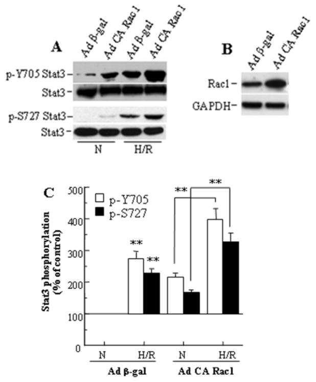
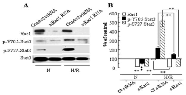
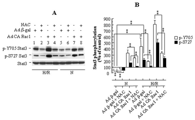
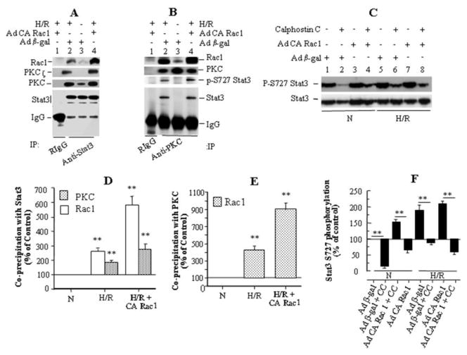
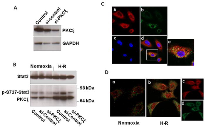
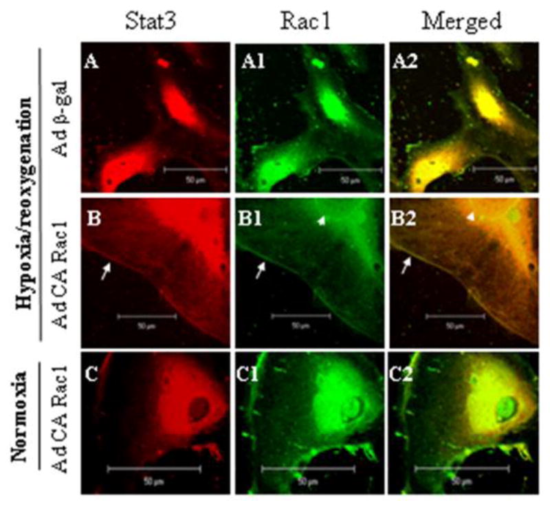
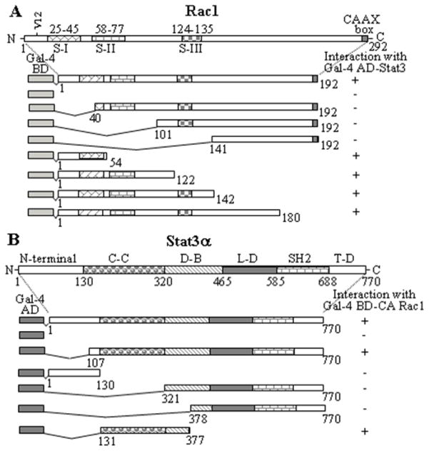
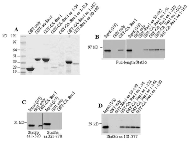
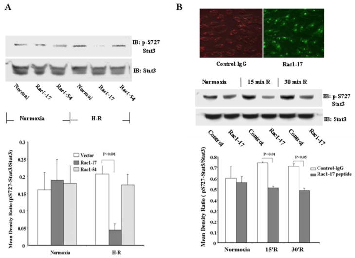
Stat3 is an important transcription factor that regulates both proinflammatory and anit-apoptotic pathways in the heart. This study examined the mechanisms of activation of Stat3 in human endothelial cells following hypoxia/reoxygenation (H/R). By expression of constitutively active Rac1 mutant protein, and by RNA silencing of Rac1, we found that Stat3 forms a multiprotein complex with Rac1 and PKC in an H/R-dependent manner, which at least in part, appears to regulate Stat3 S727 phosphorylation. Selective inhibition of PKC with calphostin C produces a marked suppression of Stat3 S727 phosphorylation. The association of Stat3 with Rax1 occurs predominantly at the cell membrane, but also inside the nucleus, and occurs through the binding of the coiled-coil domain of Stat3 to the 54 NH(2)-terminal residues of Rac1. Transfection with a peptide comprising the NH(2)-terminal 17 amino acid residues of Rac1-dependent signaling pathways resulting in physical association between Rac1 and Stat3 and the formation of a novel multiprotein complex with PKC.

Keywords: Stat3; endothelial cells; inflammation; protein kinase C; protein-protein interaction; reactive oxygen species.


Figures

Figure 1
Figure 1
Stat3 phosphorylation following hypoxia-reoxygenation is increased by CA Rac1 in HUVECs. (A) CA Rac1 increases Y705 and S727 Stat3 phosphorylation, both in normoxia (N) and after H/R. (B) Expression of CA Rac1 after 48 h infection. (C) Densitometry of Western blots in (A). Values are mean ± SEM, expressed relative to the blot for Ad β-gal under normoxic conditions and averaged for 3 independent experiments. ** p<0.01 vs N for Ad β-gal or vs N for Ad CA Rac1 as indicated.
Figure 2
Figure 2
(A) Stat3 phosphorylation following hypoxia-reoxygenation is inhibited by siRNA for Rac1. (B) Densitometry of 3 blots, with data normalized for Stat3 (loading control). Values represent mean ± SEM. ** p<0.01 for indicated comparison or comparison to Control siRNA during N.
Figure 3
Figure 3
(A) N-acetyl cysteine (NAC) reduces phosphorylation of both Y705 and S727 Stat3 in HUVECs, both in normoxia (N), and following H/R. (B) Densitometry of 3 blots, with data normalized for Stat3 (loading control). Values represent mean ± SEM. ** p<0.01 for indicated comparison.
Figure 4
Figure 4
Stat3 activation following H/R is regulated by association of Stat3 with Rac1 and PKC. (A) In COS-7 cells stably expressing Stat3, the amount of Rac1 coprecipitating with Stat3 increased during H/R (lane 3 vs 2) and following infection with Ad CA Rac1 (lane 4), and the immunoprecipitate also contained PKC and its ζ isoform. (B) In COS-7/Stat3 stable cells, the amounts of Stat3, p-S727 Stat3, and Rac1 co-precipitating with PKC increased during H/R and following infection with Ad CA Rac1. (C) In HUVECs, pretreatment with the specific PKC inhibitor Calphostin C (1 μM) reduced S727 Stat3 phosphorylation during normoxia and H/R, with or without Ad CA Rac1 infection. (D), (E), (F) Densitometry of (A), (B) and (C), respectively, on 3 blots for each panel. Normalization was for the amount of Stat3 in (D), PKC in (E), and for Stat3 (loading control) in (F). Values are mean ± SEM. ** p<0.01 for indicated comparison, or comparison to N.
Figure 5
Figure 5
(A,B) Knockdown of PKCζ with siRNA reduces S727 Stat3 phosphorylation in HUVECs during normoxia and following hypoxia-reoxygenation. (A) HUVECs were transfected with control siRNA (si-control) or PKCζ siRNA (si-PKCζ), or with no siRNA (Control), lysed, and immunoblotted. (B) 48 h after transfection HUVECs were exposed to hypoxia for 2h and reoxygenation for 30 min, lysed, and immunoblotted. The experiment was done twice with similar results. (C,D) Following hypoxia-reoxygenation, PKCζ colocalizes with Stat3 in the cytoplasm, but after phosphorylation of S727, Stat3 is found in the nucleus without PKCζ. (C) HUVECs were exposed to hypoxia for 2 h and reoxygenation for 15 min. Cells were fixed and incubated with rabbit anti-Stat3 and goat anti-PKCζ antibodies with secondary anti-rabbit (red) and anti-goat (green) antibodies, and examined by confocal microscopy (63X). a) Stat3, b) PKCζ, c) nuclei (blue), d) merged image, e) enlargement of cell in box to demonstrate cytoplasmic colocalization of Stat3 and PKCζ. (D) HUVECs under normoxia or exposed to hypoxia for 2h and reoxygenation for 60 min were fixed and incubated with rabbit anti-p-S727 Stat3 and goat anti-PKCζ antibodies and detected by secondary antibodies (p-S727 Stat3, red; PKCζ, green), and examined by confocal microscopy (63X). H-R increases cytoplasmic and nuclear p-S727 Stat3, but there is no colocalization with PKCζ. b) merged image showing lack of colocalization, c) separated image of p-S727 Stat3, d) separated image of PKCζ. The confocal microscopy experiments in C and D were each performed twice with similar results.
Figure 6
Figure 6
Stat3 and CA Rac1 colocalize following H/R. HUVECs were infected with adenoviruses as in Fig 1, exposed to 2 h hypoxia and 5 min reoxygenation or a corresponding period of normoxia, fixed and processed for dual immunofluorescent labeling with Stat3 (red) and Rac1 (green). Images of 5 nm thick sections were taken by confocal microscopy. Colocalization (yellow color in right panels) is seen predominantly on the cell membrane (arrow) and nuclear membrane (arrow head), and inside the nucleus following H/R and infection with Ad CA Rac1.
Figure 7
Figure 7
The amino acids that sustain Stat3 and Rac1 interaction reside within the coiled-coil domain of Stat3 and the NH2-terminal 54 amino acids of Rac1. (A) CA Rac1 or its segments and Stat3 were coexpressed in S. cerevisiae and assayed for expression of reporter genes. (+) indicates reporter gene expression (interaction between the proteins/peptides) and (−) indicates non-expression (no interaction). S-I, S-II and S–III and the corresponding numbers represent position of molecular switches in the Rac1 protein. (B) Stat3 or its different segments were coexpressed with CA Rac1 in S. cerevisiae and assayed for expression of reporter genes as in (A). C-C = coiled-coil domain, D-B = DNA-binding domain, L-D = linker domain, SH2 = src homology domain 2, T-D = transactivation domain of Stat3.
Figure 8
Figure 8
Stat3 binds to Rac1 in vitro. (A) Purified GST-Rac1 fusion proteins are shown by Coomassie Blue staining. (B) Ten μg of GST alone or GST-fusion proteins of CA Rac1 or their segments were incubated with in vitro translated [35S]methionine-labeled Stat3α(B), or its amino acid segments (C), (D). All Rac1 fragments bound to Stat3 except for Rac1 aa 50–192. Similarly, Stat3 fragments 1-320 and 131–377 bound to GST/CA Rac1, but fragment 321–770 did not.
Figure 9
Figure 9
Transfection of 293 or HUVECs with a Rac1 NH2-terminal peptide blocks Stat3 phosphorylation following H/R. (A) 293 cells were transfected with Rac1-17 (amino acids 1-17) or Rac1-54 (amino acids 23–54). Densitometry of the bands is based on 3 independent observations. H/R increased S727 Stat3 phosphorylation (p = 0.05), and this was inhibited by Rac1-17, but not Rac1-54. (B) HUVECs were transfected directly with Rac1-17 peptide. The upper right panel shows the results of transfection (IgG: red fluorescent dye localized in cytoplasm); FITC-labeled Rac1-17 peptide: green fluorescence localized in nuclei and cytoplasm). The lower panel shows quantitation of 3 experiments. H/R increased S727 Stat3 phosphorylation at 15 min and 30 min (p = 0.014 and 0.035, respectively), and this increase was inhibited by Rac1-17. Mattagajasingh SN et al, “Activation of Stat3 in Endothelial Cells Following Hypoxia-reoxygenation is Mediated by Rac1 and Protein Kinase C”

Similar articles

Cited by

References

    1. Das DK. Redox regulation of cardiomyocyte survival and death. Antioxid Redox Signal. 2001;3:23–37. - PubMed
    1. Stein AB, Tang XL, Guo Y, Xuan YT, Dawn B, Bolli R. Delayed adaptation of the heart to stress: late preconditioning. Stroke. 2004;35(11 Suppl 1):2676–9. - PMC - PubMed
    1. Valen G. Signal transduction through nuclear factor kappa B in ischemia-reperfusion and heart failure. Basic Res Cardiol. 2004;99:1–7. - PubMed
    1. Soond SM, Latchman DS, Stephanou A. STAT signalling in the heart and cardioprotection. Expert Rev Mol Med. 2006;8:1–16. - PubMed
    1. Yang XP, Irani K, Mattagajasingh S, Dipaula A, Khanday F, Ozaki M, Fox-Talbot K, Baldwin WM, 3rd, Becker LC. Signal transducer and activator of transcription 3alpha and specificity protein 1 interact to upregulate intercellular adhesion molecule-1 in ischemic-reperfused myocardium and vascular endothelium. Arterioscler Thromb Vasc Biol. 2005;25:1395–400. - PubMed

Publication types

MeSH terms