

Spectral domain optical coherence tomography study of pearl-like lesions in the anterior chamber
- PMID: 22791970
- PMCID: PMC3392923
- DOI: 10.2147/OPTH.S30531
Spectral domain optical coherence tomography study of pearl-like lesions in the anterior chamber
Abstract
Aim: To study the morphological pattern of pearl-like lesions in the anterior chambers of children before and after management using anterior segment spectral domain optical coherence tomography (SD-OCT).
Patients and methods: This was a prospective, observational cross-sectional study of patients presenting with peculiar pearl-like lesions in the anterior chamber of their eyes. 1 mL of betamethasone sodium phosphate (2 mg/mL) and betamethasone dipropionate (5 mg/mL) was injected subconjunctivally. Follow-ups of all patients were conducted for a period of 6 weeks. Anterior segment imaging was done using SD-OCT and also photo slit lamp before and after management.
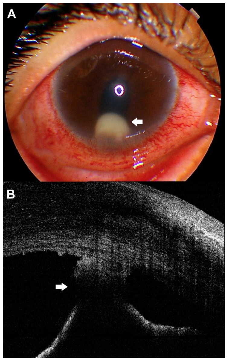
Results: Twelve eyes of 12 patients were included in this study. These patients presented with pearl-like lesions in the anterior chamber with signs of anterior uveitis. There was no history of ocular injury or tuberculosis in any patients. Six weeks after subconjunctival steroid injection, all patients achieved mean best-corrected visual acuity, which changed from 0.2 (range 0.1-0.4) to 0.5 (range 0.4-0.8), and the severity of iritis decreased. SD-OCT showed that the lesions at presentation appeared as a globular noncystic mass attached to the back of the cornea, encroaching on the angle of the eye, and attached to the anterior surface of the iris at some points.
Conclusion: SD-OCT for imaging the anterior segment allowed us to exclude the cystic nature of this pearly lesion. Some similarities may exist between these pearly lesions and superficial phlyctenular keratitis, which may support the immunological and inflammatory origin of these lesions.
Keywords: anterior chamber; anterior segment OCT; pearl-like lesions; phlycten.
Figures


Similar articles
-
The Feasibility of Spectral-Domain Optical Coherence Tomography Grading of Anterior Chamber Inflammation in a Rabbit Model of Anterior Uveitis.Invest Ophthalmol Vis Sci. 2016 Jul 1;57(9):OCT184-8. doi: 10.1167/iovs.15-18883. Invest Ophthalmol Vis Sci. 2016. PMID: 27409471 Free PMC article.
-
Optical Coherence Tomography and ImageJ Software for objective assessment of optical density of anterior chamber iris cyst.Rom J Ophthalmol. 2021 Jul-Sep;65(3):255-258. doi: 10.22336/rjo.2021.50. Rom J Ophthalmol. 2021. PMID: 35036646 Free PMC article.
-
The effect of internal fixation lamp on anterior chamber angle width measured by anterior segment optical coherence tomography.Jpn J Ophthalmol. 2018 Jan;62(1):48-53. doi: 10.1007/s10384-017-0533-x. Epub 2017 Nov 1. Jpn J Ophthalmol. 2018. PMID: 29094326
-
Anterior Segment Optical Coherence Tomography Changes to the Anterior Chamber Angle in the Short-term following Laser Peripheral Iridoplasty.J Curr Glaucoma Pract. 2014 Jan-Apr;8(1):1-6. doi: 10.5005/jp-journals-10008-1152. Epub 2014 Jan 16. J Curr Glaucoma Pract. 2014. PMID: 26997799 Free PMC article. Review.
-
[Optical coherence tomography: from retina imaging to intraoperative use - a review].Klin Monbl Augenheilkd. 2009 Dec;226(12):958-64. doi: 10.1055/s-0028-1109939. Epub 2009 Dec 15. Klin Monbl Augenheilkd. 2009. PMID: 20108189 Review. German.
Cited by
-
Surgical outcomes of complicated cataract with pediatric trematode granulomatous uveitis.Int J Ophthalmol. 2023 Mar 18;16(3):354-360. doi: 10.18240/ijo.2023.03.04. eCollection 2023. Int J Ophthalmol. 2023. PMID: 36935794 Free PMC article.
References
-
- Ahmed AA, Ali OM. Management of health tumour like lesion of the anterior chamber. New Egypt J Med. 1994;10:1613–1616.
-
- Kroosh S, Ahmed A. Histopathology and cytology anterior chamber pearl tumour like lesion. New Egypt J Med. 1994;10:1609–1612.
-
- Gibson GG. Posterior phlyctenular keratitis. Society transactions: College of Physicians of Philadelphia, section on ophthalmology. Arch Ophthal. 1942;29:159–166.
-
- Wojtkowski M, Leitgeb R, Kowalczyk A, Bajraszewski T, Fercher AF. In vivo human retinal imaging by Fourier domain optical coherence tomography. J Biomed Opt. 2002;7:457–463. - PubMed
-
- Wojtkowski M, Bajraszewski T, Targowski P, Kowalczyk A. Real-time in vivo imaging by high-speed spectral optical coherence tomography. Opt Lett. 2003;28:1745–1747. - PubMed
LinkOut - more resources
Full Text Sources
