

Changes in functional properties of A-type but not C-type sensory neurons in vivo in a rat model of peripheral neuropathy
- PMID: 22792004
- PMCID: PMC3392709
- DOI: 10.2147/JPR.S26367
Changes in functional properties of A-type but not C-type sensory neurons in vivo in a rat model of peripheral neuropathy
Abstract
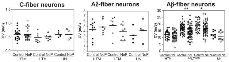
Background: The aim of this study was to compare primary sensory neurons in controls and in an animal neuropathic pain model in order to understand which types of neurons undergo changes associated with peripheral neuropathy. On the basis of intracellular recordings in vivo from somata, L4 sensory dorsal root ganglion neurons were categorized according to action potential configuration, conduction velocity, and receptive field properties to mechanical stimuli.
Methods: Intracellular recordings were made from functionally identified dorsal root ganglion neurons in vivo in the Mosconi and Kruger animal model of peripheral neuropathic pain.
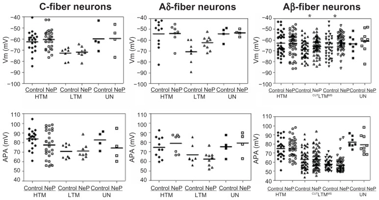
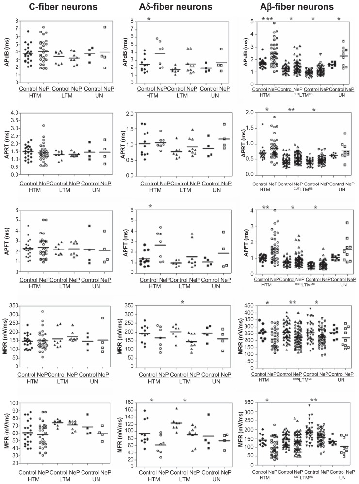
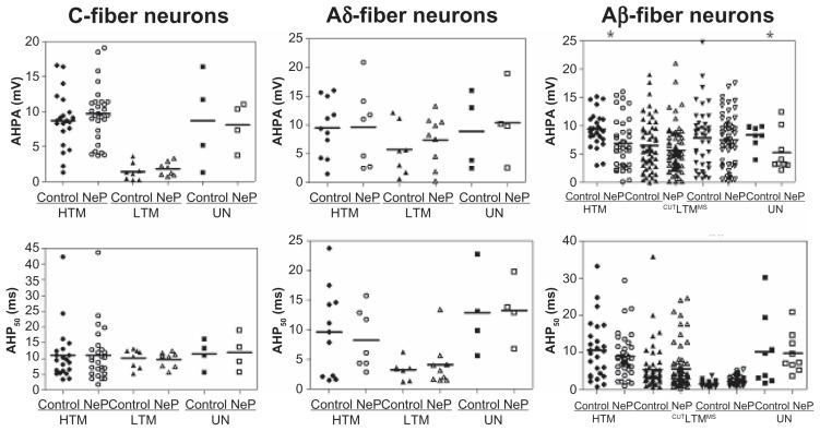
Results: In this peripheral neuropathy model, a specific population of Aβ-fiber low threshold mechanoreceptor neurons, which respond normally to innocuous mechanical stimuli, exhibited differences in action potential configuration and conduction velocity when compared with control animals. No abnormal conduction velocity, action potential shapes, or tactile sensitivity of C-fiber neurons were encountered.
Conclusion: This study provides evidence for defining a potential role of Aβ-fiber low threshold mechanoreceptor neurons that might contribute to peripheral neuropathic pain.
Keywords: action potential configuration; animal model; dorsal root ganglion; in vivo recording; neuropathic pain; peripheral neuropathy; primary sensory neuron.
Figures
Similar articles
-
Cancer pain and neuropathic pain are associated with A β sensory neuronal plasticity in dorsal root ganglia and abnormal sprouting in lumbar spinal cord.Mol Pain. 2018 Jan-Dec;14:1744806918810099. doi: 10.1177/1744806918810099. Epub 2018 Oct 16. Mol Pain. 2018. PMID: 30324862 Free PMC article.
-
Excitability of Aβ sensory neurons is altered in an animal model of peripheral neuropathy.BMC Neurosci. 2012 Jan 30;13:15. doi: 10.1186/1471-2202-13-15. BMC Neurosci. 2012. PMID: 22289651 Free PMC article.
-
L5 spinal nerve axotomy induces sensitization of cutaneous L4 Aβ-nociceptive dorsal root ganglion neurons in the rat in vivo.Neurosci Lett. 2016 Jun 15;624:72-7. doi: 10.1016/j.neulet.2016.05.008. Epub 2016 May 9. Neurosci Lett. 2016. PMID: 27173166
-
[Contribution of primary sensory neurons and spinal glial cells to pathomechanisms of neuropathic pain].Brain Nerve. 2008 May;60(5):483-92. Brain Nerve. 2008. PMID: 18516970 Review. Japanese.
-
Peripheral mechanisms of neuropathic pain - involvement of lysophosphatidic acid receptor-mediated demyelination.Mol Pain. 2008 Apr 1;4:11. doi: 10.1186/1744-8069-4-11. Mol Pain. 2008. PMID: 18377664 Free PMC article. Review.
Cited by
-
Cancer pain and neuropathic pain are associated with A β sensory neuronal plasticity in dorsal root ganglia and abnormal sprouting in lumbar spinal cord.Mol Pain. 2018 Jan-Dec;14:1744806918810099. doi: 10.1177/1744806918810099. Epub 2018 Oct 16. Mol Pain. 2018. PMID: 30324862 Free PMC article.
-
Rat model of cancer-induced bone pain: changes in nonnociceptive sensory neurons in vivo.Pain Rep. 2017 Jun 22;2(4):e603. doi: 10.1097/PR9.0000000000000603. eCollection 2017 Jul. Pain Rep. 2017. PMID: 29392218 Free PMC article.
-
Revisiting PNS Plasticity: How Uninjured Sensory Afferents Promote Neuropathic Pain.Front Cell Neurosci. 2020 Dec 10;14:612982. doi: 10.3389/fncel.2020.612982. eCollection 2020. Front Cell Neurosci. 2020. PMID: 33362476 Free PMC article. Review.
-
Functional up-regulation of Nav1.8 sodium channel in Aβ afferent fibers subjected to chronic peripheral inflammation.J Neuroinflammation. 2014 Mar 7;11:45. doi: 10.1186/1742-2094-11-45. J Neuroinflammation. 2014. PMID: 24606981 Free PMC article.
-
An evaluation of the anti-hyperalgesic effects of cannabidiolic acid-methyl ester in a preclinical model of peripheral neuropathic pain.Br J Pharmacol. 2020 Jun;177(12):2712-2725. doi: 10.1111/bph.14997. Epub 2020 Mar 8. Br J Pharmacol. 2020. PMID: 31981216 Free PMC article.
References
-
- Finnerup NB, Sindrup SH, Jensen TS. Chronic neuropathic pain: mechanisms, drug targets and measurement. Fundam Clin Pharmacol. 2007;21(2):129–136. - PubMed
-
- Hawksley H. Managing pain after shingles: a nursing perspective. Br J Nurs. 2006;15(15):814–818. - PubMed
-
- Ro LS, Chang KH. Neuropathic pain: mechanisms and treatments. Chang Gung Med J. 2005;28(9):597–605. - PubMed
-
- Richards RL. Causalgia. A centennial review. Arch Neurol. 1967;16(4):339–350. - PubMed
-
- Devor M. Ectopic discharge in A-beta afferents as a source of neuropathic pain. Exp Brain Res. 2009;196(1):115–128. - PubMed
LinkOut - more resources
Full Text Sources
Other Literature Sources
