. 2012;8(7):e1002747.
doi: 10.1371/journal.ppat.1002747. Epub 2012 Jul 5.

Proteomic profiling of the TRAF3 interactome network reveals a new role for the ER-to-Golgi transport compartments in innate immunity

Affiliations

Proteomic profiling of the TRAF3 interactome network reveals a new role for the ER-to-Golgi transport compartments in innate immunity

Wendy J van Zuylen et al. PLoS Pathog. 2012.

Abstract

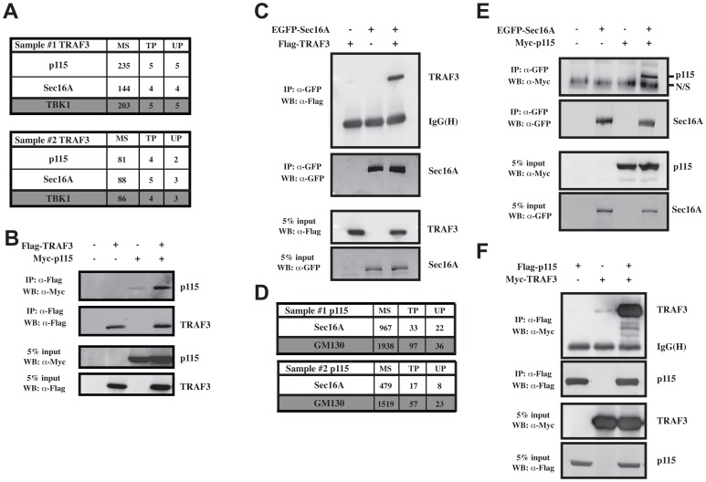
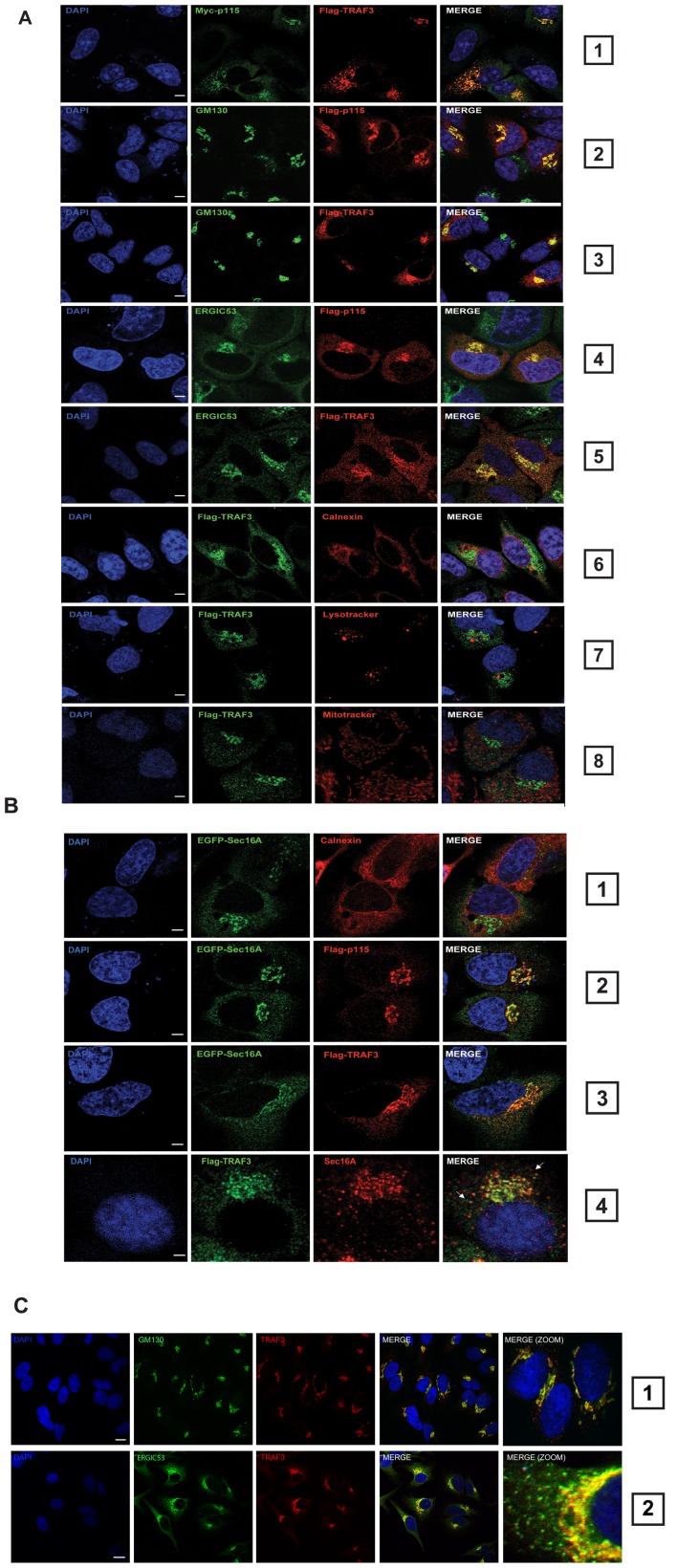
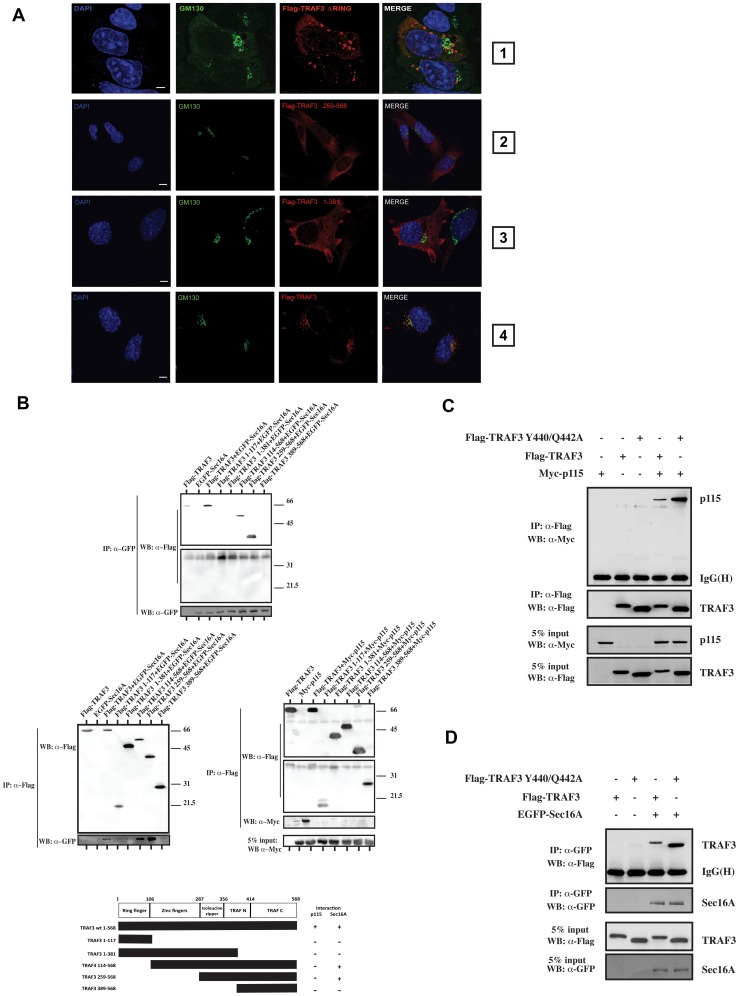
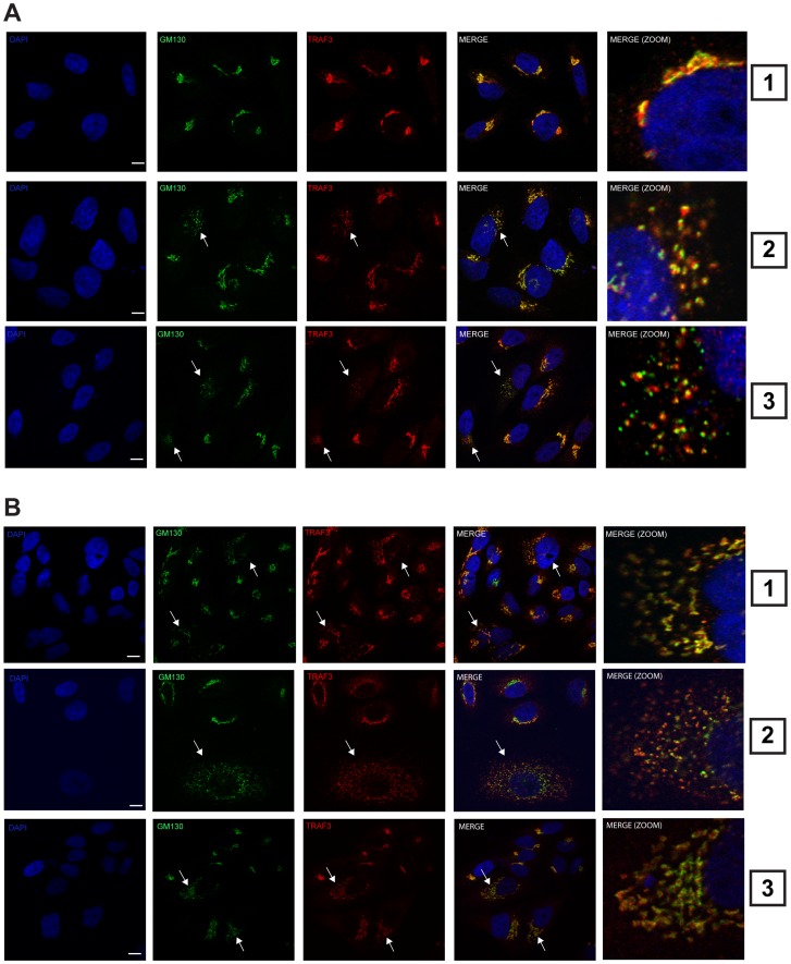
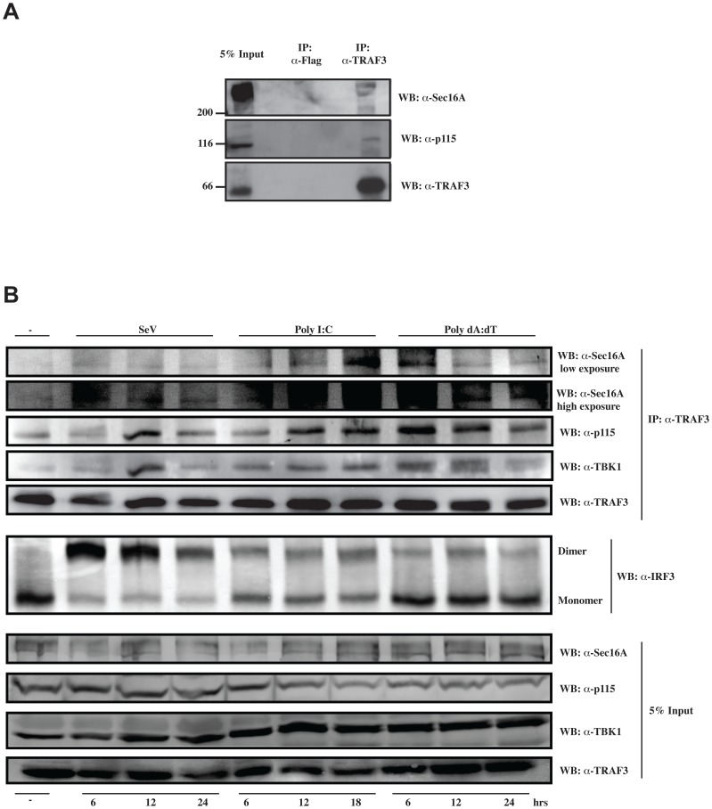
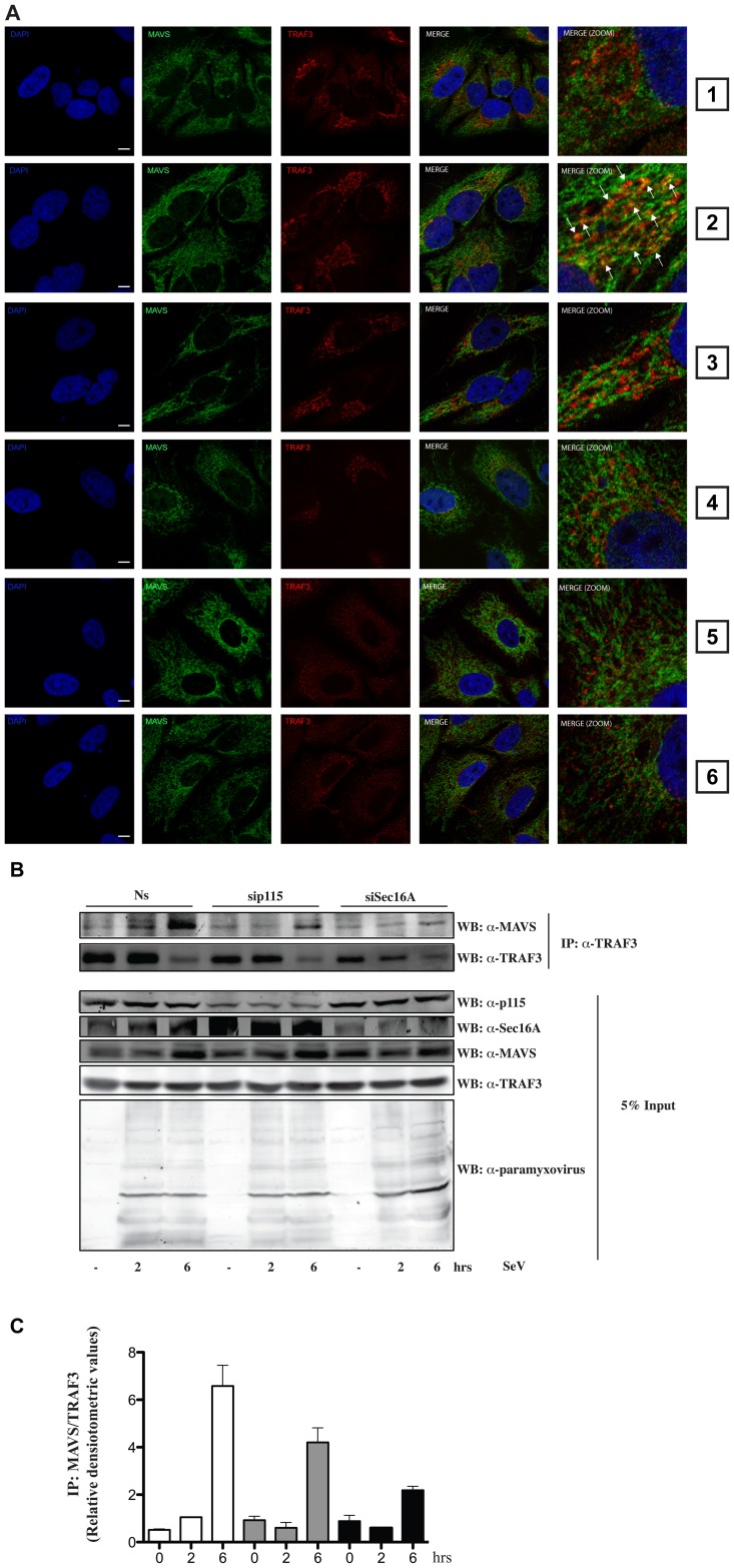
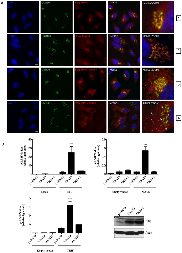
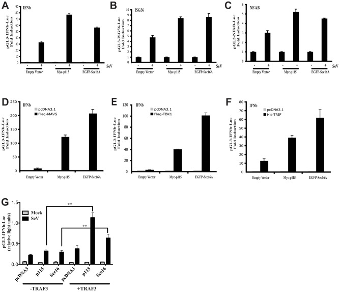
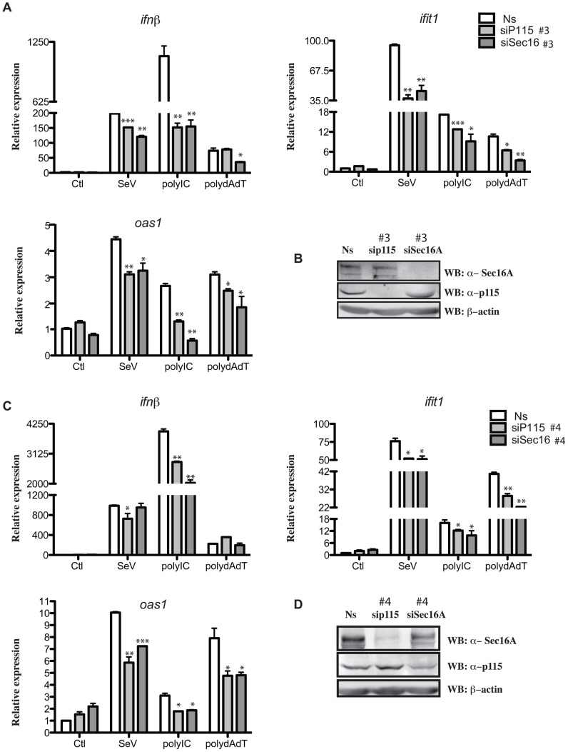
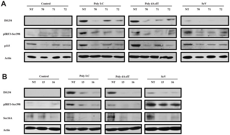
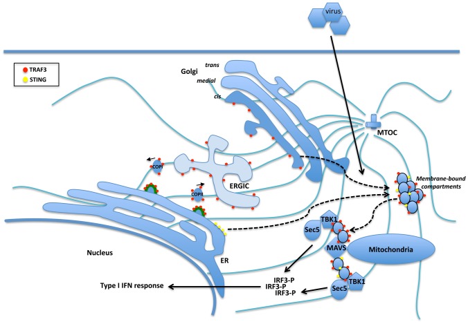
Tumor Necrosis Factor receptor-associated factor-3 (TRAF3) is a central mediator important for inducing type I interferon (IFN) production in response to intracellular double-stranded RNA (dsRNA). Here, we report the identification of Sec16A and p115, two proteins of the ER-to-Golgi vesicular transport system, as novel components of the TRAF3 interactome network. Notably, in non-infected cells, TRAF3 was found associated with markers of the ER-Exit-Sites (ERES), ER-to-Golgi intermediate compartment (ERGIC) and the cis-Golgi apparatus. Upon dsRNA and dsDNA sensing however, the Golgi apparatus fragmented into cytoplasmic punctated structures containing TRAF3 allowing its colocalization and interaction with Mitochondrial AntiViral Signaling (MAVS), the essential mitochondria-bound RIG-I-like Helicase (RLH) adaptor. In contrast, retention of TRAF3 at the ER-to-Golgi vesicular transport system blunted the ability of TRAF3 to interact with MAVS upon viral infection and consequently decreased type I IFN response. Moreover, depletion of Sec16A and p115 led to a drastic disorganization of the Golgi paralleled by the relocalization of TRAF3, which under these conditions was unable to associate with MAVS. Consequently, upon dsRNA and dsDNA sensing, ablation of Sec16A and p115 was found to inhibit IRF3 activation and anti-viral gene expression. Reciprocally, mild overexpression of Sec16A or p115 in Hec1B cells increased the activation of IFNβ, ISG56 and NF-κB -dependent promoters following viral infection and ectopic expression of MAVS and Tank-binding kinase-1 (TBK1). In line with these results, TRAF3 was found enriched in immunocomplexes composed of p115, Sec16A and TBK1 upon infection. Hence, we propose a model where dsDNA and dsRNA sensing induces the formation of membrane-bound compartments originating from the Golgi, which mediate the dynamic association of TRAF3 with MAVS leading to an optimal induction of innate immune responses.


Conflict of interest statement

The authors have declared that no competing interests exist.

Figures

Figure 1
Figure 1. Identification of p115 and Sec16A as new TRAF3 interacting proteins.
(A) HEK293 cells were stably transfected with pcDNA3-FLAG-TRAF3 or pcDNA3-FLAG alone. After G418 selection, the cells were lysed and subjected to AP/MS as described in Methods. The complete list of interactors is shown in Figure S1A. Data for p115 and Sec16A, which were not detected in immunoprecipitates of control cells, are shown here. MS; mascot score, TP; average total number of peptides (spectral counts) identified, UP; number of unique peptides observed; data from two biological replicates are shown. (B) Co-immunoprecipitation experiments showing the association of Myc-p115 to FLAG-TRAF3 when coexpressed in 293T cells. One of three independent experiments with similar results is shown. (C) Co-immunoprecipitation experiments showing the association of FLAG-TRAF3 to EGFP-Sec16A when coexpressed in 293T cells. One of three independent experiments with similar results is shown. (D) AP/MS analysis from cells stably transfected with pcDNA3-FLAG-p115 or pcDNA3-FLAG vector and analyzed as described in A). (E) Co-immunoprecipitation experiments showing the association of Myc-p115 to EGFP-Sec16A and (F) Myc-TRAF3 to FLAG-p115 when coexpressed in 293T cells. One of three independent experiments with similar results is shown.
Figure 2
Figure 2. TRAF3 localizes to the ER-to-Golgi transport compartments and behaves like a cis-Golgi protein.
(A) Confocal microscopy performed in HeLa cells on FLAG-TRAF3 and Myc-p115 (panel 1), FLAG-p115 and GM130 (panel 2), FLAG-TRAF3 and GM130 (panel 3), FLAG-p115 and ERGIC53 (panel 4), FLAG-TRAF3 and ERGIC53 (panel 5), FLAG-TRAF3 and Calnexin (panel 6), FLAG-TRAF3 and lysotracker (panel 7) or mitotracker (panel 8). The nuclei were stained utilizing DAPI. One of three independent experiments with similar results is shown. Bars represent 10 µm. (B) Confocal microscopy performed in HeLa cells on EGFP-Sec16A and calnexin (panel 1), EGFP-Sec16A and FLAG-p115 (panel 2), EGFP-Sec16A and FLAG-TRAF3 (panel 3) and FLAG-TRAF3 and endogenous Sec16A (panel 4). (C) HeLa cells were stained for endogenous TRAF3 and GM130 (panel 1) or endogenous TRAF3 and ERGIC53 (panel 2) before analysis via confocal microscopy. The nuclei were stained utilizing DAPI. Bars represent 5 µm. One of three independent experiments with similar results is shown.
Figure 3
Figure 3. TRAF3 interaction with sec16A or p115 requires its protein integrity.
(A) Confocal microscopy analysis of HeLa cells expressing FLAG-TRAF3 deltaRING (panel 1). Additionally, TRAF3 knockout MEF cells were transfected with FLAG-TRAF3 259–568 (panel 2), FLAG-TRAF3 1–381 (panel 3), or FLAG-TRAF3 WT (panel 4) and stained for GM130, FLAG-tag, and DAPI (nucleus) before analysis via confocal microscopy. Bars represent 5 µm. One of three independent experiments with similar results is shown. (B) Co-immunoprecipitation experiments showing the association of EGFP-Sec16A and Myc-p115 to FLAG-TRAF3 deletion mutants when coexpressed in 293T cells (upper and lower parts from the Western blot are from the same gel). One of three independent experiments with similar results is shown. The bottom panel is a linear representation of TRAF3 deletion mutants and their capacity to interact with p115 or Sec16A. Numbers indicate the position of amino acids in TRAF3. (C–D) Co-immunoprecipitation experiments showing the association of FLAG-TRAF3 WT and Y440/Q442A with Myc-p115 or EGFP-Sec16A immunocomplexes when coexpressed in 293T cells.
Figure 4
Figure 4. Activation of intracellular RNA and DNA sensors leads to the formation of TRAF3-contraining Golgi fragments.
Confocal microscopy of HeLa cells stained for endogenous TRAF3, GM130, and the nucleus (DAPI) upon no treatment (A, panel 1), poly I:C treatment (A, panel 2) or poly dA:dT for 4 h (A, panel 3), infection with SeV (200 HAU/ml) (B, panel 1), RSV (MOI = 3) (B, panel 2), or Influenza A virus for 4 h (B, panel 3). Arrows indicate the relocalization and the colocalization of the Golgi apparatus with TRAF3 upon treatment. Bars represent 5 µm. One of three independent experiments with similar results is shown.
Figure 5
Figure 5. p115 and Sec16A associate with TRAF3 following cytosolic RNA and DNA sensor activation.
(A) Whole-cell lysates (HeLa cells) were prepared and subjected to immunoprecipitation assays using TRAF3 (H-20) or isotype control antibodies followed by immunoblotting for the presence of p115, Sec16A, and TRAF3. (B) HeLa cells were treated as indicated for different periods of time. Whole-cell lysates were prepared and subjected to immunoprecipitation assays using TRAF3 (H-20) antibody followed by immunoblotting for the presence of p115, Sec16A, TBK1 and TRAF3. The Native-PAGE assay was conducted on the same cellular extracts to demonstrate the dimerization and activation of IRF-3 upon indicated treatments. One of three independent experiments with similar results is shown.
Figure 6
Figure 6. Sec16A and p115 are required for the proper positioning of TRAF3 along the mitochondrial network.
(A) Confocal microscopy of HeLa cells transfected with 40 nM nonsilencing RNA duplexes (panels 1 and 2) or 40 nM siRNA duplexes that specifically target Sec16A (panels 3 and 4) or p115 (panels 5 and 6) and stained for MAVS and endogenous TRAF3 upon no treatment (panels 1, 3 and 5) or SeV infection (200 HAU/ml) for 4 h (panels 2, 4 and 6). Arrows indicate the colocalization of TRAF3 with MAVS. Bars represent 5 µm. One of three independent experiments with similar results is shown. (B) p115 and Sec16A were silenced in HeLa cells as described in (A) and infected with SeV for indicated periods of time. Whole-cell lysates were subjected to immunoprecipitation using an anti-TRAF3 (H-20) antibody followed by immunoblotting for the presence of MAVS and TRAF3. Immunoblot analysis against p115, Sec16A, TRAF3 and SeV proteins are also shown (Input). One of two independent experiments with similar results is shown. (C) Densitometric analysis of the binding activity of MAVS to TRAF3 presented in Figures 6B. Data represent the ratio of immunoprecipitated MAVS over immunoprecipitated TRAF3 and are means +/− S.D. of two experiments.
Figure 7
Figure 7. Enforced retention of TRAF3 at the ER-to-Golgi compartment negatively regulates type I IFN response.
(A) Confocal microscopy analysis of FLAG-tag and GM130 performed in HeLa cells expressing FLAG-TRAF3 (panels 1 and 2) or FLAG-TRAF3-AKKFF (panels 3 and 4) upon no infection (panels 1 and 3) or SeV infection (200 HAU/ml) (panels 2 and 4) for 4 h. The nuclei were stained with DAPI. Arrows indicate TRAF3 aggregates. One of two independent experiments with similar results is shown. Bars represent 10 µm. (B) Hec1B cells were co-transfected with luciferase reporter plasmid pGL3-IFNβ (250 ng) and indicated plasmids (250 ng) for 24 h and infected with SeV (200 HAU/ml) for 16 h. Hec1B cells were also co-transfected with luciferase reporter plasmid pGL3-IFNβ (250 ng), empty vector or MAVS or TRIF (25 ng) along with indicated plasmids (250 ng) for 24 h. Relative luciferase activity was measured as described in Materials and Methods. Mean values +/− S.D. of triplicate determinations are shown (*** P<0.001). One of four independent experiments with similar results is shown. Cellular extracts from transfected Hec1B cells were also prepared and subjected to immunoblot analysis using indicated antibodies (right lower panel).
Figure 8
Figure 8. Sec16A and p115 influence the type I IFN antiviral response at the transcriptional level.
(A–C) Hec1B cells were co-transfected with the indicated luciferase reporter genes together with 125 ng of empty vector, Myc-p115 or EGFP-Sec16A. Data are expressed as fold-induction following SeV infection (16 h) over the corresponding non-infected condition. (D–F) Hec1B cells were co-transfected with pGL3-IFNβ-luciferase reporter gene together with 125 ng of empty vector, Myc-p115 or Sec16A and 15 ng of FLAG-MAVS (D), 100 ng of FLAG-TBK1 (E), or 15 ng of His-TRIF (F). Data represent the fold-activation over the corresponding vector control. Each value represents the mean +/− S.D. of triplicate determinations. The data are representative of at least four different experiments with similar results. (G) TRAF3 knockout MEF cells were co-transfected with 250 ng of luciferase reporter plasmid pGL3-IFNβ, 500 ng of pcDNA3 or FLAG-TRAF3 plasmid and 375 ng of indicated plasmids. At 24 h post-transfection, cells were left uninfected or infected with SeV (200 HAU/ml) for 16 h and relative luciferase activity was measured as described in Materials and Methods. Mean values +/− S.D. of triplicate determinations are shown (** P<0.01). One of three independent experiments with similar results is shown.
Figure 9
Figure 9. Requirement of Sec16A and p115 for optimal type I IFN innate immune response in cells exposed to cytosolic DNA and RNA sensor ligands.
(A–D) HeLa cells were transfected with nonsilencing (Ns) RNA duplexes or two different sets of siRNA duplexes that specifically target p115 or Sec16A as indicated. 72 h post-transfection, cells were left untreated (Ctl) or stimulated with poly I:C (2.5 µg/ml), poly dA:dT (1 µg/ml) or SeV (200 HAU/ml) for 6 h to 8 h. RNA was extracted and analyzed by RT-qPCR using primers for ifnβ, ifit1, oas1. Data are means +/− S.D. (n = 3). * Significantly below the induction response; * P<0.05, ** P<0.01, *** P<0.001. (B and D) Cellular extracts were also prepared and subjected to immunoblot analysis using indicated antibodies. One of three independent experiments with similar results is shown.
Figure 10
Figure 10. p115 and Sec16A are required for optimal IRF-3 activation in response to activation of cytosolic RNA and DNA sensors.
HeLa cells were infected with lentiviral vectors encoding shRNA targeting p115 (A) or Sec16A (B) and non-targeting (NT) control shRNA for 24 h followed by puromycin selection (1.5 µg/ml) for 5 days. Cells were left untreated or stimulated with poly I:C (1 µg/ml), poly dA:dT (1 µg/ml) or SeV (200 HAU/ml) for 16 h. Whole-cell lysates were prepared and subjected to immunoblot analysis with indicated antibodies. One of two independent experiments with similar results is shown.
Figure 11
Figure 11. Through its ability to interact and colocalize with components of the ERES (Sec16A, depicted as thick green lines), ERGIC (ERGIC53 and p115) and the cis-Golgi apparatus (p115 and GM130), a subpopulation of TRAF3 (red circles) resides in the ER-to-Golgi vesicular compartment in non-infected cells.
Upon dsRNA and dsDNA sensing, the cis-Golgi disorganizes into punctate structures, giving rise to membrane-bound compartments composed of at least GM130 and TRAF3 (dashed line). We propose that these membrane-bound compartments allow the proper positioning of TRAF3 with MAVS at Mitochondrial-Associated endoplasmic reticulum Membranes (MAM) . There, being in close proximity with a component of the exocyst (sec5) and the translocon (Sec61β), TRAF3 allows the activation of TBK1 and IRF3 leading to activation of the type I IFN response. A similar scenario was recently proposed for STING (yellow circles) where in response to DNA virus infection, it traffics from the ER to the cis-Golgi apparatus and finally to a distinct perinuclear region for the activation of TBK1 , . MTOC: microtubule-organizing center.

References

    1. Barbalat R, Ewald SE, Mouchess ML, Barton GM. Nucleic acid recognition by the innate immune system. Annu Rev Immunol. 2011;29:185–214. - PubMed
    1. Unterholzner L, Keating SE, Baran M, Horan KA, Jensen SB, et al. IFI16 is an innate immune sensor for intracellular DNA. Nat Immunol. 2010;11:997–1004. - PMC - PubMed
    1. Kato H, Sato S, Yoneyama M, Yamamoto M, Uematsu S, et al. Cell type-specific involvement of RIG-I in antiviral response. Immunity. 2005;23:19–28. - PubMed
    1. Yoneyama M, Kikuchi M, Natsukawa T, Shinobu N, Imaizumi T, et al. The RNA helicase RIG-I has an essential function in double-stranded RNA-induced innate antiviral responses. Nat Immunol. 2004;5:730–737. - PubMed
    1. Gitlin L, Barchet W, Gilfillan S, Cella M, Beutler B, et al. Essential role of mda-5 in type I IFN responses to polyriboinosinic:polyribocytidylic acid and encephalomyocarditis picornavirus. Proc Natl Acad Sci U S A. 2006;103:8459–8464. - PMC - PubMed

Publication types

MeSH terms

Substances