

doi: 10.1371/journal.ppat.1002757.
Epub 2012 Jul 5.
Polydnaviruses as symbionts and gene delivery systems
Affiliations
- PMID: 22792063
- PMCID: PMC3390406
- DOI: 10.1371/journal.ppat.1002757
Item in Clipboard
Polydnaviruses as symbionts and gene delivery systems
PLoS Pathog.
2012.
No abstract available
Conflict of interest statement
The authors have declared that no competing interests exist.
Figures
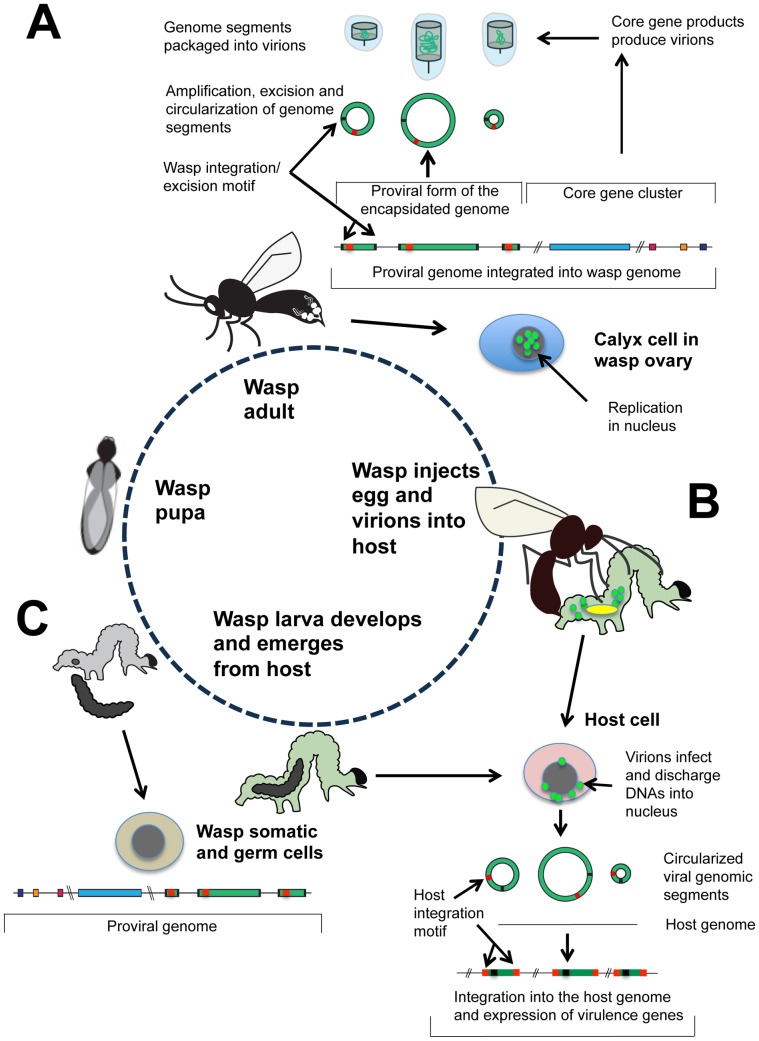
(A) In pupal and adult stage wasps, the proviral genome consists of two components: a domain of core genes (red, yellow, purple) and a domain of tandemly arrayed proviral DNAs (green) that encode virulence genes. The borders of these proviral DNAs are identified by conserved, flanking excision motifs (black). The specific location of BV core genes and proviral DNAs in wasp genomes is currently unknown and is thus indicated by double slash marks. BV replication to produce virions occurs in the nuclei of calyx cells of the female ovary. Coordinate expression of the core genes begins during the wasp pupal stage and continues in the adult stage. This results in the assembly of virions, and the replication, excision, and circularization of proviral DNAs, which are packaged into virions. (B) Wasps inject virions plus one or more eggs containing the proviral genome into the host insect. The egg hatches into a wasp larva that feeds on the host. Virions infect and discharge their DNAs into host cell nuclei, which then rapidly integrate into the genome of the host cell via a second domain present on the viral DNAs named the host integration motif (red). Virulence genes are then transcribed in host cells over the duration required for the wasp larvae to complete their development. The location(s) in the host genome where each viral DNA integrates is currently unknown as indicated by double slash marks. (C) Upon completing development, the wasp larva emerges from the host to pupate, while the host larva dies. Each germ line and somatic cell of the wasp contains the two-component proviral genome.
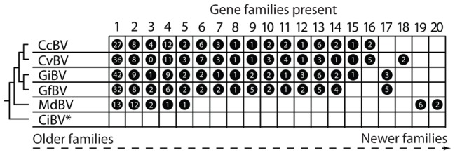
Each BV isolate is indicated to the left in relation to phylogenetic placement of its associated wasp. The isolates are: Cotesia congregata bracovirus (CcBV), C. vestalis bracovirus (CvBV), Glyptapanteles indiensis bracovirus (GiBV), G. flavicoxis bracovirus (GfBV), Microplitis demolitor bracovirus (MdBV), and Chelonus inanitus bracovirus (CiBV). C. inanitus resides in the subfamily Cheloninae while the other wasp species shown are in the subfamily Microgastrinae. The 20 gene families identified from BVs are listed above the figure. Family names derive from predicted function or the presence of a distinguishing motif: 1) protein tyrosine phosphatases; 2) ankyrin-repeat; 3) cysteine-rich; 4) BEN domain; 5) BV4; 6) EP-1 like, homologs of “early expressed protein 1” of CcBV; 7) cystatins; 8) histone-like; 9) C-type lectin; 10) ribonuclease T2; 11) 94K-like, related to baculovirus 94K protein; 12) CrV1-like, homologs of a gene in CrBV; 13) BV2; 14) BV3; 15) Duffy binding-like; 16) BV1; 17) sugar transporter; 18) serine rich; 19) Egf, epidermal growth factor-like; 20) Glc, glycosylated central domain proteins. A circle to the right of an isolate indicates that the gene family is present, while the number inside the circle indicates the number of family members encoded by that isolate. Phylogenetic evidence further suggests families to the left are more ancient, while families to the right have been more recently acquired by particular BV isolates. *The encapsidated genome of CiBV is only partially sequenced. To date, 16 single copy genes are described in the CiBV genome but none of these genes are present in BV isolates outside the genus Chelonus.
References
-
- Luria SE, Darnell JE, Baltimore D, Campbell A. General virology. 3rd edition. New York: John Wiley & Sons; 1978. 578
-
- Cann AJ. Principles of molecular virology. 3rd edition. San Diego: Academic Press; 2001. 337
-
- Strand MR, Drezen J-M. Family polydnaviridae. In: King AMQ, Adams MJ, Carstens EB, Lefkowitz EJ, editors. Virus taxonomy: ninth report of the international committee on taxonomy of viruses. Amsterdam: Elsevier; 2012. pp. 237–248.
-
- Strand MR. Polydnaviruses. In: Asgari S, Johnson KN, editors. Insect virology. Norfolk, UK: Caister Academic Press; 2010. pp. 171–197.
-
- Beckage NE. Polydnaviruses as endocrine regulators. In: Beckage NE, Drezen J-M, editors. Parasitoid viruses symbionts and pathogens. San Diego: Academic Press; 2012. pp. 163–168.
Publication types
MeSH terms
Grants and funding
LinkOut - more resources
Full Text Sources
