The role of rice HEI10 in the formation of meiotic crossovers
- PMID: 22792078
- PMCID: PMC3390396
- DOI: 10.1371/journal.pgen.1002809
The role of rice HEI10 in the formation of meiotic crossovers
Abstract
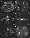
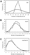
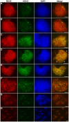
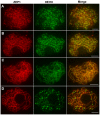
HEI10 was first described in human as a RING domain-containing protein that regulates cell cycle and cell invasion. Mice HEI10(mei4) mutant displays no obvious defect other than meiotic failure from an absence of chiasmata. In this study, we characterize rice HEI10 by map-based cloning and explore its function during meiotic recombination. In the rice hei10 mutant, chiasma frequency is markedly reduced, and those remaining chiasmata exhibit a random distribution among cells, suggesting possible involvement of HEI10 in the formation of interference-sensitive crossovers (COs). However, mutation of HEI10 does not affect early recombination events and synaptonemal complex (SC) formation. HEI10 protein displays a highly dynamic localization on the meiotic chromosomes. It initially appears as distinct foci and co-localizes with MER3. Thereafter, HEI10 signals elongate along the chromosomes and finally restrict to prominent foci that specially localize to chiasma sites. The linear HEI10 signals always localize on ZEP1 signals, indicating that HEI10 extends along the chromosome in the wake of synapsis. Together our results suggest that HEI10 is the homolog of budding yeast Zip3 and Caenorhabditis elegans ZHP-3, and may specifically promote class I CO formation through modification of various meiotic components.
Conflict of interest statement
The authors have declared that no competing interests exist.
Figures



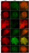
 is the ν value (estimated SE) for which the best fit of the observed distances to the gamma model was got. Here, the estimated
  is 8.39, which indicates a strong interference among HEI10 bright foci on the shortest chromosome in rice (if there is no interference, ν is 1).

Similar articles
-
The Arabidopsis HEI10 is a new ZMM protein related to Zip3.PLoS Genet. 2012;8(7):e1002799. doi: 10.1371/journal.pgen.1002799. Epub 2012 Jul 26. PLoS Genet. 2012. PMID: 22844245 Free PMC article.
-
The E3 ubiquitin ligase DESYNAPSIS1 regulates synapsis and recombination in rice meiosis.Cell Rep. 2021 Nov 2;37(5):109941. doi: 10.1016/j.celrep.2021.109941. Cell Rep. 2021. PMID: 34731625
-
HEIP1 regulates crossover formation during meiosis in rice.Proc Natl Acad Sci U S A. 2018 Oct 16;115(42):10810-10815. doi: 10.1073/pnas.1807871115. Epub 2018 Oct 1. Proc Natl Acad Sci U S A. 2018. PMID: 30275327 Free PMC article.
-
HEI10 coarsening, chromatin and sequence polymorphism shape the plant meiotic recombination landscape.Curr Opin Plant Biol. 2024 Oct;81:102570. doi: 10.1016/j.pbi.2024.102570. Epub 2024 Jun 4. Curr Opin Plant Biol. 2024. PMID: 38838583 Review.
-
ASY1 coordinates early events in the plant meiotic recombination pathway.Cytogenet Genome Res. 2008;120(3-4):302-12. doi: 10.1159/000121079. Epub 2008 May 23. Cytogenet Genome Res. 2008. PMID: 18504359 Review.
Cited by
-
Joint control of meiotic crossover patterning by the synaptonemal complex and HEI10 dosage.Nat Commun. 2022 Oct 12;13(1):5999. doi: 10.1038/s41467-022-33472-w. Nat Commun. 2022. PMID: 36224180 Free PMC article.
-
Manipulation of crossover frequency and distribution for plant breeding.Theor Appl Genet. 2019 Mar;132(3):575-592. doi: 10.1007/s00122-018-3240-1. Epub 2018 Nov 27. Theor Appl Genet. 2019. PMID: 30483818 Free PMC article. Review.
-
Crossover interference mechanism: New lessons from plants.Front Cell Dev Biol. 2023 May 19;11:1156766. doi: 10.3389/fcell.2023.1156766. eCollection 2023. Front Cell Dev Biol. 2023. PMID: 37274744 Free PMC article. Review.
-
The F-Box Protein ZYGO1 Mediates Bouquet Formation to Promote Homologous Pairing, Synapsis, and Recombination in Rice Meiosis.Plant Cell. 2017 Oct;29(10):2597-2609. doi: 10.1105/tpc.17.00287. Epub 2017 Sep 22. Plant Cell. 2017. PMID: 28939596 Free PMC article.
-
HEIP1 is required for efficient meiotic crossover implementation and is conserved from plants to humans.Proc Natl Acad Sci U S A. 2023 Jun 6;120(23):e2221746120. doi: 10.1073/pnas.2221746120. Epub 2023 May 30. Proc Natl Acad Sci U S A. 2023. PMID: 37252974 Free PMC article.
References
-
- Youds JL, Boulton SJ. The choice in meiosis - defining the factors that influence crossover or non-crossover formation. J Cell Sci. 2011;124:501–513. - PubMed
-
- Borner GV, Kleckner N, Hunter N. Crossover/noncrossover differentiation, synaptonemal complex formation, and regulatory surveillance at the leptotene/zygotene transition of meiosis. Cell. 2004;117:29–45. - PubMed
Publication types
MeSH terms
Substances
Associated data
- Actions
- Actions
- Actions
- Actions
- Actions
- Actions
- Actions
LinkOut - more resources
Full Text Sources
Molecular Biology Databases

