

Melanonychia
- PMID: 22792094
- PMCID: PMC3390039
- DOI: 10.1155/2012/952186
Melanonychia
Abstract
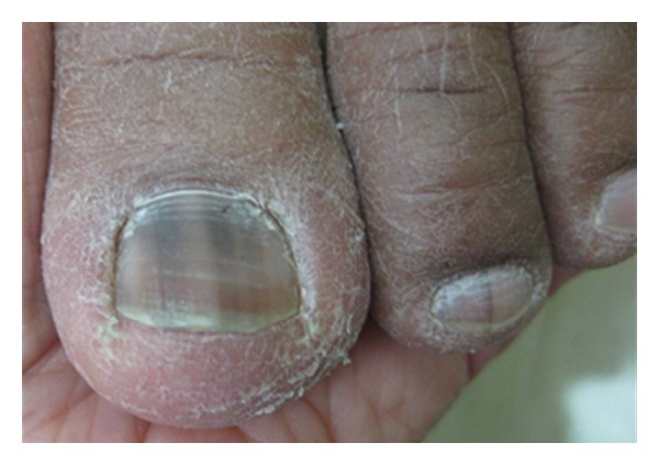
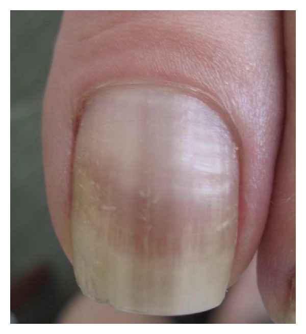
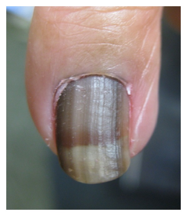
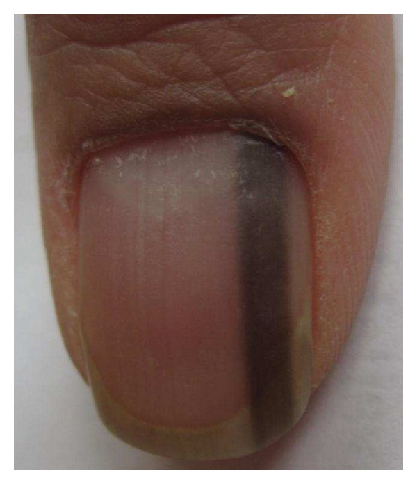
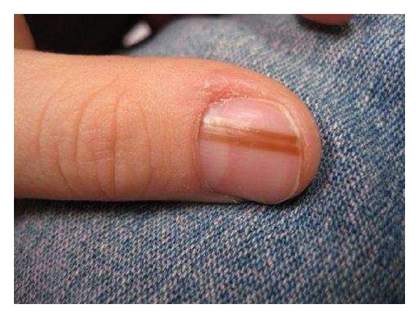
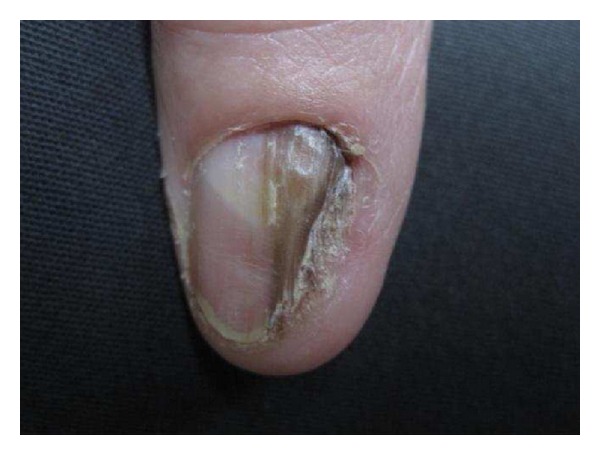
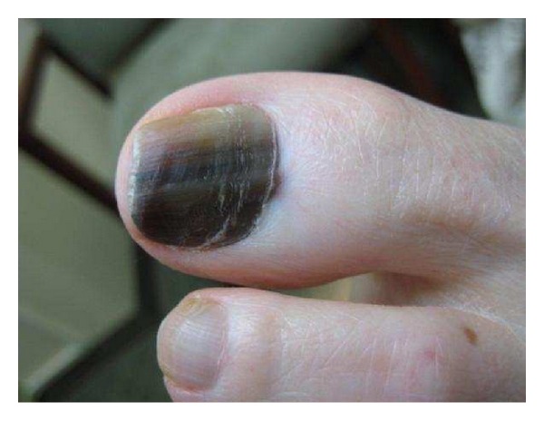
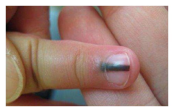
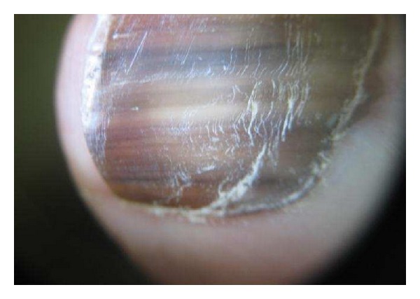
Melanonychia, or melanin-derived brown-to-black nail pigmentation, is a diagnostic challenge for clinicians. The most serious disease of the nail unit, melanoma, primarily presents with melanonychia. However, melanonychia most often occurs as a result of benign etiologies such as nail matrix melanocytic activation, nail matrix melanocytic hyperplasia, and nail invasion by melanin-producing pathogens. Regrettably, patients with nail apparatus melanoma are often initially misdiagnosed, and due to diagnostic delays of an average of 2 years, melanoma of the nail unit carries a poor prognosis. Having a thorough knowledge of the various causes of melanonychia and using a systematic approach when evaluating brown-to-black nail pigmentation may help prevent misdiagnosis and thereby improve prognosis.
Figures
Similar articles
-
Melanonychia striata: clarifying behind the Black Curtain. A review on clinical evaluation and management of the 21st century.Int J Dermatol. 2019 Nov;58(11):1239-1245. doi: 10.1111/ijd.14464. Epub 2019 Apr 21. Int J Dermatol. 2019. PMID: 31006857 Review.
-
Key point in dermoscopic differentiation between early nail apparatus melanoma and benign longitudinal melanonychia.J Dermatol. 2011 Jan;38(1):45-52. doi: 10.1111/j.1346-8138.2010.01175.x. J Dermatol. 2011. PMID: 21175755
-
Melanonychia, melanocytic hyperplasia, and nail melanoma in a Hispanic population.J Am Acad Dermatol. 2008 Nov;59(5):785-91. doi: 10.1016/j.jaad.2008.07.012. Epub 2008 Sep 19. J Am Acad Dermatol. 2008. PMID: 18804895
-
Automated evaluation system of dermoscopic images of longitudinal melanonychia: proposition of a discrimination index for detecting early nail apparatus melanoma.J Dermatol. 2014 Oct;41(10):867-71. doi: 10.1111/1346-8138.12593. Epub 2014 Sep 9. J Dermatol. 2014. PMID: 25200569
-
Melanonychia - Clues for a Correct Diagnosis.Cureus. 2020 Jan 10;12(1):e6621. doi: 10.7759/cureus.6621. Cureus. 2020. PMID: 32064201 Free PMC article. Review.
Cited by
-
Dark fingernails.Malays Fam Physician. 2015 Dec 31;10(3):40-2. eCollection 2015. Malays Fam Physician. 2015. PMID: 27570609 Free PMC article. No abstract available.
-
[Longitudinal melanonychia as a clinical manisfestation of Addison's disease].Aten Primaria. 2019 Mar;51(3):193-194. doi: 10.1016/j.aprim.2018.05.006. Epub 2018 Aug 28. Aten Primaria. 2019. PMID: 30170759 Free PMC article. Spanish. No abstract available.
-
Exposome Impact on Nail Health.Skin Appendage Disord. 2024 Jun;10(3):186-198. doi: 10.1159/000536573. Epub 2024 Mar 11. Skin Appendage Disord. 2024. PMID: 38835707 Free PMC article. Review.
-
Transient neonatal hyperpigmentation of the proximal nail fold in a Chinese infant: a case report.J Int Med Res. 2022 Jan;50(1):3000605211067748. doi: 10.1177/03000605211067748. J Int Med Res. 2022. PMID: 35023378 Free PMC article.
-
POSSIBLE NATURE OF THE RADIATION-INDUCED SIGNAL IN NAILS: HIGH-FIELD EPR, CONFIRMING CHEMICAL SYNTHESIS, AND QUANTUM CHEMICAL CALCULATIONS.Radiat Prot Dosimetry. 2016 Dec;172(1-3):112-120. doi: 10.1093/rpd/ncw216. Epub 2016 Aug 13. Radiat Prot Dosimetry. 2016. PMID: 27522053 Free PMC article.
References
-
- Haneke E, Baran R. Longitudinal melanonychia. Dermatologic Surgery. 2001;27(6):580–584. - PubMed
-
- Andre J, Lateur N. Pigmented nail disorders. Dermatologic Clinics. 2006;24(3):329–339. - PubMed
-
- Jellinek N. Nail matrix biopsy of longitudinal melanonychia: diagnostic algorithm including the matrix shave biopsy. Journal of the American Academy of Dermatology. 2007;56(5):803–810. - PubMed
-
- Rich P. Nail surgery. In: Bolognia JL, Jorizzo JL, Rapini RP, editors. Dermatology. 2nd edition. chapter 149. New York, NY, USA: Mosby; 2006. pp. 2260–2268.
-
- Klausner JM, Inbar M, Gutman M, et al. Nail-bed melanoma. Journal of Surgical Oncology. 1987;34(3):208–210. - PubMed
LinkOut - more resources
Full Text Sources
