. 2012:2012:971963.
doi: 10.1155/2012/971963. Epub 2012 Jun 25.

Regional blockade of the shoulder: approaches and outcomes

Affiliations

Regional blockade of the shoulder: approaches and outcomes

Clifford Bowens Jr et al. Anesthesiol Res Pract. 2012.

Abstract

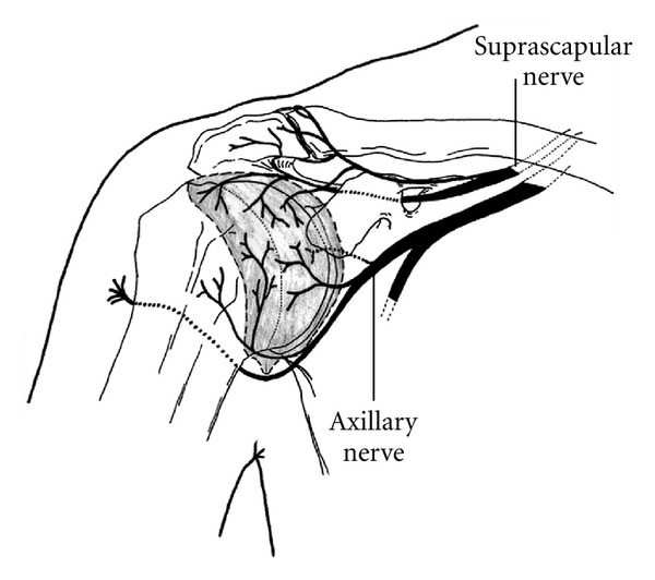
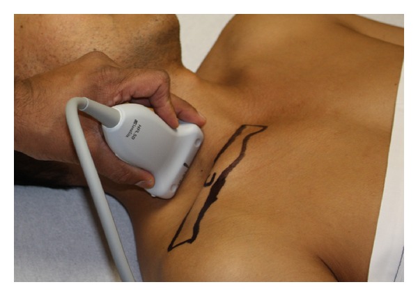
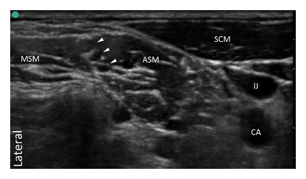
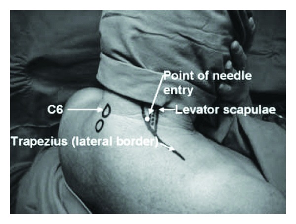
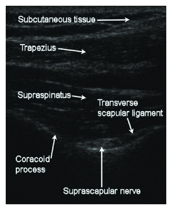
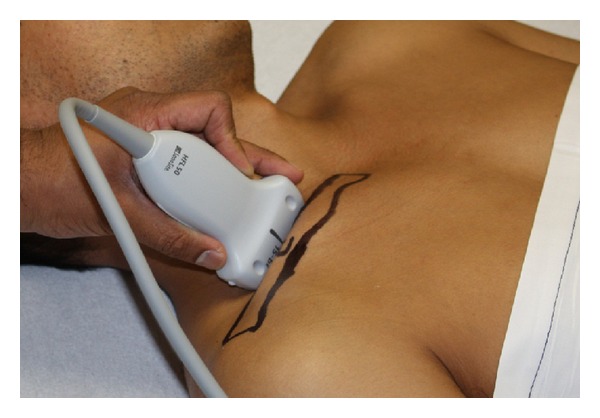
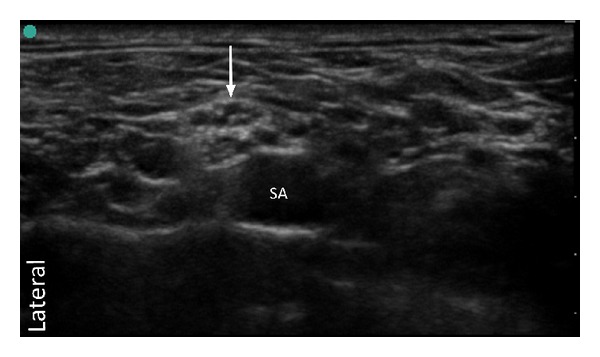
The article reviews the current literature regarding shoulder anesthesia and analgesia. Techniques and outcomes are presented that summarize our present understanding of regional anesthesia for the shoulder. Shoulder procedures producing mild to moderate pain may be managed with a single-injection interscalene block. However, studies support that moderate to severe pain, lasting for several days is best managed with a continuous interscalene block. This may cause increased extremity numbness, but will provide greater analgesia, reduce supplemental opioid consumption, improve sleep quality and patient satisfaction. In comparison to the nerve stimulation technique, ultrasound can reduce the volume of local anesthetic needed to produce an effective interscalene block. However, it has not been shown that ultrasound offers a definitive benefit in preventing major complications. The evidence indicates that the suprascapular and/or axillary nerve blocks are not as effective as an interscalene block. However in patients who are not candidates for the interscalene block, these blocks may provide a useful alternative for short-term pain relief. There is substantial evidence showing that subacromial and intra-articular injections provide little clinical benefit for postoperative analgesia. Given that these injections may be associated with irreversible chondrotoxicity, the injections are not presently recommended.


Figures

Figure 1
Figure 1
Anterior innervation of the shoulder joint. The suprascapular nerve and axillary nerve are the primary nerves supplying the capsule and the glenohumeral joint (Borgeat and Ekatodramis [1]). (Reprinted with permission from Elsevier.)
Figure 2
Figure 2
Posterior innervation of the shoulder joint. The suprascapular nerve and axillary nerve are the primary nerves supplying this region (Borgeat and Ekatodramis [1]). (Reprinted with permission from Elsevier.)
Figure 3
Figure 3
Ultrasound probe position for the interscalene brachial plexus block.
Figure 4
Figure 4
Transverse ultrasound image of interscalene brachial plexus. Arrowheads outline brachial plexus roots within the interscalene groove. SCM: sternocleidomastoid muscle, IJ: internal jugular vein, CA: carotid artery, ASM: anterior scalene muscle, MSM: middle scalene muscle.
Figure 5
Figure 5
Cervical paravertebral block. Needle entry is at the level of C6, anterolateral to the trapezius muscle and posteromedial to the levator scapulae muscle (Boezaart [2]). (Reprinted with permission from Elsevier.)
Figure 6
Figure 6
Meier technique for the suprascapular nerve block. Needle insertion is 2 cm cephalad and 2 cm medial to the midpoint of a line connecting the lateral acromion and medial border of the spine of the scapula. The needle is angled 45° in the coronal plane, with 30° of ventral inclination (Price [3]). (Reprinted with permission.)
Figure 7
Figure 7
Anatomic representation of the superior view of the Meier technique. Note the needle is in the supraspinous fossa, demonstrating the 30° of ventral inclination. The suprascapular nerve enters the groove at the suprascapular notch (SSN) and winds laterally around the greater scapular notch (GSN) (Price [3]). (Reprinted with permission.)
Figure 8
Figure 8
Price technique for the axillary nerve block. A line connects the anterior acromion (1) with the inferior angle of the scapula (2). The midpoint of the line represents the level of the horizontal axis (H) of the quadrilateral space. The line representing the vertical axis (V) is drawn down from the posterolateral aspect of the acromion (3) (Price [3]). (Reprinted with permission.)
Figure 9
Figure 9
Anatomic representation of the Price technique. The horizontal axis (H) lies at the level of the quadrilateral space, where the axillary nerve passes beneath the glenohumeral joint capsule. The interception of the vertical axis (V) with the horizontal axis (H) allows location of the axillary nerve as it crossing the posterior neck of the humerus (Price [3]). (Reprinted with permission.)
Figure 10
Figure 10
Checcucci technique for the suprascapular nerve block. Needle insertion is 2 cm medial to the medial border of the acromion and 2 cm cephalad to the superior margin of the scapular spine (Checcucci et al. [4]). (Reprinted with permission from Elsevier.)
Figure 11
Figure 11
Checcucci technique for the axillary nerve block. A line is drawn between the posterolateral angle of the acromion and the olecranon tip of the elbow. The needle insertion is 2 cm cephalad to the convergence of this line with the perpendicular line originating from the axillary fold (Checcucci et al. [4]). (Reprinted with permission from Elsevier.)
Figure 12
Figure 12
Matsumoto technique for the suprascapular nerve block. Needle insertion point is the midpoint of a line connecting the anterolateral edge of the acromion and the superomedial angle of the scapula. The needle is advanced, at an angle 30° dorsal to the coronal plane, to make contact with the base of coracoid process (Matsumoto et al. [5]). (Reprinted with permission from Elsevier.)
Figure 13
Figure 13
Anatomic representation of the Matsumoto technique (Matsumoto et al. [5]). (Reprinted with permission from Elsevier.)
Figure 14
Figure 14
Ultrasound probe and needle position for suprascapular nerve block (Harmon and Hearty [6]). (Reprinted with permission.)
Figure 15
Figure 15
Transverse view of suprascapular fossa and suprascapular notch (Harmon and Hearty [6]). (Reprinted with permission.)
Figure 16
Figure 16
Ultrasound probe position for the supraclavicular brachial plexus block.
Figure 17
Figure 17
Transverse ultrasound image of supraclavicular brachial plexus. Arrow points toward the divisions of the brachial plexus. The location of the brachial plexus is posterolateral to the subclavian artery (SA).

References

    1. Borgeat A, Ekatodramis G. Anaesthesia for shoulder surgery. Best Practice and Research. 2002;16(2):211–225. - PubMed
    1. Boezaart AP. Continuous interscalene block for ambulatory shoulder surgery. Best Practice and Research. 2002;16(2):295–310. - PubMed
    1. Price DJ. The shoulder block: a new alternative to interscalene brachial plexus blockade for the control of postoperative shoulder pain. Anaesthesia and Intensive Care. 2007;35(4):575–581. - PubMed
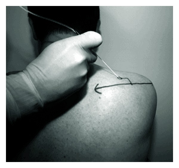
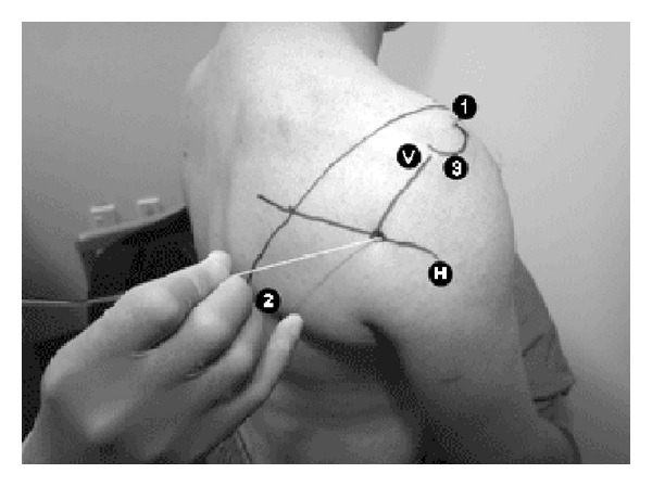
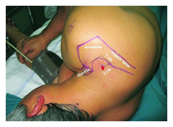
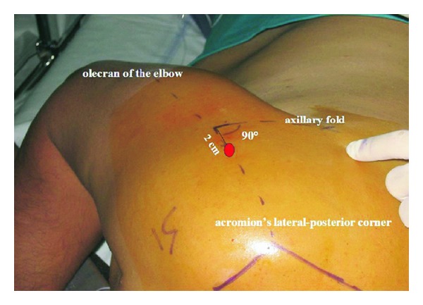
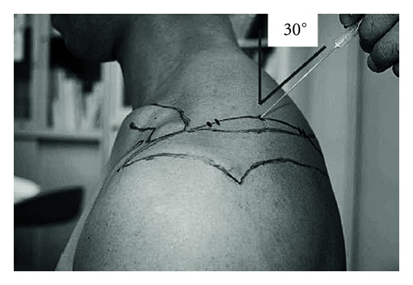
    1. Checcucci G, Allegra A, Bigazzi P, Gianesello L, Ceruso M, Gritti G. A new technique for regional anesthesia for arthroscopic shoulder surgery based on a suprascapular nerve block and an axillary nerve block: an evaluation of the first results. Arthroscopy. 2008;24(6):689–696. - PubMed
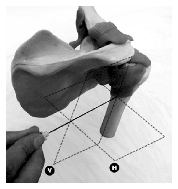
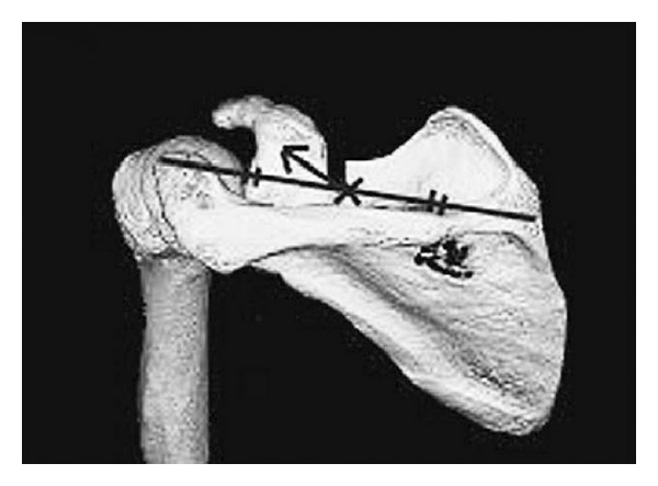
    1. Matsumoto D, Suenaga N, Oizumi N, Hisada Y, Minami A. A new nerve block procedure for the suprascapular nerve based on a cadaveric study. Journal of Shoulder and Elbow Surgery. 2009;18(4):607–611. - PubMed

LinkOut - more resources