

doi: 10.1155/2012/729290.
Epub 2012 Jun 27.
Mitochondrial dynamics in cancer and neurodegenerative and neuroinflammatory diseases
Affiliations
- PMID: 22792111
- PMCID: PMC3391904
- DOI: 10.1155/2012/729290
Item in Clipboard
Mitochondrial dynamics in cancer and neurodegenerative and neuroinflammatory diseases
Int J Cell Biol.
2012.
Abstract
Mitochondria are key organelles in the cell, hosting essential functions, from biosynthetic and metabolic pathways, to oxidative phosphorylation and ATP production, from calcium buffering to red-ox homeostasis and apoptotic signalling pathways. Mitochondria are also dynamic organelles, continuously fusing and dividing, and their localization, size and trafficking are finely regulated. Moreover, in recent decades, alterations in mitochondrial function and dynamics have been implicated in an increasing number of diseases. In this review, we focus on the relationship clarified hitherto between mitochondrial dynamics and cancer, neurodegenerative and neuroinflammatory diseases.
Figures
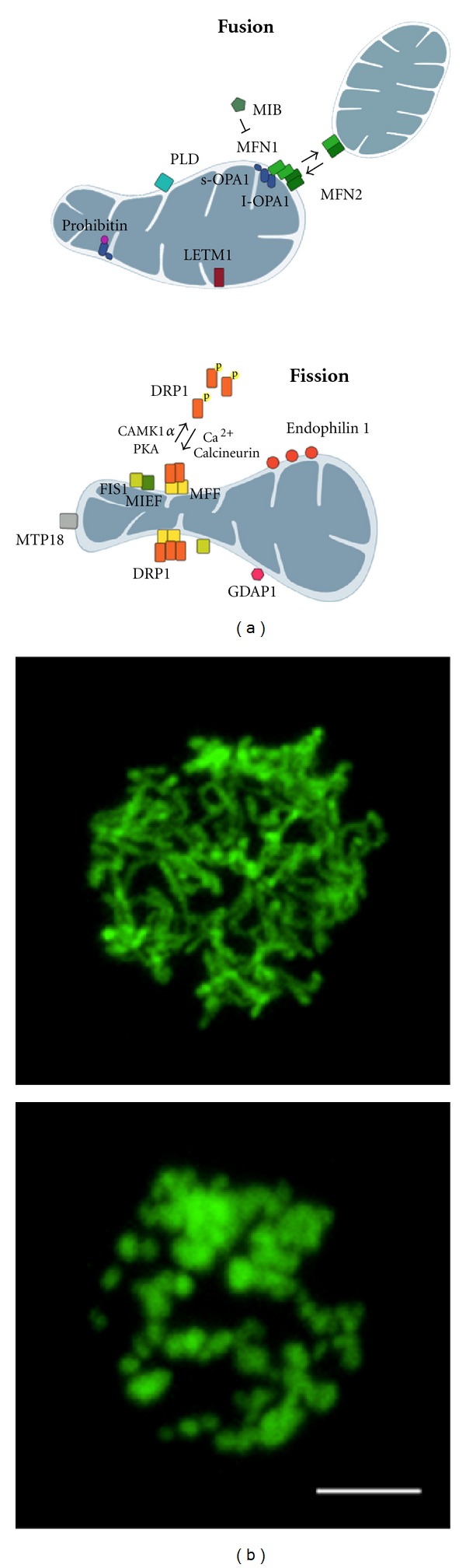
The dynamic nature of mitochondrial shape. (a) Main proteins involved in mitochondrial shape changes are depicted. In fused unopposed condition, DRP1 is phosphorylated and sequestered in the cytoplasm. Once dephosphorylated, it is recruited to the OMM where it oligomerizes and interacts with FIS1, MFF, or MIEF inducing constriction of membranes and, eventually, fission of mitochondria. MFNs homo- and heterooligomerization on the OMM and oligomerization between long and short isoform of Opa1 on the IMM control fusion of mitochondrial membranes. Additional proteins affecting mitochondrial shape are also shown. (b) Mitochondrial morphology in Jurkat cells overexpressing yellow fluorescent protein targeted to mitochondria. The upper panel shows a network of elongated and interconnected mitochondria. In the lower panel, mitochondria appear fragmented (Scale bar: 5 μm).
References
-
- Scorrano L. Opening the doors to cytochrome c: changes in mitochondrial shape and apoptosis. International Journal of Biochemistry and Cell Biology. 2009;41(10):1875–1883. - PubMed
-
- Bereiter-Hahn J, Voth M. Dynamics of mitochondria in living cells: shape changes, dislocations, fusion, and fission of mitochondria. Microscopy Research and Technique. 1994;27(3):198–219. - PubMed
LinkOut - more resources
Full Text Sources
