. 2012;7(7):e39268.
doi: 10.1371/journal.pone.0039268. Epub 2012 Jul 9.

RBPJ is a novel target for rhabdomyosarcoma therapy

Affiliations

RBPJ is a novel target for rhabdomyosarcoma therapy

Hiroko Nagao et al. PLoS One. 2012.

Abstract

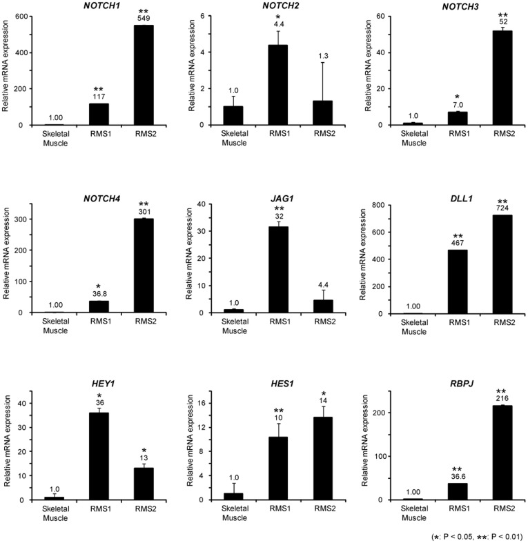
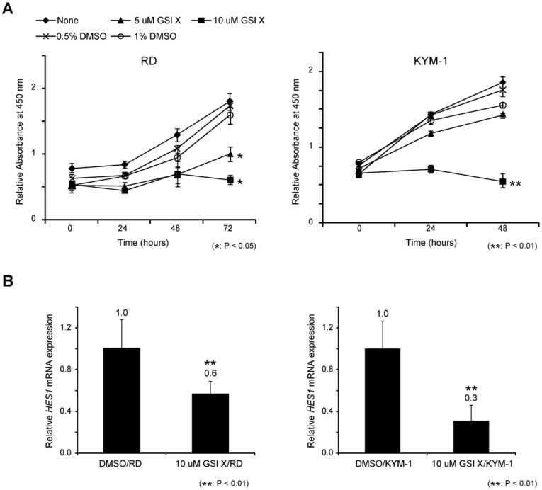
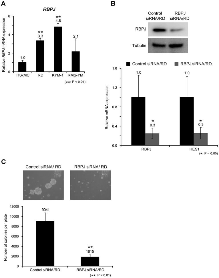
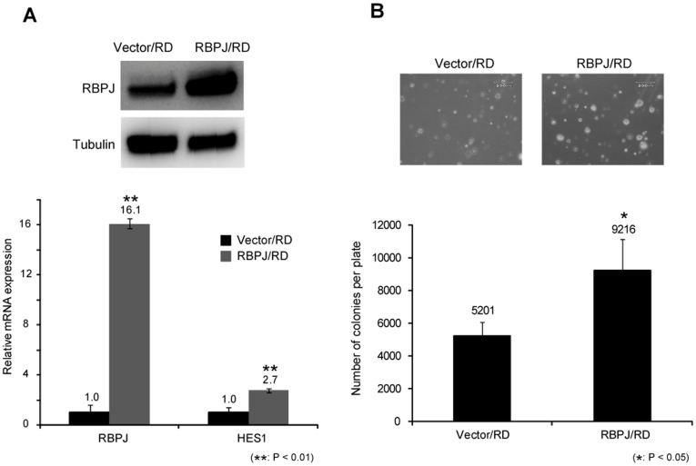
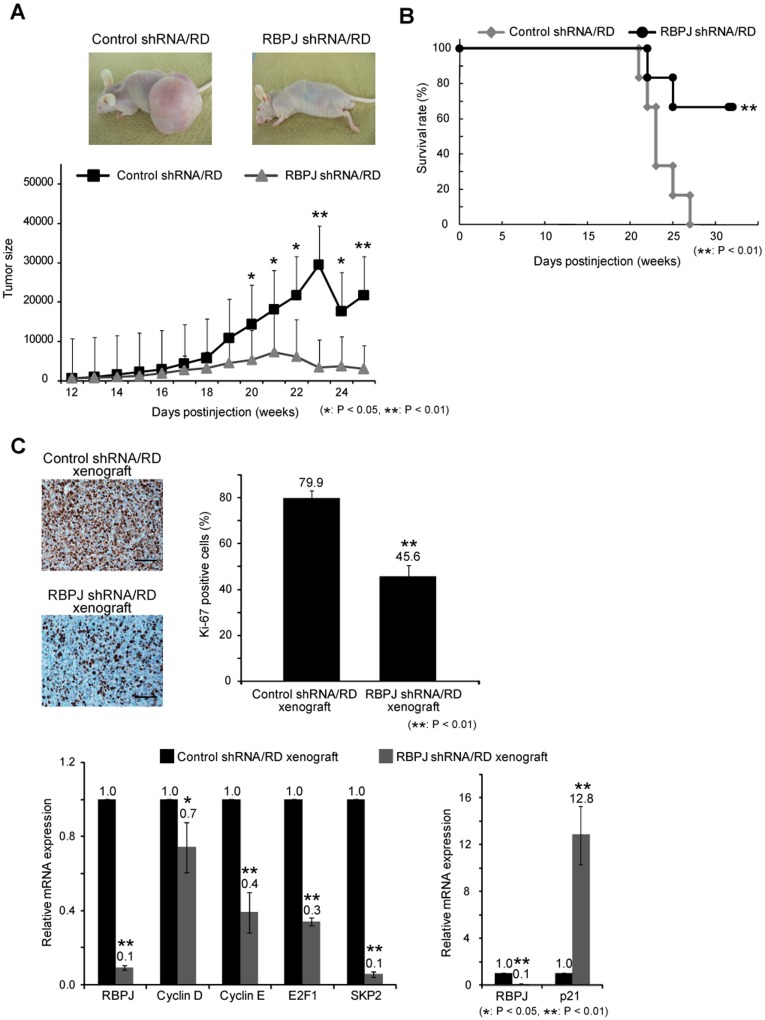
The Notch pathway regulates a broad spectrum of cell fate decisions and differentiation processes during fetal and postnatal development. In addition, the Notch pathway plays an important role in controlling tumorigenesis. However, the role of RBPJ, a transcription factor in the Notch pathway, in the development of tumors is largely unknown. In this study, we focused on the role of RBPJ in the pathogenesis of rhabdomyosarcoma (RMS). Our data showed that Notch pathway genes were upregulated and activated in human RMS cell lines and patient samples. Inhibition of the Notch pathway by a γ-secretase inhibitor (GSI) decreased the in vitro proliferation of RMS cells. Knockdown of RBPJ expression by RNAi inhibited the anchorage-independent growth of RMS cells and the growth of xenografts in vivo. Additionally, overexpression of RBPJ promoted the anchorage-independent growth of RMS cells. Further, we revealed that RBPJ regulated the cell cycle in RMS xenograft tumors and decreased proliferation. Our findings suggest that RBPJ regulates the RMS growth, and that the inhibition of RBPJ may be an effective therapeutic approach for patients with RMS.


Conflict of interest statement

Competing Interests: The authors have declared that no competing interests exist.

Figures

Figure 1
Figure 1. Notch pathway molecules are overexpressed in rhabdomyosarcoma cells.
Notch pathway genes (receptors NOTCH1-4, ligands JAG1 and DLL1, target genes HES1 and HEY1, and transcription factor RBPJ) were assessed by real-time PCR in a normal human skeletal muscle specimen and 2 human RMS biopsy specimens. The Ct values of all RMS samples were normalized to those of ACTB. The values of the human RMS specimens were compared with those of the human skeletal muscle sample, which is defined as a relative expression of 1.0. Columns, mean values of 3 independent experiments; bar, SD. *p<0.05, **p<0.01.
Figure 2
Figure 2. Effects of GSI X on the proliferation of rhabdomyosarcoma cells.
A, Proliferation of RD and KYM-1 cells treated with GSI X or equal volume of DMSO vehicle were measured by WST-1 assay. B, Expression of HES1 mRNA was assessed by real-time PCR in RD cells (left) and KYM-1 cells (right) treated with 10 µM of GSI X for 24 hours. Columns and lines, mean values of 3 independent experiments; bar, SD. *p<0.05, **p<0.01.
Figure 3
Figure 3. Knockdown of RBPJ suppresses anchorage-independent growth of rhabdomyosarcoma cells. A,
The expression of RBPJ mRNA in RD cells was assessed by real-time PCR. The Ct values of all RMS cell lines were normalized to those of ACTB. The values of the RMS cell lines were compared with HSkMc cell, which is defined as a relative expression of 1.0. B, RBPJ protein levels in RD cells transfected with control and RBPJ siRNA were examined by western blotting analysis (top). RBPJ and HES1 mRNA in RD cells transfected with control and RBPJ siRNA were assessed by real-time PCR analysis. Ct values of RBPJ and HES1 were normalized to ACTB. The values of the cells transfected with RBPJ siRNA were compared to those the RD cells transfected with control siRNA, which is defined as a relative expression of 1.0 (bottom). C, Anchorage-independent growth of RD cells transfected with control and RBPJ siRNA were evaluated by colony formation assay. After 3 weeks, each of the colonies were counted and photographed. Scale bar is 200 µM. Columns, mean values of 3 independent experiments; bar, SD. *p<0.05, **p<0.01.
Figure 4
Figure 4. Overexpression of RBPJ promotes rhabdomyosarcoma cell growth. A,
RBPJ protein levels in RD cells transfected with control vector and RBPJ overexpression vector were measured by Western blotting analysis (top). RBPJ and HES1 mRNA in RD cells transfected with control vector and RBPJ overexpression vector were assessed by real-time PCR analysis. Ct values of RBPJ and HES1 were normalized to ACTB. Comparison was made to the RD cells transfected control vector, which is defined as a relative expression of 1.0 (bottom). B, Anchorage-independent growth in RD cells transfected with control vector and RBPJ overexpression vector were evaluated by colony formation assay. Fourteen days later, the each colonies were counted and pictured. Scale bar is 200 µM. Columns, mean values of 3 independent experiments; bar, SD. *p<0.05, **p<0.01.
Figure 5
Figure 5. Knockdown of RBPJ inhibits the growth of rhabdomyosarcoma in nude mice. A,
After transfection of control shRNA or RBPJ shRNA, 1×10⁶ RD cells were subcutaneously inoculated in nude mice (n = 7). Tumor size was calculated weekly by using the formula LW2/2 (with L and W representing the length and width of tumors). B, Survival rate of the mice injected with control shRNA- or RBPJ shRNA-transfected RD cells was assessed by Kaplan–Meier analysis. C, The number of Ki67-positive cells in control shRNA- or RBPJ shRNA-transfected xenograft were accessed by immnohistochemistry. Scale bar is 100 µm. Expression of Cell cycle-related genes (CyclinD, CyclinE, E2F1, SKP2, p21) were assessed by real-time PCR in control shRNA- or RBPJ shRNA-transfected xenograft. The Ct values of xenograft samples were normalized to those of ACTB. The values of the RBPJ shRNA-transfected xenograft was compared with those of the control shRNA sample, which is defined as a relative expression of 1.0. Columns, mean values of 3 independent experiments. Bar, SD. *p<0.05, **p<0.01.

Similar articles

Cited by

References

    1. Wachtel M, Schafer BW. Targets for cancer therapy in childhood sarcomas. Cancer Treat Rev. 2010;36:318–327. - PubMed
    1. Rodeberg D, Paidas C. Childhood rhabdomyosarcoma. Semin Pediatr Surg. 2006;15:57–62. - PubMed
    1. Wachtel M, Schafer BW. Targets for cancer therapy in childhood sarcomas. Cancer Treat Rev. 2010;36:318–327. - PubMed
    1. Gupta AA, Anderson JR, Pappo AS, Spunt SL, Dasgupta R, et al. Patterns of chemotherapy-induced toxicities in younger children and adolescents with rhabdomyosarcoma: A Report From the Children’s Oncology Group Soft Tissue Sarcoma Committee. Cancer. 2011;118:1130–1137. - PMC - PubMed
    1. Perez EA, Kassira N, Cheung MC, Koniaris LG, Neville HL, et al. Rhabdomyosarcoma in Children: A SEER Population Based Study. J Surg Res. 2011;170:e243–e251. - PubMed

MeSH terms