. 2012;7(7):e39340.
doi: 10.1371/journal.pone.0039340. Epub 2012 Jul 9.

Transscleral diode photocoagulation of large retinal and choroidal vascular lesions

Affiliations

Transscleral diode photocoagulation of large retinal and choroidal vascular lesions

Yun Feng et al. PLoS One. 2012.

Abstract

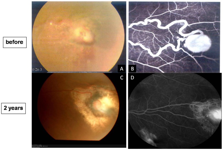
Background: Transscleral retinal photocoagulation with a diode laser is used in glaucoma refractory to medical and surgical treatment. Our main research question was how the technique performed in large vascular lesions associated with hemangiomas of the retina and choroid.

Methodology/clinical findings: Patient charts were retrieved from the hospital files for patients who underwent the procedure and were followed for at least 24 months. Five patients (6 eyes) fit the criteria. Cases included Von Hippel's disease (2 eyes), Coats' disease (1 eye) and choroidal hemangioma (3 cases). Transscleral diode laser treatment was performed under retrobulbar and topical anesthesia with a retinopexy probe (IRIS DioPexy, IRIS Medical Instruments, Mountain View, CA) applied transsclerally under indirect ophthalmoscope visualization. We found an improvement in best-corrected visual acuity at 24 months postoperatively.

Conclusions/significance: Transscleral photocoagulation may have a clinical application in these diseases as an alternate to the high cost of photodynamic therapy with photosensitizing agents.


Conflict of interest statement

Competing Interests: The authors have declared that no competing interests exist.

Figures

Figure 1
Figure 1. Fundus photographs and OCT before surgery and 2 years postoperatively in a 14-year-old girl with von Hippel’s disease in her right eye (Case 1) (A–E).
A. Fundus photograph shows that the retinal capillary hemangioma appears as a red to orange tumor arising within the retina with large-caliber, tortuous afferent and efferent retinal blood vessels. The lesion is surrounded by yellow-white retinal and subretinal exuadates that seem to have a predilection for foveal involvement. C. Cystoid macular degeneration was found in OCT, and the subretinal exuadates was noted. B.The retina is well attached two years postoperatively. Scar formation was noted at the original lesion, previously tortuous vessels were stretched. D and E. the OCT reveals that the macular area is relatively normal and the retina is flat at the region of the stretched vessel at 2 years postoperatively.
Figure 2
Figure 2. Fundus photographs and fluorescein angiography before surgery and 2 years postoperatively in the 14-year-old girl of von Hippel’s disease in her left eye (case 1) (A–D).
A. Left eye of the same patient with von Hippel’s disease (right eye shown in Figure 1). One localized abnormal bean-like lesion is noted at the temporary inferior quadrant with the toutuous afferent and efferent vessels, some yellow exudates are visible surrounding the lesion. B. Fluorescein angiography reveals the hyperfluorescein of the lesion with toutuous vessels at the same area. C. Scar and surrounding chorioretinal atrophy can be seen at 2 years postoperatively, the vessels are stretching to straight and the surrounded exudates are completely obsorbed. D. Fluorescein angriography in the same area at 2 years postoperatively. The chorioretinal atrophy is observed.
Figure 3
Figure 3. Coats’ disease in a 6-year-old boy: OCT (A–E), fundus photographs (F-I) and fluorescein angiograph at different follow-up durations (case 2).
A. OCT reveals the detachment of the neuroretina layer in fovea before the treatment of the surgery. B. the subretinal fluid was partly absorbed and some exudates are observed at one week postoperatively, C.cystoid macular edema was seen at one month postoperatively. D. the subretinal fluid was completely absorbed at 6 months postoperatively. D. the structure is relatively normal in the fovea at two years postoperatively. F. Fundus photographs shows retinal telangiectasias, “light bulb aneurysms”, beading of vessel walls, capillary dilatation, and massive retinal edema with shallow exudative retinal detachment, surrounded by massive white-yellow masses with blurred outlines. G.Fundus photograpy at 1 month postoperatively, there was no much improvement observed after the surgery, except for the dilation of the abnormal vessels. H. The detached retina was reattached with the absorption of the subretinal exudates at six months postoperatively. I. The yellow exudates almost completely absorbed at two years postoperatively. G. Fluorescein angiography at the lesion area. The remarkable retinal vascular telangiectasis, dilated, partly looped, partly beaded thickened vessels and nonperfusion were observed before the surgery. H. Fluorescein angiography at the the same area, significant chorioretinal atrophy was seen without any typical lesion left at two years postoperatively.
Figure 4
Figure 4. Fundus photography (A, D), Fluorescein angiography (B, E) and OCT (C,F,G) of a 29-year-old man with choroidal hemangioma (Case 3) before our treatment (A–C) and two years postoperatively (D–G).
A. massive round orange-colored lesion, slightly elevated at the posterior pole before our treatment. B. Fluorescein angiography reveals that the lesion rapidly develop diffuse hyperfluorescence and do not tend to show the same retinal pigment epithelium changes as melanomas. C. OCT showed the shallow serous retinal detachment. D. Large chorioretinal atrophy and scar were noted two yearspostoperatively. The vessels in the macular area are a bit stretched. E&F, horizontal and vertical scan of OCT revealed an epiretinal membrane (arrow).
Figure 5
Figure 5. Fundus photography, Fluorescein angiography and OCT of a 52-year-old female with previously failed PDT treatment (Case 4) (choroidal hemangioma) before our treatment and two years postoperatively.
A. The lesion appears as a massive red tumor located in the postequatorial zone of the fundus, close to the macular area. B. fluorescein angiography shows the rapid diffuse hyperluorescence at the corresponding area. C. The tumor regressed 6 months postoperatively with a well reattached retina. D.Pigmented chorioretinal scar can be seen at 2 years postoperatively.
Figure 6
Figure 6. Fundus photograph and corresponding OCT of a 36-year-old male without any previously treatment (Case 5) (choroidal hemangioma) before the surgery and two years postoperatively.
A & B. There is a localized red tumor located at the postequatorial zone of the fundus and a secondary retinal detachment that extends into the foveal region.C&D. horizontal and vertical scan of OCT at macular area shows detachment of the outer retinal layers with fovea involved. E&F showed thereattachment of the retina at 2 years postoperatively. The tumor regressed after diode laser treatment, leaving a pigmented chorioretinal scar. The arrow shows a little bit stretching of the retina. G&H. horizontal and vertical scan of OCT at macular area reveals a relatively normal fovea at two years postoperatively.
Figure 7
Figure 7. Fluorescein angiography and corresponding ICG findings of Case 5 before the surgery (A–D) and two years postoperatively (E–H).
A & B, at early stage, the rapid developed hyperfluorecence of a small localized round lesion reveals a choroidal angioma. C&D. at late stage the secondary serous retinal detachment can be observed. E-G. chorioretinal scar can be noted without any recurrent lesion.

Similar articles

Cited by

References

    1. Turell ME, Singh AD. Vascular tumors of the retina and choroid: Diagnosis and treatment. Middle East Afr J Ophthalmol. 2010;17:191–200. - PMC - PubMed
    1. McHugh DA, Schwartz S, Dowler JG, Ulbig M, Blach RK, et al. Diode laser contact transscleral retinal photocoagulation: a clinical study. Br J Ophthalmol. 1995;79:1083–1087. - PMC - PubMed
    1. McHugh JD, Marshall J, Ffytche TJ, Hamilton AM, Raven A, et al. Initial clinical experience using a diode laser in the treatment of retinal vascular disease. Eye (Lond) 3. 1989;(5):516–527. - PubMed
    1. Abramson DH, Servodidio CA, Nissen M. Treatment of retinoblastoma with the transscleral diode laser. Am J Ophthalmol. 1998;126:733–735. - PubMed
    1. Parvaresh MM, Modarres M, Falavarjani KG, Sadeghi K, Hammami P. Transscleral diode laser retinal photocoagulation for the treatment of threshold retinopathy of prematurity. J AAPOS. 2009;13:535–538. - PubMed

MeSH terms