. 2012;7(7):e39499.
doi: 10.1371/journal.pone.0039499. Epub 2012 Jul 6.

Blockage of spontaneous Ca2+ oscillation causes cell death in intraerythrocitic Plasmodium falciparum

Affiliations

Blockage of spontaneous Ca2+ oscillation causes cell death in intraerythrocitic Plasmodium falciparum

Masahiro Enomoto et al. PLoS One. 2012.

Abstract

Malaria remains one of the world's most important infectious diseases and is responsible for enormous mortality and morbidity. Resistance to antimalarial drugs is a challenging problem in malaria control. Clinical malaria is associated with the proliferation and development of Plasmodium parasites in human erythrocytes. Especially, the development into the mature forms (trophozoite and schizont) of Plasmodium falciparum (P. falciparum) causes severe malaria symptoms due to a distinctive property, sequestration which is not shared by any other human malaria. Ca(2+) is well known to be a highly versatile intracellular messenger that regulates many different cellular processes. Cytosolic Ca(2+) increases evoked by extracellular stimuli are often observed in the form of oscillating Ca(2+) spikes (Ca(2+) oscillation) in eukaryotic cells. However, in lower eukaryotic and plant cells the physiological roles and the molecular mechanisms of Ca(2+) oscillation are poorly understood. Here, we showed the observation of the inositol 1,4,5-trisphospate (IP(3))-dependent spontaneous Ca(2+) oscillation in P. falciparum without any exogenous extracellular stimulation by using live cell fluorescence Ca(2+) imaging. Intraerythrocytic P. falciparum exhibited stage-specific Ca(2+) oscillations in ring form and trophozoite stages which were blocked by IP(3) receptor inhibitor, 2-aminoethyl diphenylborinate (2-APB). Analyses of parasitaemia and parasite size and electron micrograph of 2-APB-treated P. falciparum revealed that 2-APB severely obstructed the intraerythrocytic maturation, resulting in cell death of the parasites. Furthermore, we confirmed the similar lethal effect of 2-APB on the chloroquine-resistant strain of P. falciparum. To our best knowledge, we for the first time showed the existence of the spontaneous Ca(2+) oscillation in Plasmodium species and clearly demonstrated that IP(3)-dependent spontaneous Ca(2+) oscillation in P. falciparum is critical for the development of the blood stage of the parasites. Our results provide a novel concept that IP(3)/Ca(2+) signaling pathway in the intraerythrocytic malaria parasites is a promising target for antimalarial drug development.


Conflict of interest statement

Competing Interests: The authors have declared that no competing interests exist.

Figures

Figure 1
Figure 1. Life cycle of Plasmodium. falciparum.
Schematic illustration of the life cycle of P. falciparum. The blood stages on which this study is focused are shown in detail.
Figure 2
Figure 2. Cytosolic calcium (Ca2+) dynamics in the early ring forms (ERf) (A) and early trophozoites (ET) (B) and effects of 2-aminoethyl diphenylborinate (2-APB).
Each colour represents cytosolic Ca2+ dynamics acquired from individual parasites in the presence (right columns) or absence (left columns) of 100 µM 2-APB. Embedded images in left panels are representative images of Fluo-4-loaded P. falciparum during each intraerythrocytic stage (indicated by arrowheads). Scale bars, 5 µm.
Figure 3
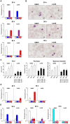
Figure 3. Inhibition of intraerythrocytic P. falciparum development by 2-APB.
(A) The FCR-3 strain was cultured for 40 h of the intraerythrocytic development cycle. Cultures were terminated at 20, 30 and 40 h of the assay after synchronization, and thin smears of erythrocytes were prepared for parasite counting. Representative results of 3 independent experiments are shown. (B) Morphology of intraerythrocytic parasites cultured with DMSO or 100 µM 2-APB at 20, 30 and 40 h of the assay after synchronization. (C) 100 µM 2-APB significantly decreased the area, perimeter and maximum diameter of intraerythrocytic parasites at 15, 30 and 40 h of the assay after synchronization. Columns and error bars represent the mean + S.D. Fifty parasites were measured at each time point. The P values compared with DMSO controls are given below each figure (two-tailed unpaired t test with Welch’s correction). (D) Cultures were terminated at 40 and 70 h of the assay after synchronization for parasite counting. Culture medium with DMSO or 2-APB was replaced at 40 h. Representative results of 3 independent experiments are shown. (E) The chloroquine-resistant strain K1 was cultured for 72 h of the intraerythrocytic development cycle. Cultures were terminated at 24, 48 and 72 h of the assay after synchronization, and thin smears of erythrocytes were prepared for parasite counting. Culture medium with DMSO or 2-APB was replaced at 24 and 48 h. Representative results of 3 independent experiments are shown. Parasiaemia of ring form (Rf), trophozoites (T), early schizonts (ES) and late schizonts (LS) is shown as mean + S.D. of 3 independent counts of a single well (A) or 3 wells (D, E). Stages with parasitaemia of less than 0.1% are not shown (A, D and E).
Figure 4
Figure 4. Time window for inhibition of intraerythrocytic P. falciparum development by 2-APB.
(A) 2-APB was removed at 10 h of the assay. (B) 2-APB was removed at 21 h of the assay. (C) 2-APB was added at 21 h of the assay. (D) Percentages of T and S in 2 independent experiments shown in (C) just before 2-APB was added at 21 h of the assay. (E) 2-APB was added at 28 h of the assay. Parasitaemia at 40 h of the assay is shown as mean + S.D. of 3 independent counts of 3 wells. Stages with parasitaemia of less than 0.1% are not shown. The difference in Rf parasitaemia between the DMSO and 2-APB groups was analysed statistically (two-tailed unpaired t test ) and P values are given in each panel (A, B, C and E). (F) Effects of 100 µM 2-APB on the area, perimeter and maximum diameter of parasites. Three experimental groups were tested as follows. 2-APB was added at the beginning of the assay during synchronization, and cell size was analysed at 10 or 20 h of the assay. 2-APB was added at the beginning of the assay, removed at 10 h of the assay and cell size was measured at 20 h of the assay. Fifty parasites were measured in each experimental group. P values compared with DMSO controls are given in each panel (two-tailed unpaired t test with Welch’s correction).
Figure 5
Figure 5. Electron micrographs of parasites treated with 2-APB.
(A) After 30 h of DMSO administration (control group; original magnification, ×30,000). (B) After 30 h of 100 µM 2-APB administration (original magnification, ×30,000). (C) After 30 h of 100 µM 2-APB administration (original magnification, ×30,000). (D) After 30 h of 100 µM 2-APB administration. Reticular ER structure is observed (arrow) (original magnification, ×50,000). (E) After 40 h of 100 µM 2-APB administration. The nuclear envelope (NE) surrounded by ribosomal granules (Ri) is observed (arrow) (original magnification, ×50,000). (F) After 40 h of 100 µM 2-APB administration. Formation of rhoptries (Rh) and other micro-organelles is observed (original magnification, ×50,000). N, nucleus; MP, malaria pigment; MC, Maurer’s cleft.
Figure 6
Figure 6. Effect of 2-APB on ER structure.
The parasite nucleus and ER were stained simultaneously with Hoechst 33342 (blue) and ER-Tracker (red) after 30 h of DMSO (upper panels) or 2-APB (lower panels) administration. Merged images are shown in the right columns (Merge).

References

    1. World Health Organization. World Malaria Report 2010. WHO. 2011.
    1. Haldar K, Mohandas N. Erythrocyte remodeling by malaria parasites. Curr Opin Hematol. 2007;14:203–209. doi: 10.1097/MOH.0b013e3280f31b2d. - DOI - PubMed
    1. Maier AG, Cooke BM, Cowman AF, Tilley L. Malaria parasite proteins that remodel the host erythrocyte. Nat Rev Microbiol. 2009;7:341–354. doi: 10.1038/nrmicro2110. - DOI - PubMed
    1. Dondorp AM, Pongponratn E, White NJ. Reduced microcirculatory flow in severe falciparum malaria: pathophysiology and electron-microscopic pathology. Acta Trop. 2004;89:309–317. - PubMed
    1. Berridge MJ, Lipp P, Bootman MD. The versatility and universality of calcium signalling. Nat Rev Mol Cell Biol. 2000;1:11–21. doi: 10.1038/35036035. - DOI - PubMed

Publication types