. 2012;7(7):e40038.
doi: 10.1371/journal.pone.0040038. Epub 2012 Jul 6.

Peptide YY regulates bone remodeling in mice: a link between gut and skeletal biology

Affiliations

Peptide YY regulates bone remodeling in mice: a link between gut and skeletal biology

Iris P L Wong et al. PLoS One. 2012.

Abstract

Background & aims: Gastrointestinal peptides are increasingly being linked to processes controlling the maintenance of bone mass. Peptide YY (PYY), a gut-derived satiety peptide of the neuropeptide Y family, is upregulated in some states that also display low bone mass. Importantly, PYY has high affinity for Y-receptors, particularly Y1R and Y2R, which are known to regulate bone mass. Anorexic conditions and bariatric surgery for obesity influence circulating levels of PYY and have a negative impact on bone mass, but the precise mechanism behind this is unclear. We thus examined whether alterations in PYY expression affect bone mass.

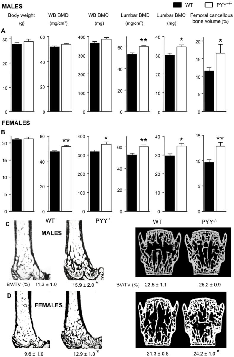
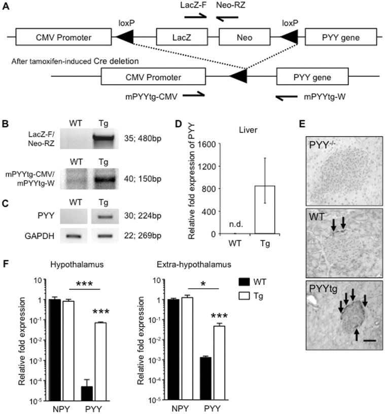
Methods: Bone microstructure and cellular activity were analyzed in germline PYY knockout and conditional adult-onset PYY over-expressing mice at lumbar and femoral sites using histomorphometry and micro-computed tomography.

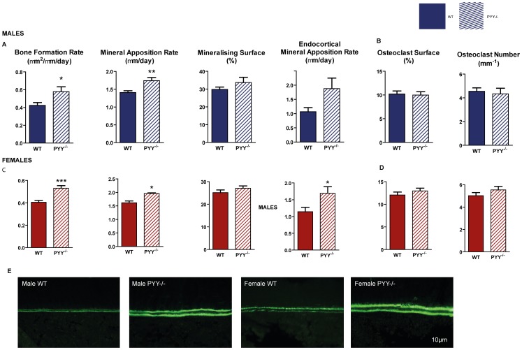
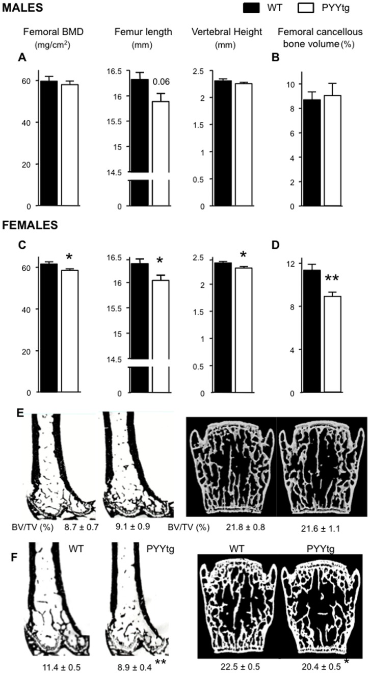
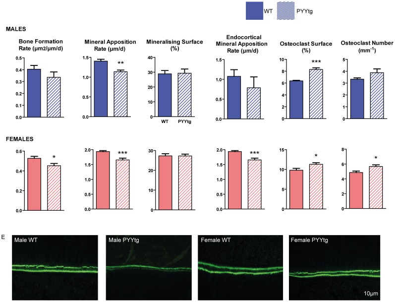
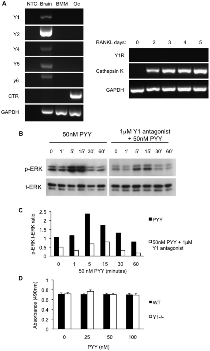
Results: PYY displayed a negative relationship with osteoblast activity. Male and female PYY knockout mice showed enhanced osteoblast activity, with greater cancellous bone mass. Conversely, PYY over-expression lowered osteoblast activity in vivo, via a direct Y1 receptor mediated mechanism involving MAPK stimulation evident in vitro. In contrast to PYY knockout mice, PYY over expression also altered bone resorption, as indicated by greater osteoclast surface, despite the lack of Y-receptor expression in osteoclastic cells. While evident in both sexes, cellular changes were generally more pronounced in females.

Conclusions: These data demonstrate that the gut peptide PYY is critical for the control of bone remodeling. This regulatory axis from the intestine to bone has the potential to contribute to the marked bone loss observed in situations of extreme weight loss and higher circulating PYY levels, such as anorexia and bariatric obesity surgery, and may be important in the maintenance of bone mass in the general population.


Conflict of interest statement

Competing Interests: The authors have declared that no competing interests exist.

Figures

Figure 1
Figure 1. Greater bone mass in PYY−/− mice.
Male (A) and female (B) PYY−/− mice display no difference in body weight, but greater whole body BMD, whole body BMC in female mice. Regional analysis revealed greater lumbar BMD and BMC in PYY−/− of both genders. Representative images of distal femur histological sections and 3D surface rendered models of µCT scanned lumbar vertebra of male (C) and female (D) mice showing greater cancellous bone volume in the distal femoral metaphysis of PYY−/− mice in both genders and in the lumbar vertebral body of female PYY−/− mice (D). Mean ± SEM of 7–18 mice per group are shown. *p<0.05, **88p<0.005 versus wild-type (WT).
Figure 2
Figure 2. Bone cell activities in the femora of PYY−/− mice.
In the distal femoral metaphysis, lack of PYY signaling results in greater bone formation rate (A,C) due to greater mineral apposition rate (MAR) with no change in mineralizing surface (C,I) in both genders. Endocortical MAR at the mid femur was also greater in PYY−/− compared to wild-type. Parameters of cancellous bone resorption as indicated by osteoclast surface and number (B,D) are similar between PYY−/− and wild-type controls. Representative images of endocortical MAR are shown in E. Scale bar represents 10 µm. Mean ± SEM of 5–18 mice per group are shown. *p<0.05, **p<0.005, ***p<0.001 versus wild-type (WT).
Figure 3
Figure 3. Evaluation of PYY overproduction.
As shown in the schematic diagram of the PYY transgene construct (A), liver genomic DNA was analyzed for presence of PYY transgene construct (B) and deletion of stop cassette (hence activation of PYY transgene) by southern analysis. Hepatic PYY mRNA expression was detected only in PYYtg mice and not littermate controls as shown by RT?PCR (C) and qPCR (D). PYY staining (brown) is more intense in the islets of Langerhans in the pancreas of PYY transgenic than wild-type littermates (E). PYY mRNA levels are greater in PYYtg hypothalamus and the rest of the brain, compared to wild-type littermates, but are lower compared to NPY mRNA levels of the same genotype (F). Number of PCR cycles is indicated at right, followed by size of PCR product (B-C). Values shown are normalised to ribosomal protein L13 (RPL-13) levels and relative to wild-type expression (D, F). n.d., not detected (E). Scale bar represents 50 µm (E). Pictures are representative of staining observed in tissues obtained from 16-week-old mice (2?3 mice per group) (E). *p<0.05, ***p<0.001 versus wild-type (WT).
Figure 4
Figure 4. Lower bone mass and reduced bone size in female PYYtg mice.
Although male PYYtg mice are similar to wild-type littermates (A-B), female PYYtg mice have lower femoral BMD, femur length, vertebral height and cancellous bone volume of distal femoral metaphysis (C-D); representative images from male (E) and female (F) mice of distal femoral histological sections and 3D model images of µCT scanned lumbar vertebra. Mean ± SEM of 4–12 mice per group are shown. *p<0.05, **p<0.05 versus wild-type (WT).
Figure 5
Figure 5. Bone cell activities in the femora of PYYtg mice.
Although cancellous bone volume is lower in female but not male PYYtg, alterations in bone cell activities are observed in both genders. Mineral apposition rate (MAR) was reduced in both genders (A,C), with bone formation rate reduced only in females, with no change in mineralizing surface. Endocortical MAR at the mid femur was reduced in female PYYtg compared to wild-type. Osteoclast surface was greater in both genders of PYYtg and osteoclast number only greater in females. Representative images of endocortical MAR are shown in E. Scale bar represents 10 µm. Mean ± SEM of 4–12 mice per group are shown. *p<0.05, **p<0.005, ***p<0.001 versus wild-type (WT).
Figure 6
Figure 6. PYY signalling in osteoblasts via Y1 receptors.
Y receptor gene expression was evident in brain tissue, but was absent in bone marrow macrophages (BMM) or cultured osteoclasts (Oc) from wild type mice(A). Y1R was not detected at any time point throughout RANKL-induced osteoclast differentiation (A). Calcitonin Receptor (CTR) and Cathepsin K gene expression were used as osteoclast markers. NTC, no template control. PYY induced signaling in primary calvarial osteoblasts through strong phosphorylation of ERK, a response which was blunted in the presence of Y1 receptor specific antagonist (1229U91). (B) p-ERK, phosphorylated ERK; t-ERK, total ERK from western blots of calvarial cultures. (C) Western blots were quantified by densitometry of p-ERK and t-ERK bands. Proliferation of osteoblastic cultures from wild type and Y1 receptor null mice were not altered by PYY (D). Mean ± SEM of 4 replicates per group are shown.

Similar articles

Cited by

References

    1. Nguyen ND, Ahlborg HG Center JR, Eisman JA, Nguyen TV. Residual lifetime risk of fractures in women and men. J Bone Miner Res. 2007;22:781–788. - PubMed
    1. Wong IP, Baldock PA, Herzog H. Gastrointestinal peptides and bone health. Curr Opin Endocrinol Diabetes Obes. 2010;17:44–50. - PubMed
    1. Fukushima N, Hanada R, Teranishi H, Fukue Y, Tachibana T, et al. Ghrelin directly regulates bone formation. J Bone Miner Res. 2005;20:790–798. - PubMed
    1. Henriksen DB, Alexandersen P, Hartmann B, Adrian CL, Byrjalsen I, et al. Four-month treatment with GLP-2 significantly increases hip BMD A randomized, placebo-controlled, dose-ranging study in postmenopausal women with low BMD. Bone. 2009. - PubMed
    1. Ding KH, Shi XM, Zhong Q, Kang B, Xie D, et al. Impact of glucose-dependent insulinotropic peptide on age-induced bone loss. J Bone Miner Res. 2008;23:536–543. - PMC - PubMed

Publication types