. 2012;7(7):e40124.
doi: 10.1371/journal.pone.0040124. Epub 2012 Jul 5.

Estrogen leads to reversible hair cycle retardation through inducing premature catagen and maintaining telogen

Affiliations

Estrogen leads to reversible hair cycle retardation through inducing premature catagen and maintaining telogen

Hui-min Hu et al. PLoS One. 2012.

Abstract

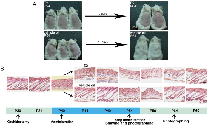
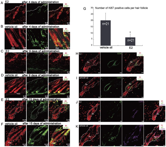
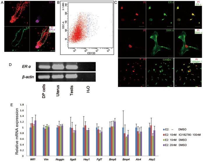
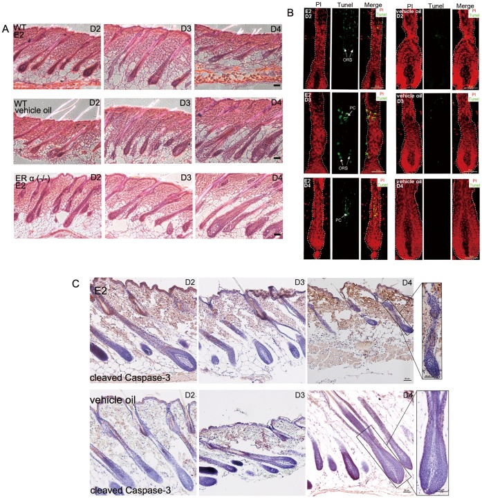
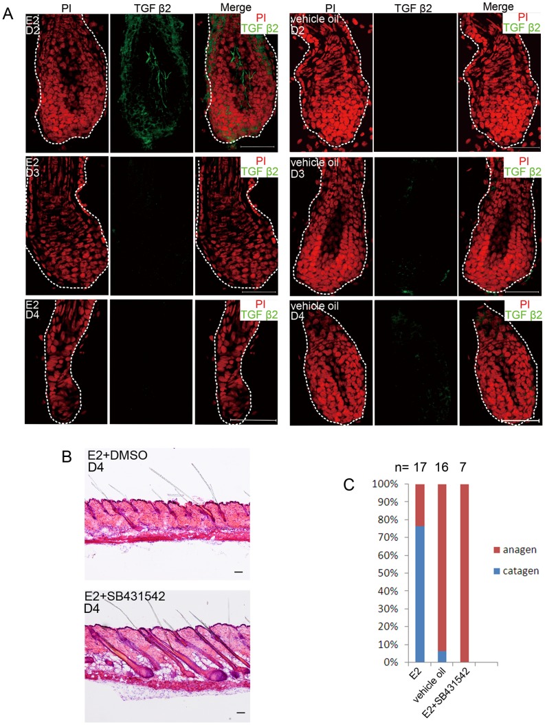
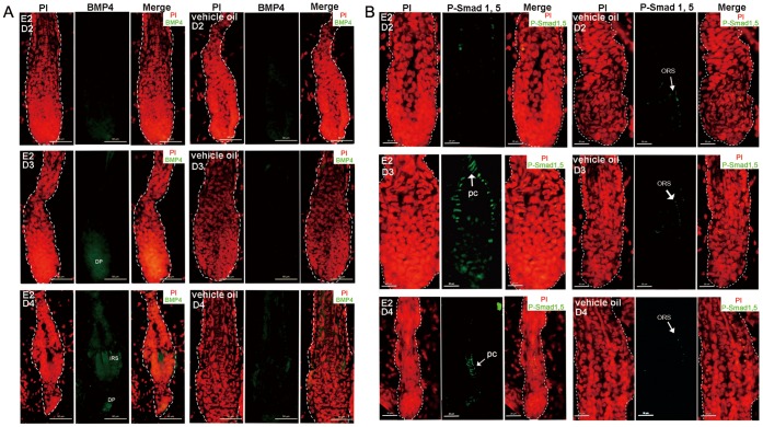
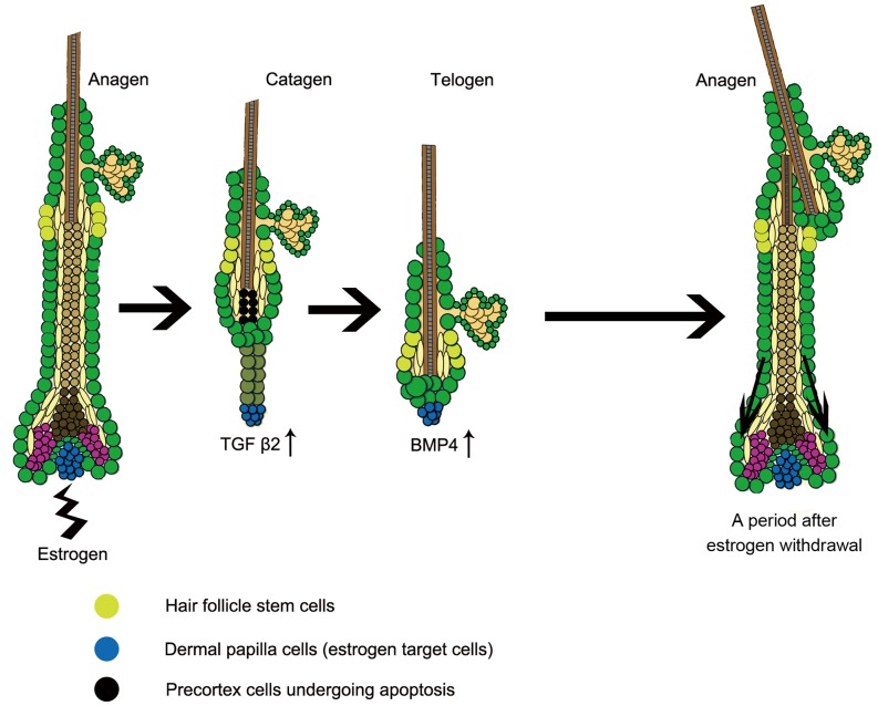
Estrogen dysregulation causes hair disorder. Clinical observations have demonstrated that estrogen raises the telogen/anagen ratio and inhibits hair shaft elongation of female scalp hair follicles. In spite of these clinical insights, the properties of estrogen on hair follicles are poorly dissected. In the present study, we show that estrogen induced apoptosis of precortex cells and caused premature catagen by up-regulation of TGF β2. Immediately after the premature catagen, the expression of anagen chalone BMP4 increased. The up-regulation of BMP4 may further function to prevent anagen transition and maintain telogen. Interestingly, the hair follicle stem cell niche was not destructed during these drastic structural changes caused by estrogen. Additionally, dermal papilla cells, the estrogen target cells in hair follicles, kept their signature gene expressions as well as their hair inductive potential after estrogen treatment. Retention of the characteristics of both hair follicle stem cells and dermal papilla cells determined the reversibility of the hair cycle suppression. These results indicated that estrogen causes reversible hair cycle retardation by inducing premature catagen and maintaining telogen.


Conflict of interest statement

Competing Interests: The authors have declared that no competing interests exist.

Figures

Figure 1
Figure 1. Estrogen induced premature catagen and suppressed HFs at telogen in a reversible manner.
(A) Estrogen administration inhibited hair regrowth. At the tenth day after hair shaving (P64), the estrogen treated mice remained bald, whereas the backs of the vehicle oil treated mice were fully covered by hairs. (B) Estrogen induced premature catagen and arrested HFs at telogen. After orchidectomy, HFs entered a long-lasting anagen (P30–49). In contrast, just after four days of 17β-estradiol injection (P44), the hairs were obliged to enter catagen from anagen, and stayed at telogen until at least ten days after stopping injection (P54–64). However, 15 days after withdrawal of the 17β-estradiol treatment (P69), the mice had their hairs regrown. The period of 17β-estradiol or vehicle oil treatment is highlighted by blue. E2: 17β-estradiol; HF: hair follicle. Scale bar: 100 µm.
Figure 2
Figure 2. Estrogen inhibited proliferation of hair matrix cells, while HFSCs were reserved.
HFs of the estrogen treated mice with a typical catagen phenotype (A) and telogen phenotype (C) contained just several Ki67 positive cells after 4 (A) and 9 (C) days of treatment. HFs of the vehicle oil treated mice manifested typical anagen phenotype with lengthening hair fibers and large bulb after 4 (B) and 9 (D) days of treatment, and abundant Ki67 positive cells were detected in hair matrix. (E) After another 6 days of continuous treatment, few cells were activated in HFs of the estrogen treated mice. (F) HFs of the vehicle oil treated mice had gone through a whole hair cycle at the fifteenth day of treatment. They stayed at telogen, yet still contained a mass of Ki67 positive cells in their distal ORS. (G) The histogram gives a quantitative view over the different count of proliferating cells in differently treated HFs at P54. 21 HFs of each treatment were investigated (*, P<0.05, independent sample T test). (H) At the fifteenth day of treatment, the HFSC marker CD34 expressed in the bulge of estrogen treated mice in a similar manner as vehicle oil treated mice (I). Integrin α6 and LGR5 were detected both in HFs of the estrogen treated mice (J) and the vehicle oil treated mice (K). Hm: hair matrix; Bu: bulge. Dotted line indicates epithelium (A–D) or the boundary of HF (E–F, H–K). Propidium iodide-labeled nuclei are in red. E2: 17β-estradiol; HFSCs: hair follicle stem cell; HF: hair follicle; ORS: outer root sheath; LGR5: Leucine-rich G protein-coupled receptor 5. Scale bars: 50 µm (A–C); 100 µm (D); 20 µm (E–F); 10 µm (H–K).
Figure 3
Figure 3. The hair inductive potential of DP cells was not compromised upon estrogen.
(A) ER α located at DP of adult HF. (B) DP cells were isolated from neonatal mice dermis following published protocol. CD133 positive cells were collected for estrogen treatment and further assay. (C) Sorted DP cells expressed ER α. As a mesenchymal cell population, they also expressed SMA α and collagen I. (D) Sorted DP cells expressed ER α on mRNA level. Mice uterus and testis were employed as positive control while H2O as negative control. (E) Real-time PCR was conducted on a battery of DP signature genes. After exposed respectively to 10 nM, 20 nM of 17β-estradiol or 10 nM of 17β-estradiol and 100 nM ICI 182780, the expression levels of these DP signature genes were not significantly altered (P<0.01 was considered to be significant, one-way ANOVA analysis). Error bars indicate SD of triplicate independent experiments. E2: 17β-estradiol; DP: dermal papilla; ER: estrogen receptor; HF: hair follicle; SMA α: smooth muscle actin α. Scale bars: 20 µm (A); 50 µm (C).
Figure 4
Figure 4. The premature catagen induced by estrogen was accompanied with advance apoptosis of precortex cells.
(A) Premature catagen happened to the WT mice HFs as soon as the 4th day of estrogen treatment (P44). Yet estrogen treatment showed no effect on HFs of ER α (−/−) ones. (B) After 3 days or 4 days of estrogen treatment, precortex cells of the HFs showed Tunel-positive. The days of treatment are indicated. (C) Compared with HFs of the vehicle oil treated mice, the HFs upon estrogen showed cleaved caspase-3 expression in precortex region. Boxed areas are magnified in right-hand panels. Propidium iodide-labeled nuclei are in red. PC: precortex; ORS: outer root sheath; E2: 17β-estradiol; HF: hair follicle; ER: estrogen receptor. Scale bar: 50 µm.
Figure 5
Figure 5. TGF β2 acted as a downstream effector in the estrogen induced hair cycle arrest.
(A) TGF β2 expressed in HFs of the estrogen treated mice. The skin was derived after 2, 3 and 4 days of estrogen or vehicle oil administration. Note TGF β2 up-regulation in DP of the estrogen treated mice HF after 2 days of administration. The days of treatment are indicated. Propidium iodide-labeled nuclei are in red. (B) The pretreatment with a specific TGF β signalling inhibitor SB431542 rescued the hair cycle arrest caused by estrogen. The estrogen+DMSO mice were with catagen HFs, whereas the estrogen+SB431542 ones with anagen HFs. (C) Statistics of phenotypes of differently treated mice. Numbers above the bars indicate number of mice used for each assay. E2: 17β-estradiol; HF: hair follicle; DP: dermal papilla. Scale bar: 50 µm (A), 100 µm (B).
Figure 6
Figure 6. HFs of the estrogen treated mice showed activation of BMP pathway.
(A) Increase expression of BMP4 in hair keratinocytes and DP cells after 3 and 4 days of estrogen treatment vs. HFs of the oil treatment mice. (B) After 3 and 4 days of estrogen treatment, a high phosphorylation level of BMP signaling effector, Smad 1/5, was detected in precortex, while the control ones had negligible P-Smad 1/5 expression in ORS. The days of treatment are indicated. Propidium iodide-labeled nuclei are in red. ORS: outer root sheath; IRS: inner root sheath; E2: 17β-estradiol; HF: hair follicle; DP: dermal papilla. Scale bar: 50 µm (A); 20 µm (B).
Figure 7
Figure 7. Model of the mechanism of estrogen induces hair cycle arrest.
The intrinsic or extrinsic estrogen activates ER α on DP cells. Then the expression of TGF β2 is up-regulated and the precortex cells undergo apoptosis, which triggers premature onset of catagen. Immediately after the catagen, the anagen chalone BMP4 increases its expression in DP and hair matrix. As the result, telogen is sustained. Although estrogen plays a suppressive role on hair growth, the HFSCs are kept from damage. After estrogen treatment withdrawal, the reserved HFSCs were reactivated and the hairs could regenerate. ER: estrogen receptor; DP: dermal papilla; HFSC: hair follicle stem cell.

Similar articles

Cited by

References

    1. Paus R, Cotsarelis G. The biology of hair follicles. New Engl J Med. 1999;341:491. - PubMed
    1. Alonso L, Fuchs E. The hair cycle. J Cell Sci. 2006;119:391–393. - PubMed
    1. Blanpain C, Fuchs E. Epidermal homeostasis: a balancing act of stem cells in the skin. Nat Rev Mol Cell Bio. 2009;10:207. - PMC - PubMed
    1. Jung K, Simon T, Darryl S. Human hair genealogies and stem cell latency. BMC Biol. 2006;4:2. - PMC - PubMed
    1. Lin KK, Chudova D, Hatfield G, Smyth P, Andersen B. Identification of hair cycle-associated genes from time-course gene expression profile data by using replicate variance. Proc Natl Acad Sci U S A. 2004;101:15955. - PMC - PubMed

Publication types

MeSH terms

Substances