. 2012;7(7):e40255.
doi: 10.1371/journal.pone.0040255. Epub 2012 Jul 5.

Drosophila motor neuron retraction during metamorphosis is mediated by inputs from TGF-β/BMP signaling and orphan nuclear receptors

Affiliations

Drosophila motor neuron retraction during metamorphosis is mediated by inputs from TGF-β/BMP signaling and orphan nuclear receptors

Ana Boulanger et al. PLoS One. 2012.

Abstract

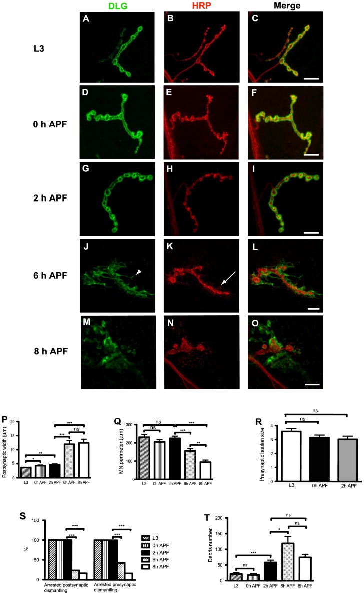
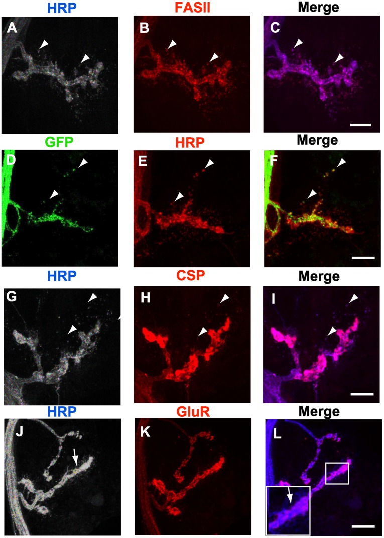
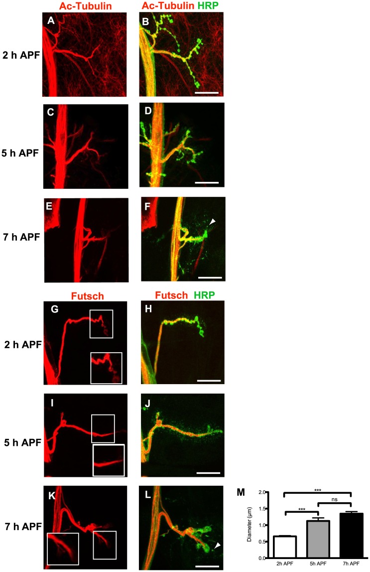
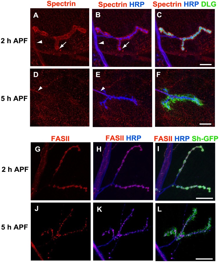
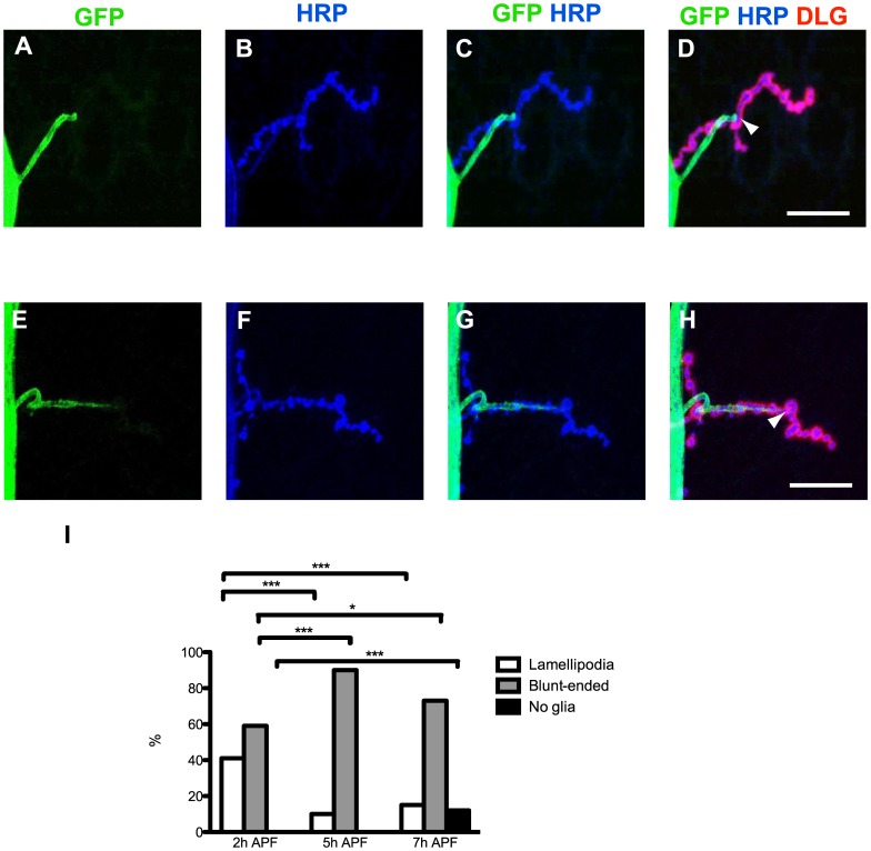
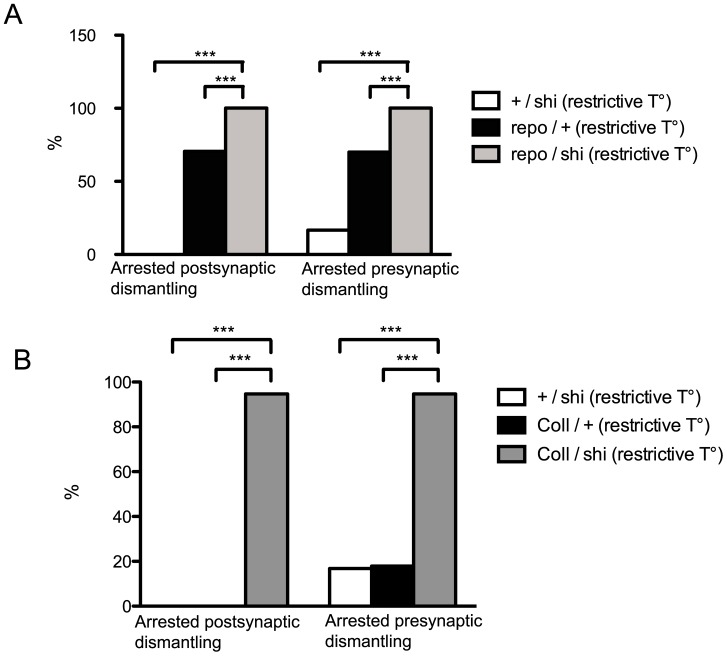
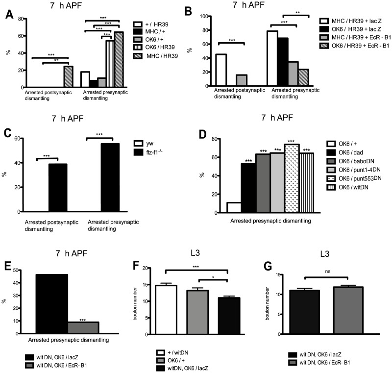
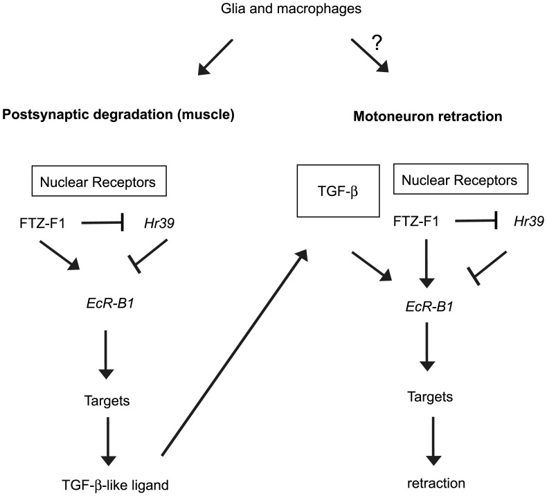
Larval motor neurons remodel during Drosophila neuro-muscular junction dismantling at metamorphosis. In this study, we describe the motor neuron retraction as opposed to degeneration based on the early disappearance of β-Spectrin and the continuing presence of Tubulin. By blocking cell dynamics with a dominant-negative form of Dynamin, we show that phagocytes have a key role in this process. Importantly, we show the presence of peripheral glial cells close to the neuro-muscular junction that retracts before the motor neuron. We show also that in muscle, expression of EcR-B1 encoding the steroid hormone receptor required for postsynaptic dismantling, is under the control of the ftz-f1/Hr39 orphan nuclear receptor pathway but not the TGF-β signaling pathway. In the motor neuron, activation of EcR-B1 expression by the two parallel pathways (TGF-β signaling and nuclear receptor) triggers axon retraction. We propose that a signal from a TGF-β family ligand is produced by the dismantling muscle (postsynapse compartment) and received by the motor neuron (presynaptic compartment) resulting in motor neuron retraction. The requirement of the two pathways in the motor neuron provides a molecular explanation for the instructive role of the postsynapse degradation on motor neuron retraction. This mechanism insures the temporality of the two processes and prevents motor neuron pruning before postsynaptic degradation.


Conflict of interest statement

Competing Interests: The authors have declared that no competing interests exist.

Figures

Figure 1
Figure 1. NMJ dismantling during Drosophila metamorphosis.
A-O, Composite confocal images of muscle 4 NMJ in wild type larvae (L3) and pupae, labeled with anti-DLG antibody (green) and anti-HRP antibody (red). (A-C). At L3, the anti-DLG staining surrounds the bouton membranes as a thin layer. (D-I) Between 0 and 2 h APF the overall NMJ morphology was quite similar to L3. (J-L) 6 h APF the NMJ is disorganized, a high number of filopodia (arrowhead in J) and HRP-labelled debris were observed (arrow in K). (M-O) 8 h APF the anti-DLG staining became discontinuous and fragmented. Individual synaptic boutons are no longer observed with the HRP-staining. The motor neuron became shorter and swollen at the synapse. Bars, 20 µm. P, This histogram shows the width of the postsynapse. Results are means and s.e.m. n represents hemisegments in all our quantifications. L3 (n = 18), 0 h (n = 20), 2 h (n = 20), 6 h (n = 21) and 8 h (n = 23) APF. Note the statistically significant increase of the postsynaptic width at 0 h APF. Q, Histogram representing the perimeter size of the presynapse at different developmental stages. Results are means and s.e.m. The number of NMJ quantified was the following: L3 (n = 20), 0 h (n = 20), 2 h (n = 21), 6 h (n = 21) and 8 h (n = 24) APF. Note the statistically significant decrease in the motoneuron perimeter between 2 h and 6 h APF and between 6 h and 8 h. R, The histogram shows the diameter of the synaptic boutons (µm) at L3 (n = 19), 0 h (n = 18) and 2 h APF (n = 18). The synaptic boutons diameter was similar at the three stages analyzed. S, Percentage of post and presynapses displaying arrested dismantling at L3 (n = 20), 0 h APF (n = 21), 2 h APF (n = 13), 6 h APF (n = 21) and 8 h APF (n = 24). Both, the arrested post and presynaptic dismantling phenotypes decreased at 6 h APF. T, The number of debris particles was scored. L3 (n = 19), 0 h (n = 20), 2 h (n = 17), 6 h (n = 18) and 8 h (n = 19) APF. The amount of debris significantly increases at 2 h APF and then at 6 h APF. Results are means and s.e.m. The differences are significant in a t-test (*:P<0.05, **:P<0.01, ***:P<0.001) or not (ns: no statistical difference) in P-R and T. ***: P value <0.001 (χ2 test) in S. Genotype: y w67c23.
Figure 2
Figure 2. HRP-labeled debris are presynaptically derived.
Composite confocal images correspond to 6 h APF pupae. A-C, The anti-HRP labeled puncta (grey, blue in the merge) colocalized with anti-FASII (red) (arrowheads). D-F, UAS-mCD8-GFP (green) was driven in motor neurons with OK6-GAL4. Most of the anti-HRP labeled debris (red) were GFP-positive (arrowheads). G-I, The anti-HRP labeled puncta (grey, blue in the merge) colocalized with CSP (red) (arrowheads). Note the pink color in the merge marked by arrowheads in C and I. J-L, The HRP-positive puncta (grey, blue in the merge) did not co-localize with D-GluRIIC (red), (arrow in J and L). The inset in L shows a high magnification (2×). Bars, 20 µm. Genotypes: (A-C, G-L) MHC-mGFP-Shaker, (D-F) OK6-GAL4/2×UAS-mGFP.
Figure 3
Figure 3. Microtubule disorganization and retraction but not degeneration take place during NMJ remodeling. A-F
, Anti-Acetylated Tubulin (red) and anti-HRP (green) staining at 2 h, 5 h and 7 h APF. Tubulin staining was present and continuous at the NMJ in all the stages analyzed, and was absent in the HRP-labeled debris (arrowhead in F). G-L, Anti-Futsch (red) and anti-HRP (green) staining. The insets correspond to high magnifications. (I, J) 5 h APF, the presynaptic microtubules become thicker. (K, L) 7 h APF, the thickness of the microtubule bundle is maintained. Futsch was absent in HRP-labelled debris (arrowhead in L). Bars, 20 µm. M, Diameter of the distal end of the axons at 2 h (n = 17), 5 h (n = 16) and 7 h (n = 18) APF. Results are mean and s.e.m. ***: P<0.001, ns: no statistical difference (t-test). Genotype: MHC-mGFP-Shaker.
Figure 4
Figure 4. NMJ destabilization is correlated with the loss of β-Spectrin and the disorganization of the cell adhesion molecule FASII.
A-F, anti-β-Spectrin (red), anti-HRP (blue) and anti-DLG (green). (A-C) 2 h APF β-Spectrin was detected in the motor nerve (arrowhead in A and B), in the synapse (arrow in A and B) and in the muscle (see also the co-localization with DLG as yellow in C). (D-F) β-Spectrin is lost in 5 h APF NMJ. Only traces were observed in axons (arrowhead in D and E). Bars, 20 µm. G-L, anti-FASII (red), anti-HRP (blue) and muscle-driven GFP by MHC-mGFP-Shaker (green). (G-I) The anti-FASII, anti-HRP and GFP co-localized throughout the NMJ 2 h APF. (J-L) The FASII staining was disorganized 5 h APF. The co-localization between FASII and HRP at the synapse is lost in K. Bars, 40 µm. Genotypes : (A-F) y w67c23, (G-L) MHC-mGFP-Shaker.
Figure 5
Figure 5. Glia invade NMJ early in metamorphosis and dynamically retracts before motoneuron pruning. A-H,
Glial extensions were observed (green) at 2 h APF by expressing UAS-mCD8-GFP with the repo-GAL4 driver. The NMJ was also labeled with HRP (blue) and DLG (red). (A-D) A blunt-ended glial process is seen in A. A lamellipodium process is shown in E. Additional merges including the anti-DLG staining are shown in D and H. Here, the arrowheads point to the end of the glial extension. Bars, 30 µm. I, Glial projections were scored at 2 h (n = 17), 5 h (n = 20) and 7 h (n = 26) APF. The percentages of lamellipodia, blunt-ended and no glial extensions are represented. *: P<0.05, ***: P<0.001 (χ2 test). Genotype: 2× UAS-mGFP/+; repo-GAL4/+.
Figure 6
Figure 6. Disrupting glial and phagocyte function blocks NMJ dismantling.
Percentage of 7 h APF NMJ showing arrested dismantling at restrictive temperature in A: +/shi (n = 36), repo/+ (n = 95) and repo/shi (n = 40). B: +/shi (n = 36), Coll/+ (n = 28) and Coll/shi (n = 57). Expression of shits in glia and in phagocytes significantly increased the percentage of arrested post and presynaptic dismantling phenotypes. ***: P<0.001 (χ2 test). Genotypes: A: +/shi  =  +/UAS-shits1. repo/+  =  repo-GAL4/+. repo/shi  =  repo-GAL4/UAS-shits1 B: +/shi  =  +/UAS-shits1. Coll/+  =  Collagen-GAL4/+. Coll/shi  =  Coll-GAL4/UAS-shits1.
Figure 7
Figure 7. The nuclear receptor pathway regulates EcR-B1 expression in both muscles and motor neurons but the TGF-β pathway regulates EcR-B1 expression only in motor neurons.
A, Percentages of 7 h APF NMJ showing arrested post and presynaptic dismantling in: +/Hr39 (n = 56), MHC/+ (n = 38), OK6/+ (n = 37), OK6/Hr39 (n = 35) and MHC/Hr39 (n = 45) pupae. B, Percentages of 7 h APF NMJ showing arrested post and presynaptic dismantling in: MHC/Hr39+ lacZ (n = 42), OK6/Hr39+ lacZ (n = 19), MHC/Hr39+ EcR-B1 (n = 127) and OK6/Hr39+ EcR-B1 (n = 32) pupae. C, Percentages of 7 h APF NMJ showing arrested post and presynaptic dismantling phenotypes in: ftz-f1−/− (n = 36) and yw (n = 18) pupae. D, Percentages of 7 h APF NMJ showing arrested post and presynaptic dismantling in different TGF-β signaling mutant backgrounds: OK6/+ controls (n = 37), OK6/dad (n = 17), OK6/baboDN (n = 19), OK6/punt1-4DN (n = 31) and OK6/punt553DN (n = 23), OK6/witDN (n = 14). E, The arrested presynaptic dismantling phenotype obtained in witDN, OK6/lacZ individuals (n = 41) was rescued in witDN, OK6/EcRB1 in 7 h APF motor neurons (n = 56). F, At larval stages, bouton number in witDN, OK6/lacZ individuals (n = 18) was lower than in +/witDN (n = 18) and in OK6/+ (n = 20). G, At larval stages, the bouton number in witDN, OK6/lacZ individuals (n = 18) was similar than in witDN, OK6/EcR-B1 (n = 19). Results are means and s.e.m. The differences are significant in a χ2 test (**: P value is <0.01, ***: P<0.001) or not statistically different (ns) for A-E. The differences are significant in a t-test (*: the two-tailed P value is <0.05, ***: P<0.001) or not statistically different (ns) for F and G. Genotypes: A: +/HR39 =  +/UAS-Hr39. MHC/+  =  MHC-GAL4/+. OK6/+  =  OK6-GAL4/+. OK6/HR39 =  OK6-GAL4/UAS-Hr39. MHC/HR39 =  +/UAS-Hr39; MHC-GAL4/+. B: MHC/HR39+ lacZ  =  +/UAS-Hr39, UAS-lacZ; MHC-GAL4/+. OK6/HR39+ lacZ  =  OK6-GAL4/UAS-Hr39, UAS-lacZ. MHC/HR39+ EcR-B1 =  MHC-GAL4/UAS-Hr39, UAS-EcR-B1. OK6/HR39+ EcR-B1 = OK6-GAL4/+; UAS-Hr39, UAS-EcR-B1/+. C: yw  =  y w67c23. ftz-f1−/−  =  ftz-f117/ftz-f119. D: OK6/+  =  OK6-GAL4/+. OK6/dad  =  OK6-GAL4/UAS-dad. OK6/baboDN  =  OK6-GAL4/2xUAS-baboΔI; +/2xUAS-baboΔI. OK6/punt1-4DN  =  OK6-GAL4/2xUAS-puntΔI. OK6/punt553DN  =  OK6-GAL4/2xUAS-puntΔI. OK6/witDN  =  OK6-GAL4/UAS-witDN. E and G : witDN, OK6/lacZ  =  UAS-witDN, OK6-GAL4/+; UAS-lacZ/+. witDN, OK6/EcR-B1 =  UAS-witDN, OK6-GAL4/+; UAS-EcR-B1/+. (F): +/witDN  =  +/UAS-witDN. OK6/+  =  OK6-GAL4/+. witDN, OK6/lacZ  =  UAS-witDN, OK6-GAL4/+; +/UAS-lacZ.
Figure 8
Figure 8. NMJ dismantling model.
The nuclear receptor pathway involving FTZ-F1 and HR39 regulates the EcR-B1 expression in muscle and initiate dismantling. FTZ-F1 and HR39 positively and negatively respectively control the EcR-B1 expression at the postsynapse. FTZ-F1-mediated EcR-B1 expression leads to transcriptional regulation of target genes involved in postsynaptic degradation. Then, TGF-β/BMP signaling is supposed to be induced in response to postsynaptic degradation. We propose that this signal produced by the dismantling muscle is received by the motor neuron where activation of EcR-B1 expression (by TGF-β signaling and nuclear receptor) triggers the axon retraction. In addition, glial cells and macrophages participate to the NMJ dismantling process.

Similar articles

Cited by

References

    1. Luo L, O’Leary DD. Axon retraction and degeneration in development and disease. Annual review of neuroscience. 2005;28:127–156. - PubMed
    1. Watts RJ, Hoopfer ED, Luo L. Axon pruning during Drosophila metamorphosis: evidence for local degeneration and requirement of the ubiquitin-proteasome system. Neuron. 2003;38:871–885. - PubMed
    1. Lee T, Marticke S, Sung C, Robinow S, Luo L. Cell-autonomous requirement of the USP/EcR-B ecdysone receptor for mushroom body neuronal remodeling in Drosophila. Neuron. 2000;28:807–818. - PubMed
    1. Williams DW, Truman JW. Cellular mechanisms of dendrite pruning in Drosophila: insights from in vivo time-lapse of remodeling dendritic arborizing sensory neurons. Development. 2005;132:3631–3642. - PubMed
    1. Zheng X, Wang J, Haerry TE, Wu AY, Martin J, et al. TGF-beta signaling activates steroid hormone receptor expression during neuronal remodeling in the Drosophila brain. Cell. 2003;112:303–315. - PubMed

Publication types

MeSH terms

LinkOut - more resources