. 2012;7(7):e40272.
doi: 10.1371/journal.pone.0040272. Epub 2012 Jul 6.

Calcitonin inhibits SDCP-induced osteoclast apoptosis and increases its efficacy in a rat model of osteoporosis

Affiliations

Calcitonin inhibits SDCP-induced osteoclast apoptosis and increases its efficacy in a rat model of osteoporosis

Yi-Jie Kuo et al. PLoS One. 2012.

Abstract

Introduction: Treatment for osteoporosis commonly includes the use of bisphosphonates. Serious side effects of these drugs are caused by the inhibition of bone resorption as a result of osteoclast apoptosis. Treatment using calcitonin along with bisphosphonates overcomes these side-effects in some patients. Calcitonin is known to inhibit bone resorption without reducing the number of osteoclasts and is thought to prolong osteoclast survival through the inhibition of apoptosis. Further understanding of how calcitonin inhibits apoptosis could prove useful to the development of alternative treatment regimens for osteoporosis. This study aimed to analyze the mechanism by which calcitonin influences osteoclast apoptosis induced by a bisphosphate analog, sintered dicalcium pyrophosphate (SDCP), and to determine the effects of co-treatment with calcitonin and SDCP on apoptotic signaling in osteoclasts.

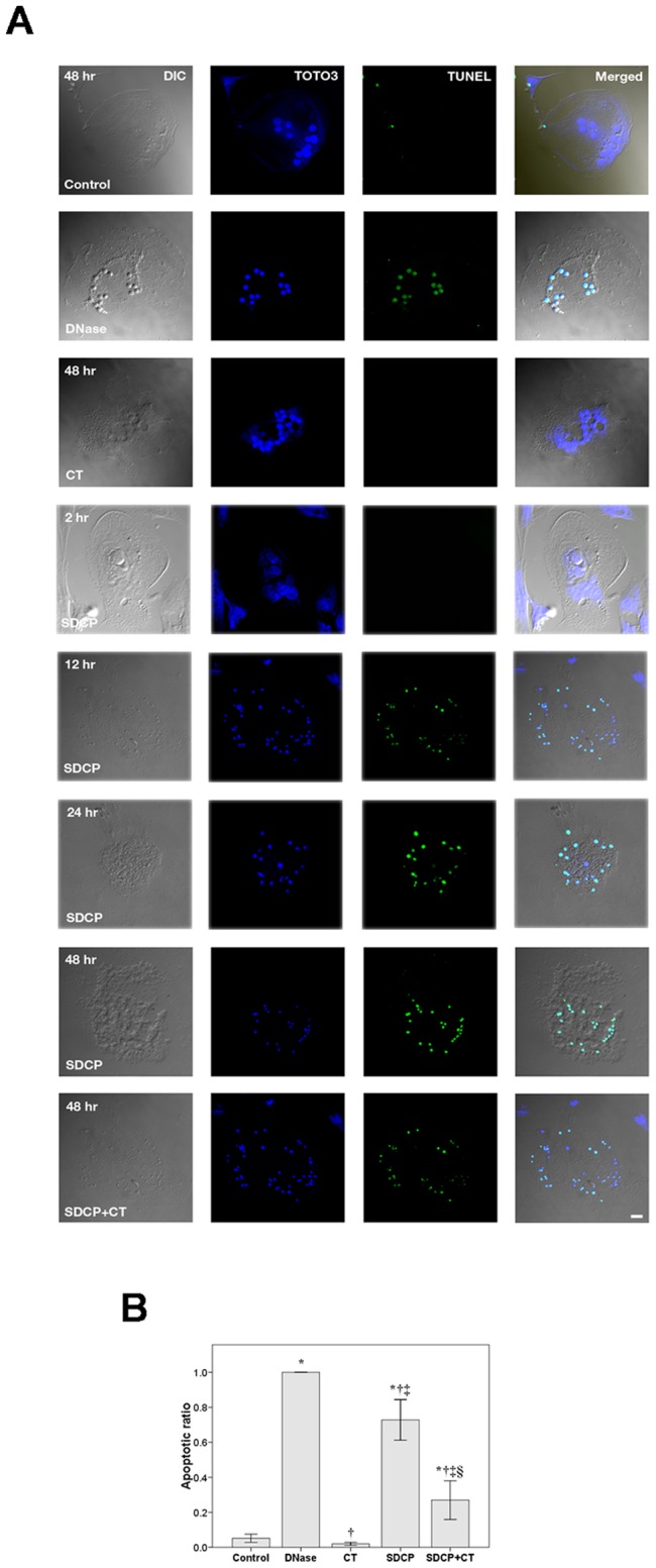
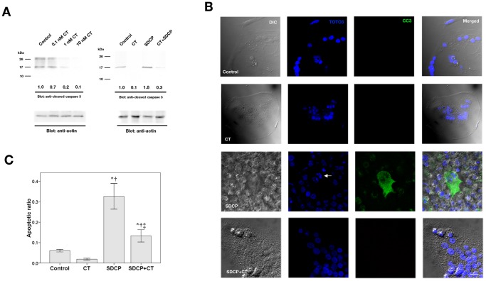
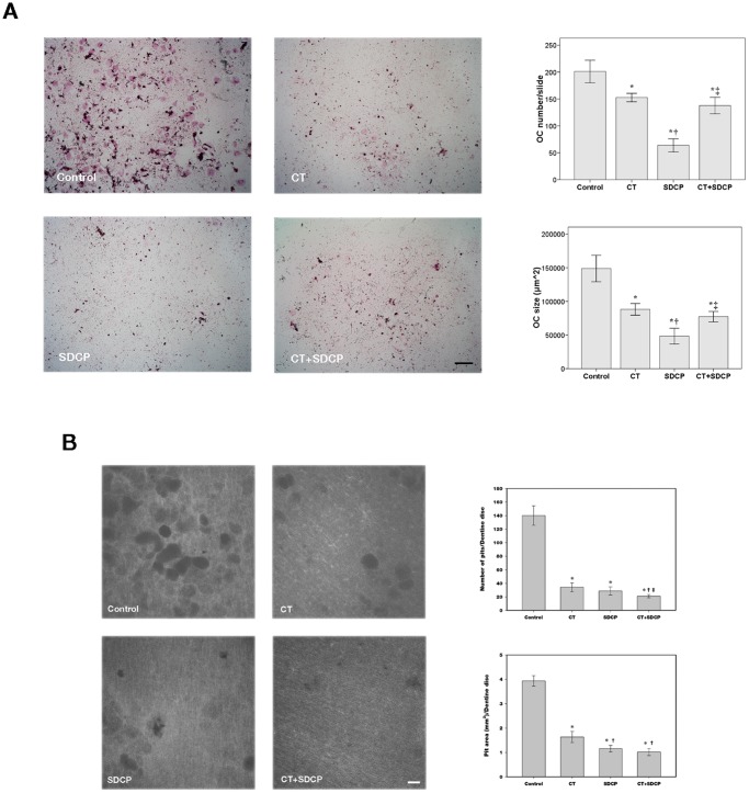
Methods: Isolated osteoclasts were treated with CT, SDCP or both for 48 h. Osteoclast apoptosis assays, pit formation assays, and tartrate-resistant acid phosphatase (TRAP) staining were performed. Using an osteoporosis rat model, ovariectomized (OVX) rats received calcitonin, SDCP, or calcitonin + SDCP. The microarchitecture of the fifth lumbar trabecular bone was investigated, and histomorphometric and biochemical analyses were performed.

Results: Calcitonin inhibited SDCP-induced apoptosis in primary osteoclast cultures, increased Bcl-2 and Erk activity, and decreased Mcl-1 activity. Calcitonin prevented decreased osteoclast survival but not resorption induced by SDCP. Histomorphometric analysis of the tibia revealed increased bone formation, and microcomputed tomography of the fifth lumbar vertebrate showed an additive effect of calcitonin and SDCP on bone volume. Finally, analysis of the serum bone markers CTX-I and P1NP suggests that the increased bone volume induced by co-treatment with calcitonin and SDCP may be due to decreased bone resorption and increased bone formation.

Conclusions: Calcitonin reduces SDCP-induced osteoclast apoptosis and increases its efficacy in an in vivo model of osteoporosis.


Conflict of interest statement

Competing Interests: The authors have declared that no competing interests exist.

Figures

Figure 1
Figure 1. Detection of osteoclast apoptosis using TUNEL analysis.
(A) Osteoclasts were treated with CT (10 nM), SDCP (10 µM), or both for the times indicated. Osteoclasts treated with DNase I (3 U/mL) were included as positive controls. Green nuclear labeling indicates apoptotic cells. Nuclei were counterstained using TOTO-3 (blue). Difference interference contrast (DIC) images show morphology of the cells. Bar = 20 µm. (B) Quantitative results of the experiment shown in panel A. P<0.05 compared to *control group, †DNase group, ‡CT group, and §SDCP group after Bonferroni adjustment, mean ± SD, n = 6 in each group.
Figure 2
Figure 2. Detection of apoptosis by annexin V labeling in osteoclasts.
(A) Osteoclasts were treated with CT (10 nM), SDCP (10 µM), or both for 18 h. Osteoclasts treated with TGF-βI (10 ng/mL) were included as positive controls. Green cellular membrane labeling indicates apoptotic cells. Only cells without propidium iodide (red) labeling were considered to be apoptotic. DIC images and nucleus stain were performed as described in Fig. 1. Bar = 10 µm. (B) Quantitative results of the experiment shown in panel A. The measurement of apoptosis was calculated as the percentage of positive annexin V labeling cells in a total of at least 500 osteoclasts. P<0.05 compared to *control group, †TGF-β1 group, ‡CT group; and §SDCP group after Bonferroni adjustment, mean ± SD, n = 6 in each group.
Figure 3
Figure 3. Calcitonin inhibits SDCP-induced expression of cleaved caspase 3 in osteoclasts.
(A) Western blot analysis of cleaved caspase 3 expression in osteoclasts treated with CT (10 nM), SDCP (10 µM), or both. Protein levels were quantified by densitometry, corrected for the sample load based on actin expression, and expressed as fold-increase or decrease relative to the control lane. Each blot is representative of at least three replicate experiments. (B) Confocal analysis of immunofluorescent labeling of cleaved caspase 3 in calcitonin- or SDCP-treated osteoclast. Green intracellular cleaved caspase 3 labeling and blue TOTO3-labeled nuclear chromatin condensation (white arrow) indicate cells that underwent apoptosis. Bar = 10 µm. (C) Quantitative results of the experiment shown in panel B. The measurement of apoptosis was calculated as a percentage of positive cleaved caspase-3–labelled cells in a total of at least 500 osteoclasts. P<0.05 compared to *control group, † CT group, and ‡SDCP group after Bonferroni adjustment, mean ± SD, n = 6 in each group.
Figure 4
Figure 4. Regulation of apoptotic signaling by calcitonin and SDCP.
(A) Western blot analysis of Bcl-2 and Mcl-1 expression in osteoclasts treated with CT (10 nM), SDCP (10 µM), or both. (B) Caspase 8 and 9 activation in response to CT (10 nM) or SDCP (10 µM) stimulation. Western blot analysis revealed the presence of two fragments (p43 and p18), corresponding to the cleaved caspase 8 under the treatment of FasL (10 ng/mL). Western blot analysis of cleaved caspase 9 was shown in the right panel. Protein levels were quantified by densitometry, corrected for the sample load based on actin expression, and expressed as fold-increase or decrease relative to the control lane. Each blot is representative of at least three replicate experiments.
Figure 5
Figure 5. Calcitonin inhibits osteoclast apoptosis through Bcl-2 and Erk signaling.
(A) Western blot analysis of expression of caspase 3 and 9. Osteoclasts were first treated with HA14-1 (50 µM) for 2 h and then CT (10 nM), SDCP (10 µM), or both were added to the culture medium. (B) Examination of apoptotic signaling regulated by calcitonin and SDCP in the presence of an Erk inhibitor. Osteoclasts were first treated with PD98059 (5 µM) for 2 h and then CT (10 nM), SDCP (10 µM), or both were added to the culture medium. Protein levels were quantified by densitometry, corrected for the sample load based on actin expression, and expressed as fold-increase or decrease relative to the control lane. Each blot is representative of at least three replicate experiments.
Figure 6
Figure 6. Calcitonin reduces inhibitory effect of SDCP on osteoclast survival but not activity.
(A) TRAP stain of osteoclasts treated with CT (10 nM), SDCP (10 µM), or both. Red intracellular stain with multiple nuclei indicates positive labeling. Quantitative results of cell number and size were shown in right panel. Bar = 500 µm. (B) Pit formation assay. Osteoclasts were cultured on dentine discs and treated with CT (10 nM), SDCP (10 µM), or both. Quantitative results of the number and area of resorption pits were shown in right panel. Bar = 50 mm. P<0.05 compared to *control group, † CT group, and ‡SDCP group after Bonferroni adjustment, mean ± SD, n = 6 in each group.
Figure 7
Figure 7. Calcitonin increases therapeutic efficacy of SDCP to treat osteoporotic rats.
(A) Micro-computed tomography analysis of 5th lumbar vertebrate in ovariectomized (OVX) rats treated with CT (5 IU/kg/day), SDCP (1 mg/kg/day), or both. Figures are representative 3D reconstruction images from each treatment groups except lower left panel which is 2D reconstruction image shows the contouring method used to delineate the trabecular bone region. (B) Quantitative results of the experiment shown in panel A. (C) Calcein double-labeling in OVX rats. Representative fluorescent micrographs show that the distance between the two labeled mineralization fronts. The quantification of the bone formation rate per bone surface is shown in the right panel. (D) Serum bone resorption marker (CTX-1) and bone formation marker (P1NP) were determined by ELISA. P<0.05 compared to *sham group, †Ovx group, ‡Ovx+CT group, and §Ovx+SDCP group after Bonferroni adjustment, mean ± SD, n = 6 in each group.

Similar articles

Cited by

References

    1. Rogers MJ, Frith JC, Luckman SP, Coxon FP, Benford HL, et al. Molecular mechanisms of action of bisphosphonates. Bone. 1999;24:73S–79S. - PubMed
    1. Drake MT, Clarke BL, Khosla S. Bisphosphonates: mechanism of action and role in clincal practice. Mayo Clin Proc. 2008;83:1032–1045. - PMC - PubMed
    1. Rogers MJ, Crockett JC, Coxon FP, Mönkkönen J. Biochemical and molecular mechanisms of action of bisphosphonates. Bone. 2011;49:34–41. - PubMed
    1. Luckman SP, Hughes DE, Coxon FP, Graham R, Russell G, et al. Nitrogen-containing bisphosphonates inhibit the mevalonate pathway and prevent post-translational prenylation of GTP-binding proteins, including Ras. J Bone Miner Res. 1998;13:581–589. - PubMed
    1. Karsdal MA, Henriksen K, Arnold M, Christiansen C. Calcitonin: a drug of the past or for the future? Physiologic inhibition of bone resorption while sustaining osteoclast numbers improves bone quality. BioDrugs. 2008;22:137–144. - PubMed

Publication types

MeSH terms