. 2012;7(7):e40437.
doi: 10.1371/journal.pone.0040437. Epub 2012 Jul 9.

NR4A1 (Nur77) mediates thyrotropin-releasing hormone-induced stimulation of transcription of the thyrotropin β gene: analysis of TRH knockout mice

Affiliations

NR4A1 (Nur77) mediates thyrotropin-releasing hormone-induced stimulation of transcription of the thyrotropin β gene: analysis of TRH knockout mice

Yasuyo Nakajima et al. PLoS One. 2012.

Abstract

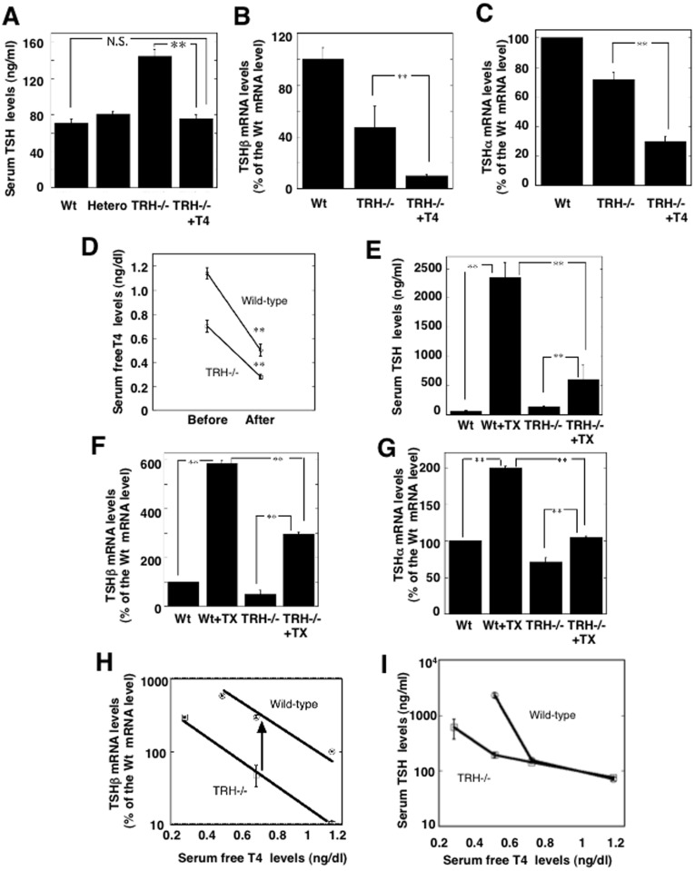
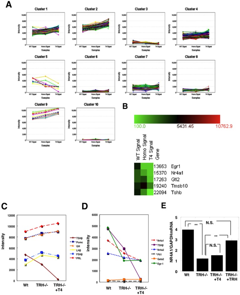
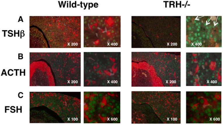
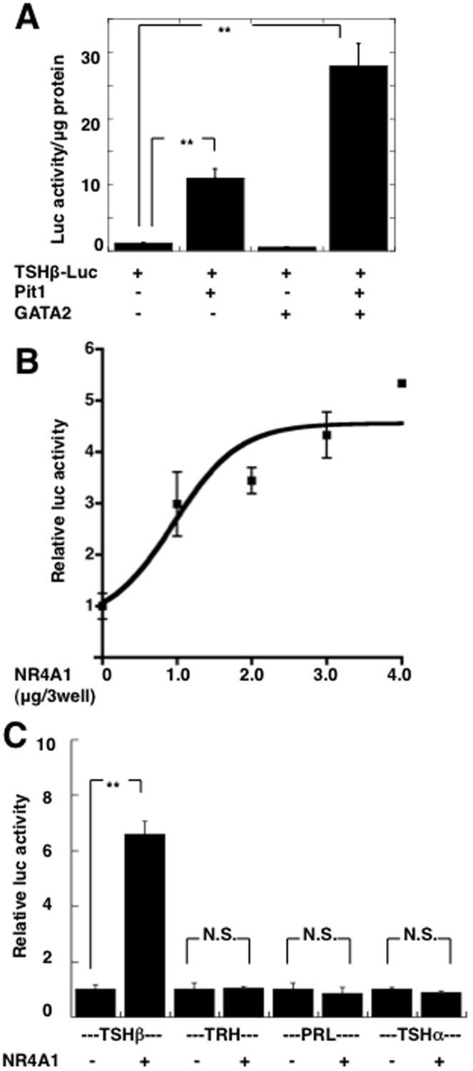
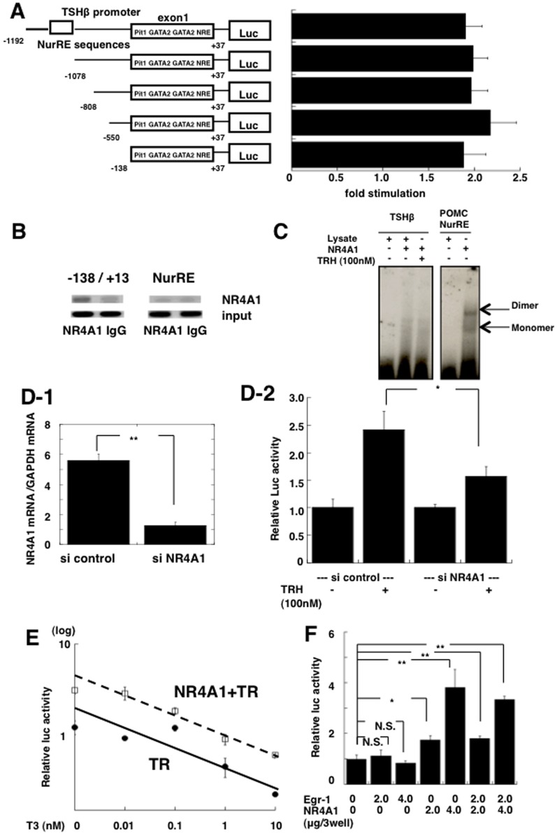
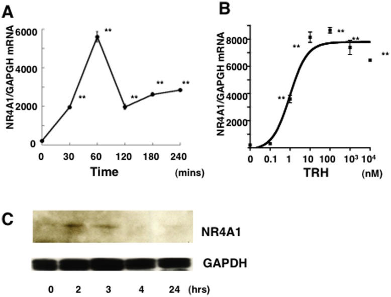
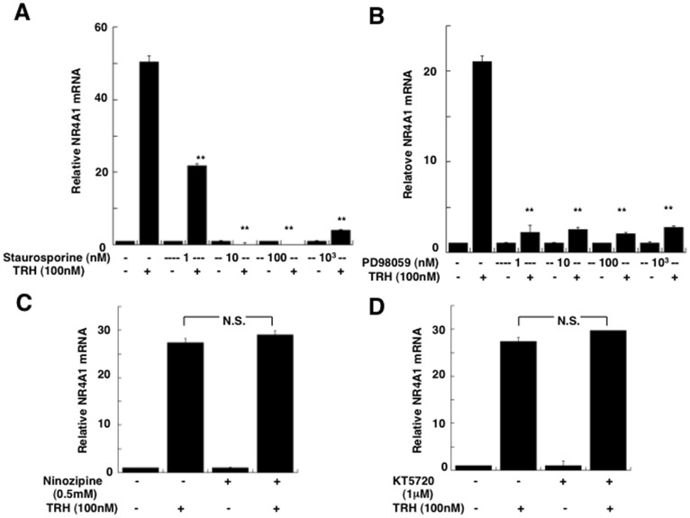
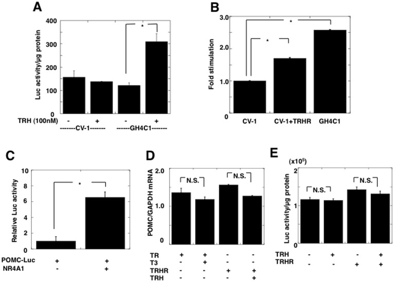
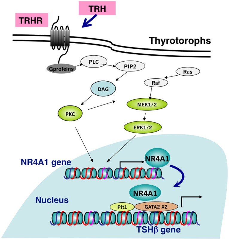
Thyrotropin-releasing hormone (TRH) is a major stimulator of thyrotropin-stimulating hormone (TSH) synthesis in the anterior pituitary, though precisely how TRH stimulates the TSHβ gene remains unclear. Analysis of TRH-deficient mice differing in thyroid hormone status demonstrated that TRH was critical for the basal activity and responsiveness to thyroid hormone of the TSHβ gene. cDNA microarray and K-means cluster analyses with pituitaries from wild-type mice, TRH-deficient mice and TRH-deficient mice with thyroid hormone replacement revealed that the largest and most consistent decrease in expression in the absence of TRH and on supplementation with thyroid hormone was shown by the TSHβ gene, and the NR4A1 gene belonged to the same cluster as and showed a similar expression profile to the TSHβ gene. Immunohistochemical analysis demonstrated that NR4A1 was expressed not only in ACTH- and FSH- producing cells but also in thyrotrophs and the expression was remarkably reduced in TRH-deficient pituitary. Furthermore, experiments in vitro demonstrated that incubation with TRH in GH4C1 cells increased the endogenous NR4A1 mRNA level by approximately 50-fold within one hour, and this stimulation was inhibited by inhibitors for PKC and ERK1/2. Western blot analysis confirmed that TRH increased NR4A1 expression within 2 h. A series of deletions of the promoter demonstrated that the region between bp -138 and +37 of the TSHβ gene was responsible for the TRH-induced stimulation, and Chip analysis revealed that NR4A1 was recruited to this region. Conversely, knockdown of NR4A1 by siRNA led to a significant reduction in TRH-induced TSHβ promoter activity. Furthermore, TRH stimulated NR4A1 promoter activity through the TRH receptor. These findings demonstrated that 1) TRH is a highly specific regulator of the TSHβ gene, and 2) TRH mediated induction of the TSHβ gene, at least in part by sequential stimulation of the NR4A1-TSHβ genes through a PKC and ERK1/2 pathway.


Conflict of interest statement

Competing Interests: The authors have declared that no competing interests exist.

Figures

Figure 1
Figure 1. The hypothalamic-pituitary-thyroid axis in TRH deficient mice.
A) Serum TSH levels were measured in wild-type mice (Wt), TRH knockout mice (TRHKO) heterozygotes (Hetero) and homozygotes (TRH−/−) and TRHKO supplemented with thyroid hormone (TRH−/− + T4). TRHKO (n = 7) were supplemented with thyroid hormone by the daily injection of T4 (1.5 μg/100 g body weight) for 14 days to be euthyroid (n = 6). The serum TSH level in TRH−/−+T4 mice was similar to that in the wild-type (n = 7) and heterozygous mice (n = 6). Values are represented as the mean ± SEM. **, p<0.01; *, N.S., not significant. B) TSHβ mRNA levels were measured in pituitaries of the wild-type mice, TRHKO, and TRHKO supplemented with thyroid hormone (TRH−/−+T4). The TSHβ mRNA level in TRH−/− was decreased to about 50% of that in the wild-type, and that of TRH−/−+T4 mice was further decreased to about 10% (n = 3, p<0.01). **, p<0.01. C) TSHα mRNA levels showed a similar profile to the TSHβ mRNA level shown in Fig 1B. However, the change in TSHβ mRNA was more profound than that in TSHα mRNA. **, p<0.01. D) Two weeks after thyroidectomy, serum free T4 levels were significantly reduced in both the wild-type mice (n = 6) and TRHKO mice (TRH−/−) (n = 5). The serum T4 levels were significantly lower in TRH−/− mice than wild-type mice. **, p<0.01. E) Reflecting hypothyroidism induced by thyroidectomy (TX), the serum TSH level in the wild-type mice showed an approximately 25-fold increase (Wt+TX, n = 6), but TRHKO mice (TRH−/−) showed only about a 6-fold increase (TRH−/−+TX, n = 6). **, p<0.01. F) The TSHβ mRNA levels in thyroidectomized wild-type mice (Wt+TX) were significantly increased after two weeks to approximately 6-fold the control value (Wt) (n = 3). However, the TRHKO mice (TRH−/−) showed a lesser increase (TRH−/−+TX)(n = 3). **, p<0.01; G) A similar profile was observed in TSHα mRNA levels in the pituitary. But a milder effect was observed compared to those of TSHβ mRNA levels. **, p<0.01; n = 3. H) Correlation between serum thyroid hormone levels and the corresponding pituitary TSHβ mRNA levels. Circles represent values for the wild-types and the squares, those for TRH knockout mice (TRH−/−). When comparing the slopes of the curve, it was demonstrated that TRH altered the basal activity and responsiveness of the TSHβ gene to thyroid hormone. The arrow indicates the effect of TRH, taking approximately 80% responsibility for the value of the TSHβ mRNA level in the wild-type pituitary. I) Correlation between serum thyroid hormone levels and the corresponding serum TSH levels. In mild hypothyroidism, the serum TSH levels in TRHKO mice (TRH−/−) were similar to those in the wild-type mice. However a lack of TRH induced a marked impairment of the serum TSH level in severe hypothyroid status.
Figure 2
Figure 2. Microarray and K-means cluster analyses of genes regulated by TRH and by thyroid hormone in TRH-deficient pituitary.
RNA samples from pituitaries of wild-type, TRHKO (TRH−/−), and euthyroid TRHKO mice with thryroid hormone replacement (TRH−/−+T4) were subjected to a whole-genome microarray using Affymetrix Mouse Gene chip 320 2.0 Array. After exclusion of absent signals (less than 100 signals), 10445 out of 45102 genes expressed in the TRHKO pituitary were subjected to the cluster analysis using a K-means algorithm. Hierarchical cluster analysis was carried out using a computer program, Genowiz™ 3.2. A) Genes were divided into ten hierarchical clusters according to changes and profiles among three groups, wild-type (WT signal), TRH−/− (Homo signal), and TRH−/− + T4 (T4 signal). B) In addition to the TSHβ genes, cluster 5 contained the Egr-1, NR4A1, Glt2, and Tmsb10 genes. C) mRNA levels of other anterior pituitary hormones including POMC, GH, LHβ, FSHβ, and prolactin (PRL) were not altered among the three groups, suggesting the expression to be TRH-independent. D) Profiles of the NR4A1, NR4A2, Egr-1, Pit1 and GATA2 genes are depicted. Expression of NR4A1, NR4A2 and Egr-1 might be TRH-dependent. E) Confirmation of results of the microarray analysis of NR4A1 mRNA levels in the TRH-deficient pituitary by real-time PCR. TRHKO mice (TRH−/−, n = 3) were supplemented with thyroid hormone by the daily injection of T4 (TRH−/−+T4, n = 3) and with TRH by 1 mg/kg body weight/day with an Alzet pomp for 7 days (TRH−/−+TRH n = 3). Total RNA obtained from these pituitaries was subjected to real-time PCR. The NR4A1 mRNA level in the TRHKO pituitary was decreased to about 25% of that of the wild-type. The decreased NR4A1mRNA level was not altered by thyroid hormone replacement, but completely reversed to the normal level by TRH administration, suggesting expression of NR4A1 mRNA in the pituitary to be TRH-, but not thyroid hormone-dependent.
Figure 3
Figure 3. Expression and regulation of NR4A1 by TRH in pituitary thyrotrophs in vivo.
The expression of NR4A1 in pituitary thyrotrophs was confirmed by double fluorescent immunohistochemistry. In numbers of cells in the anterior pituitary NR4A1 were expressed and stained as green signals in the nucleus. A) Numbers of cells expressing NR4A1 were also stained with antibody against TSHβ (red signals) in the wild-type pituitary (left panel). As shown in the right panel, the intensity and number of the TSHβ-immunopositive cells were remarkably decreased in the TRH-deficient pituitary, and at X400, most of the NR4A1 expression was reduced or lost in these cells as seen as black dots (indicated by white arrows). B) As observed in Fig 4B and C, the expression of ACTH (B) and FSH (C) was also observed as red signals in the cytoplasm in the anterior lobe. As expected, these ACTH- and FSH- positive cells expressed NR4A1, but the staining intensity was not altered in the TRH-deficient pituitary (right panel).
Figure 4
Figure 4. Stimulation of the promoter activity of the TSHβ gene by NR4A1 in vitro.
A) The human TSHβ promoter region between bp –1192 and +37 linked to a luciferase reporter gene (pA3TSHβ(−1192∼+37)-Luc) was transfected into CV-1 cells. Expression of Pit1 and GATA2 was necessary for the basal promoter activity of the TSHβ gene. When an expression vector for Pit1 was co-transfected, the promoter activity of the TSHβ was stimulated by 10-fold, but co-transfection with GATA2 did not significantly alter it. However, synergistic stimulation of the promoter activity was observed when Pit1 and GATA2 were simultaneously expressed. B) Under the above conditions expressing Pit1 and GATA2, the TSHβ promoter activity was stimulated by NR4A1 in a dose-dependent manner. When 1 μg of expression vector of NR4A1/3 wells was co-trasfected, the promoter activity was stimulated about 2-fold, and when 3 μg, it reached a plateau at about 6-fold. Relative luciferase activity against the value without expression of NR4A1 is shown. C) Stimulation of the promoter activity by NR4A1 was specific for the TSHβ gene (TSHB). The promoter activities of the thyrotropin-releasing hormone (TRH), TSHα, and prolactin (PRL) genes was not stimulated by overexpression of NR4A1. Relative luciferase activity against the value without expression of NR4A1 in each promoter is shown. Values are represented as the mean ± SEM from at least three experiments. **, p<0.01; N.S., not significant.
Figure 5
Figure 5. Characterization of NR4A1-induced stimulation of the promoter activity of the TSHβ
gene. A) To determine the region responsible for the NR4A1-induced stimulation of the TSHβ promoter activity, a series of deletion constructs of the human TSHβ promoter were established. The consensus sequence of the NR4A1 response element (NurRE) was identified in the region between -1091 and -1083 from the transcription start site. The right figure shows the fold-stimulation by NR4A in each deletion mutant of the TSHβ gene. Putative Pit1 and GATA2 binding sites, and the negative thyroid hormone response element (NRE) were indicated as pit1, GATA2 and NRE. All the deletion constructs including pA3TSHβ (−138 ∼ +37) -Luc showed similar stimulation by NR4A1, suggesting the region responsible to be within this area. Fold-increase in activity against that without TRH is shown. B) Chromatin-immunoprecipitation (ChIP) assays were performed with anti-NR4A1 antibody (NR4A1) and normal mouse IgG as a negative control (IgG). GH4C1 cells were transfected with pA3TSHβ (-1192 ∼ +37)-Luc in the presence or absence of 100 n M TRH. Amplified PCR products were stained with ethidium bromide in 2% agarose gels and scanned with a Molecular Imager FX. Chip assays demonstrated that NR4A1 was recruited to the region between -138 and +13 of the TSHβ promoter (-138/+13), but not the region containing NurRE (NuRE). Addition of TRH did not alter recruitment of NR4A1 on the gene (data not shown). All ChIP assays were repeated at least three times. C) The EMSA was performed using a fragment of the radiolabeled POMC promoter containing typical NurRE and a fragment containing the human TSHβ bp -123∼-87. There was no binding of NR4A1 to the TSHβ gene in the absence (−) or presence (+) of 100 nM TRH, while the POMC promoter fragment bound to NR4A1. Arrows indicate NR4A1 bound to the POMC promoter as monomers and dimers. NR4A1 was synthesized by the TNT-coupled reticulocyte lysate system (NR4A1). Lysate indicates un-programmed lysate. D) Effect of knockdown of NR4A1 on the TRH-induced stimulation of the promoter activity of the TSHβ gene. D-1. The NR4A1 mRNA levels were measured after transfection with siCONTROL or siNR4A1 in GH4C1 cells. Transfection of siNR4A1 led to an approximately 80% reduction of the mRNA level after 48 hr. Data are presented as the mean ± SEM from three experiments. **, p<0.01 D-2. GH4C1 cells were transfected with siNR4A1 or siCONTROL, and then TSHβ (-1192 ∼ +37)-Luc reporter, Pit1, and GATA2 expression vectors were cotransfected. After incubation with 100 nM TRH for 24 hr, the promoter activity was measured. In the vehicle transfected with siCONTROL, incubation with TRH stimulated promoter activity of the TSHβ gene showing 242±33% of that without TRH stimulation. Knockdown of NR4A1 resulted in significant reduction of TRH-induced stimulation (156% of that without TRH stimulation). Data are presented as the mean ± SEM from three experiments. *, p<0.05 E) Negative regulation of the TSHβ gene by thyroid hormone was examined in the presence or absence of NR4A1 in CV-1 cells. Expression vector for thyroid hormone receptor β 1 (TRβ1) was also co-transfected. The responsiveness of the TSHβ promoter activity to thyroid hormone (T3) without expression of NR4A1 was significantly reduced as compared to that with expression of NR4A1, suggesting NR4A1 increased the basal promoter activity and responsiveness to T3 of the TSHβ promoter. F) Effect of NR4A1 and Egr-1 on the promoter activity of the TSHβ gene. Egr-1 affected neither the basal promoter activity of the TSHβ gene nor the NR4A1-induced stimulation of the gene. The amount of vector/3 wells transfected is indicated below the graph. **, p<0.01; *, p<0.05; N.S. not significant.
Figure 6
Figure 6. Immediate-early response of NR4A1 mRNA and protein levels to TRH in GH4C1 cells.
A) Incubation with 1μM TRH led to a remarkable increase of the endogenous NR4A1 mRNA level with a peak within 1 hr (51-fold) and then an immediate reduction in 120 mins in GH4C1 cells. B) The TRH-induced increase of the NR4A1 mRNA level was observed in a dose-dependent manner. The minimum effect was observed with 0.1 nM TRH and then reached a plateau at 10 nM. C) Western blot analysis in GH4C1 cells demonstrated that incubation with 100 nM TRH led to a significant increase of NR4A1 protein (NR4A1) within 2 h, and NR4A1 was then degradated in 4 h, while the level of GAPDH was not altered.
Figure 7
Figure 7. Regulation of NR4A1 mRNA by TRH through the PKC and MAPK pathways.
The signal transduction pathways for the TRH-induced stimulation of NR4A1 mRNA expression were determined using several inhibitors in GH4C1 cells. After overnight incubation in DMEM without serum, GH4C1 cells were treated for 1 hr with a PKC inhibitor, Staurosporine (A), a MAPK inhibitor, PD98059 (B), a Ca channel inhibitor, nimozipine (C), and a PKA inhibitor, KT5720 (D). Significant inhibition of 100 nM TRH-induced stimulation of endogenous NR4A1 mRNA expression was observed with 1.0∼ nM Staurosporine and 1.0 ∼ nM PD98059, but not 0.5 nM nimozipine or 1 μM KT5720. The value relative to that without TRH is shown. Data are presented as the mean ± SEM. from three experiments. **, p<0.01.
Figure 8
Figure 8. TRH stimulates the promoter activity of the NR4A1 gene through TRH receptors.
A) A fragment spanning bp −1295 and +152 of the promoter of the human NR4A1 gene fused to a luciferase reporter gene (pA3NR4A1(−1295∼+152)-Luc) was transfected into CV-1 cells (CV-1) that express no endogenous TRH receptors (TRHRs) and GH4C1 cells (GH4C1) expressing endogenous TRHRs. In CV-1 cells, 100 nM TRH did not stimulate the promoter activity of the NR4A1 gene, in contrast, it led to a significant increase in GH4C1 cells (255±33% of the control without TRH, n = 3, p<0.05). B) When TRH receptors were expressed in CV-1 cells (CV-1+TRHR), 10−7 M TRH induced a significant increase of NR4A1 promoter activity, suggesting that TRH exhibited stimulation of the NR4A1 gene through TRH receptors. Fold-increase on incubation with 100 nM TRH is represented. The value in GH4C1 cells is also shown as a control. C) The rat POMC promoter containing the region from bp −706 to +64 fused to a luciferase reporter (POMC-Luc) was transfected into At-T20 cells expressing the endogenous POMC gene. The promoter activity was significantly increased about 6-fold by overexpression of NR4A1. D) The level of endogenous POMC mRNA was not altered either by incubation with expression of TR and T3 or by incubation with expression of TRH receptors (TRHRs) and 100 nM TRH in At-T20 cells. E) The POMC promoter activity was not stimulated by incubation with 100 nM TRH even with expression of TRH receptors (TRHRs), suggesting that the effect of TRH occurred in a promoter-specific manner. Data are presented as the mean ± SEM. from three experiments. *, p<0.05; N.S., not significant.
Figure 9
Figure 9. Proposed models of the TRH-NR4A1-TSHβ pathway.
TRH stimulates the transcription of the NR4A1 gene through TRH receptors, and PKC and MAPK pathways within 1 hr. The increased amount of NR4A1 stimulates TSHβ promoter activity, which may act cooperatively with Pit1 and GATA2 on the gene.

Similar articles

Cited by

References

    1. Boler J, Enzmann F, Folkers K, Bowers C, Schally A. The identity of chemical and hormonal properties of the thyrotropin releasing hormone and pyroglutamyl-histidyl-proline amide. Biochem Biophys Res Commun. 1969;37:705–710. - PubMed
    1. Burgus R, Dunn T, Ward D, Vale W, Amoss M, et al. [Synthetic polypeptide derivatives with thyrotropin releasing factor hypophysiotropic activity]. C R Acad Sci Hebd Seances Acad Sci D. 1969;268:2116–2118. - PubMed
    1. Jackson I. Thyrotropin-releasing hormone. N Engl J Med. 1982;306:145–155. - PubMed
    1. Carr F, Shupnik M, Burnside J, Chin W. Thyrotropin-releasing hormone stimulates the activity of the rat thyrotropin beta-subunit gene promoter transfected into pituitary cells. Mol Endocrinol. 1989;3:717–724. - PubMed
    1. Carr F, Fisher C, Fein H, Smallridge R. Thyrotropin-releasing hormone stimulates c-jun and c-fos messenger ribonucleic acid levels: implications for calcium mobilization and protein kinase-C activation. Endocrinology. 1993;133:1700–1707. - PubMed

Publication types

MeSH terms

Substances