. 2012;7(7):e40447.
doi: 10.1371/journal.pone.0040447. Epub 2012 Jul 9.

Morphological and molecular diagnosis of anisakid nematode larvae from cutlassfish (Trichiurus lepturus) off the coast of Rio de Janeiro, Brazil

Affiliations

Morphological and molecular diagnosis of anisakid nematode larvae from cutlassfish (Trichiurus lepturus) off the coast of Rio de Janeiro, Brazil

Juliana Novo Borges et al. PLoS One. 2012.

Abstract

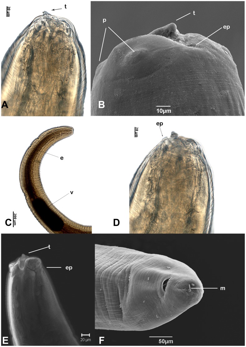
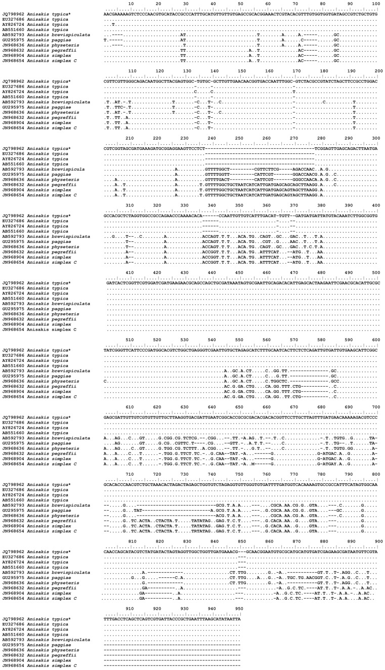
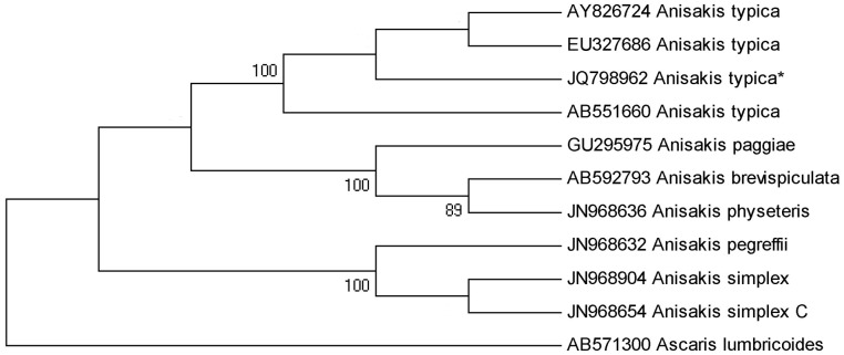
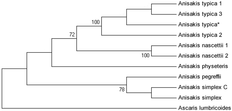
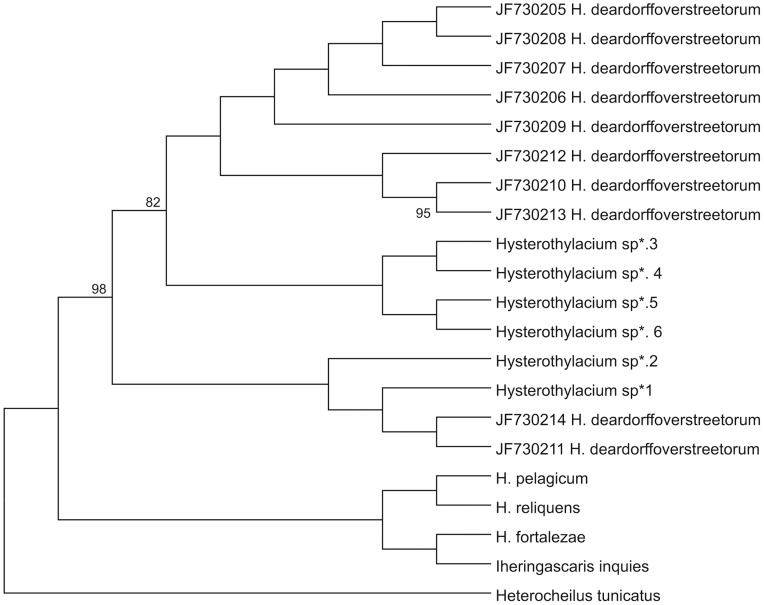
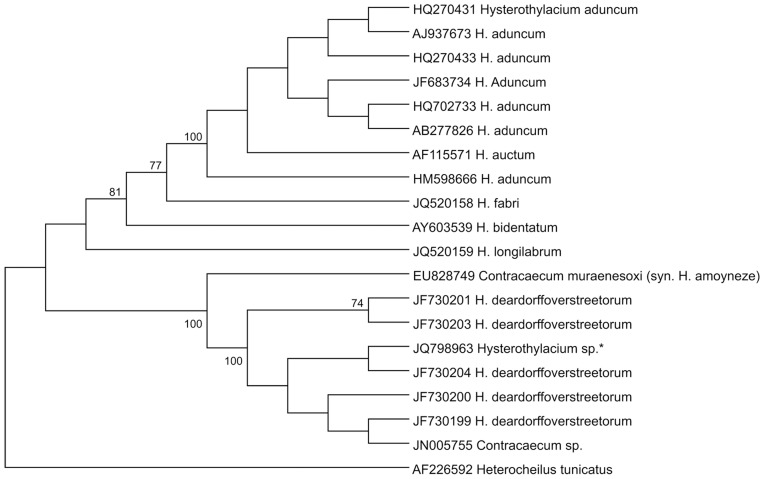
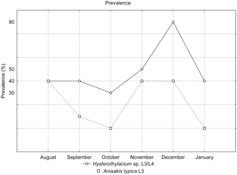
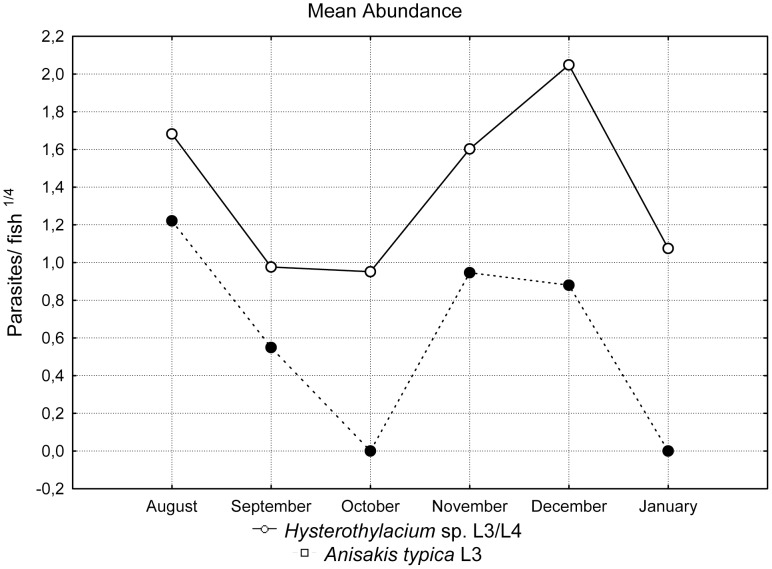
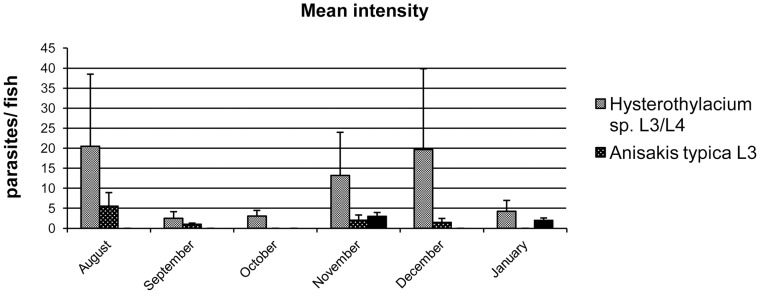
Anisakid nematode larvae from Trichiurus lepturus off coast of Rio de Janeiro were studied using light, laser confocal and scanning electron microscopy, in addition to a molecular approach. Mitochondrial cytochrome c-oxidase subunit 2 (mtDNA cox-2), partial 28S (LSU) and internal transcribed spacers (ITS-1, 5.8S, ITS-2) of ribosomal DNA were amplified using the polymerase chain reaction and sequenced to evaluate the phylogenetic relationships between the nematode taxa. The morphological and genetic profiles confirmed that, of the 1,030 larvae collected from the 64 fish examined, 398 were analysed, of which 361 were Hysterothylacium sp. and 37 were Anisakis typica. Larvae of Hysterothylacium sp. were not identified to the species level due to the absence of similar sequences for adult parasites; however, the ITS sequence clustered in the phylogenetic tree with sequences of H. deardorffoverstreetorum, whereas an mtDNA cox-2 and LSU concatenated phylogenetic analysis demonstrated the presence of two clades, both of them under the same name as the larval H. deardorffoverstreetorum. Data on the occurrence of parasites during the winter and summer months were compared using the t-test. The greatest prevalence and intensity of infection were recorded for larval Hysterothylacium, with a prevalence of 51.56% and an intensity of up to 55 parasites per fish. The larval Anisakis exhibit a higher abundance and intensity of infection in the winter months, and those of Hysterothylacium during the summer. However, the t-test indicated no significant differences between the abundance and intensity of infection recorded during the months of collection for either of these larval nematodes. All sequences generated in this study were deposited in GenBank.


Conflict of interest statement

Competing Interests: The authors have declared that no competing interests exist.

Figures

Figure 1
Figure 1. A–F: Anisakis typica larvae: light, CLSM and SEM microscopy.
A- Aanterior end with boring tooth; B- SEM of lips with papilla, boring tooth and excretory pore; C- Esophagus and ventriculus; D- Position of excretory pore; E- CLSM reconstruction with detail boring tooth and excretory pore; F- SEM of tail with mucron terminal. Abbreviations: e - esophagus; ep - excretory pore; t - tooth; p - papilla; v - ventriculus; m - mucron.
Figure 2
Figure 2. Alignment of ITS-1 and ITS-2 sequences representing Anisakis spp.
Dots indicate identity with the first sequence, dashes are inferred insertion-deletion events and * represents our sample.
Figure 3
Figure 3. Maximum likelihood reconstruction between sequences of Anisakis typica obtained in this study (*) and sequences of Anisakis species from the GenBank, with the tree inferred from the ITS data set.
The numbers on the tree branches represent the percentage of bootstrap resampling. Ascaris lumbricoides was used as an out group.
Figure 4
Figure 4. Maximum likelihood reconstruction between sequences of Anisakis typica obtained in this study (*) and sequences of Anisakis species from the GenBank, with the tree inferred from mtDNA cox-2 and LSU data sets.
The numbers on the tree branches represent the percentage of bootstrap resampling. Ascaris lumbricoides was used as an out group.
Figure 5
Figure 5. A–H: Hysterothylacium sp. larvae: SEM and CLSM microscopy.
A- SEM of anterior end with alae and excretory pore; B- Detail of L3 lips with inconspicuous boring tooth and papillae; C- Detail of lips of L4 with dorsal lip showing double papilla; D- CLSM of esophagus; E- CLSM reconstruction with ventriculus, intestinal caecum and esophagus; F: CLSM reconstruction with nerve ring and excretory pore; G- SEM of tail; H- SEM micrograph with a detail of the digitiform tip with terminal mucron. Abbreviations: a - alae; ep - excretory pore; p – papilla; t - tooth; dl - dorsal lip; e - esophagus; ic - intestinal caecum; v - ventriculus; n - nervous ring and m - mucron.
Figure 6
Figure 6. Alignment of mtDNA cox-2 sequences representing Hysterothylacium and Iheringascaris taxa.
Dots indicate identity with the first sequence, dashes are inferred insertion-deletion events and * represents our samples.
Figure 7
Figure 7. Maximum likelihood reconstruction between sequences of Hysterothylacium obtained in this study (*) and sequences of Hysterothylacium and Iheringascaris spp. from the GenBank, with the tree inferred from mtDNA cox-2 and LSU data sets.
The numbers on the tree branches represent the percentage of bootstrap resampling. Heterocheilus tunicatus was used as an out group.
Figure 8
Figure 8. Maximum likelihood reconstruction between sequences of Hysterothylacium sp. larvae obtained in this study (*) and sequences of other anisakid species from the GenBank inferred from the ITS dataset.
The numbers on the tree branches represent the percentage of bootstrap resampling. Heterocheilus tunicatus was used as an out group.
Figure 9
Figure 9. Ecological data of Anisakis typica and Hysterothylacium sp.: prevalence expressed as a percentage.
Figure 10
Figure 10. Ecological data of Anisakis typica and Hysterothylacium sp.: mean abundance (no. of parasites/fish) transformed using the fourth root.
Figure 11
Figure 11. Ecological data of Anisakis typica and Hysterothylacium sp.: mean intensity (no. of parasites/parasitized fish); the bars represent the standard deviation.

Similar articles

Cited by

References

    1. Mattiucci S, Nascetti G. Advances and trends in molecular systematics of anisakid nematodes, with implications for their evolutionary, ecology and host-parasite co-evolutionary processes. Rollinson D, Hay SI, editors. Academic Press. 2008;66:47–148. editors. Advances in Parasitology. - PubMed
    1. Klimpel S, Palm HW. Anisakid nematode (Ascaridoidea) life cycles and distribution. Increasing zoonotic potential in the time of climate change? Parasitology Research Monographs. 2001;2:201–222.
    1. Anderson RC. Nematode parasites of vertebrates: Their development and transmission. Wallingford: CABI Publishing. 672 p. 2000.
    1. Mattiucci S, Nascetti G, Cianchi R, Paggi L, Arduino P, et al. Genetic and ecological data on the Anisakis simplex complex with evidence for a new species (Nematoda, Ascaridoidea, Anisakidae). Journal of Parasitology. 1997;83:401–416. - PubMed
    1. Nadler SA, D’Amelio S, Dailey MD, Paggi L, Siu S, et al. Molecular phylogenetics and diagnosis of Anisakis, Pseudoterranova, and Contracaecum from northern Pacific marine mammals. Journal of Parasitology, 91. 2005;(6):1413–29. - PubMed

Publication types

MeSH terms

LinkOut - more resources