. 2012;7(7):e40469.
doi: 10.1371/journal.pone.0040469. Epub 2012 Jul 9.

Establishment and characterization of a buffalo (Bubalus bubalis) mammary epithelial cell line

Affiliations

Establishment and characterization of a buffalo (Bubalus bubalis) mammary epithelial cell line

Vijay Anand et al. PLoS One. 2012.

Abstract

Background: The objective of this study was to establish the buffalo mammary epithelial cell line (BuMEC) and characterize its mammary specific functions.

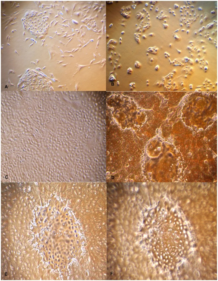
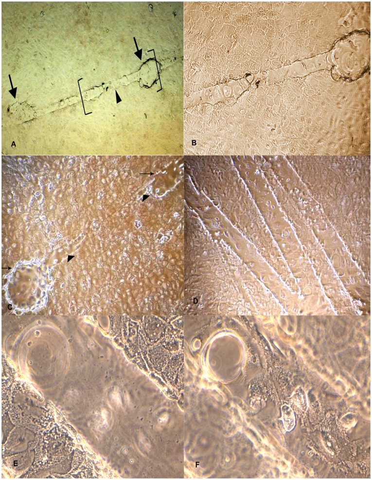
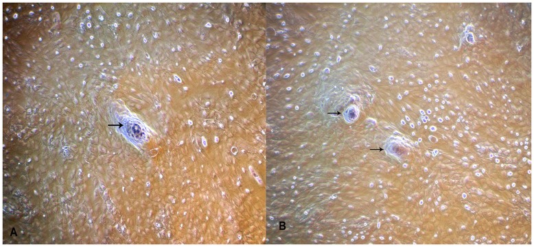
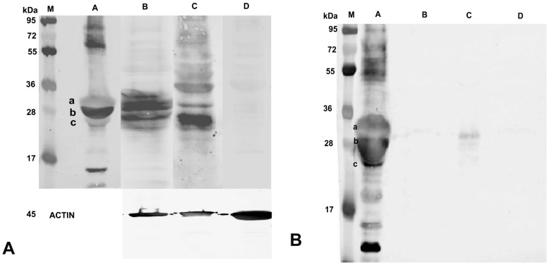
Methodology: Buffalo mammary tissue collected from the slaughter house was processed enzymatically to obtain a heterogenous population of cells containing both epithelial and fibroblasts cells. Epithelial cells were purified by selective trypsinization and were grown in a plastic substratum. The purified mammary epithelial cells (MECs) after several passages were characterized for mammary specific functions by immunocytochemistry, RT-PCR and western blot.

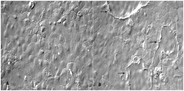
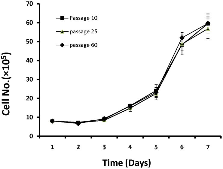
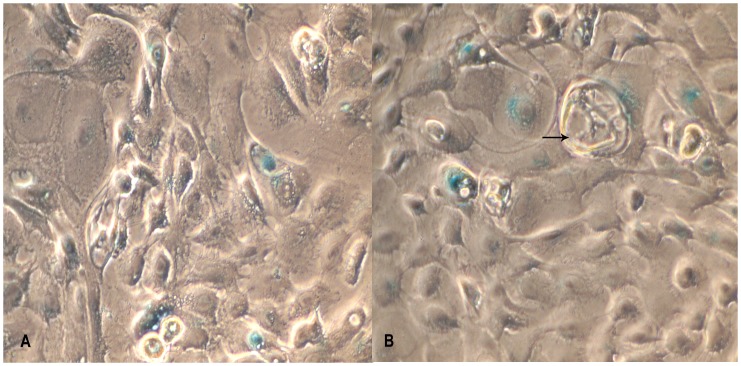
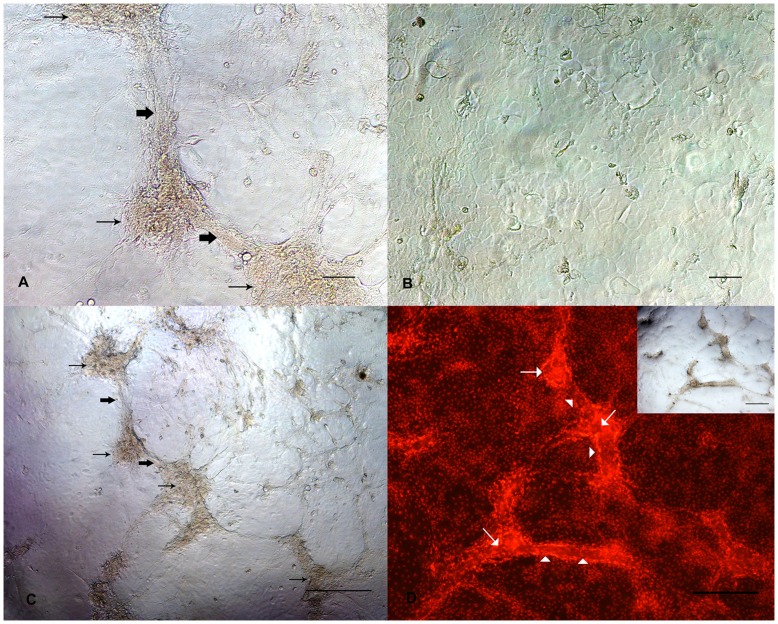
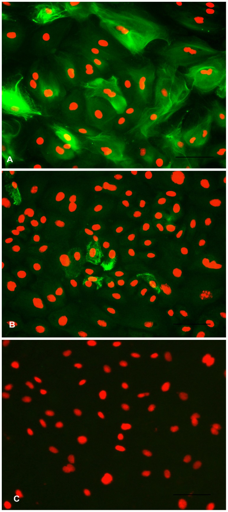
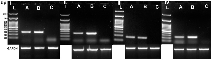
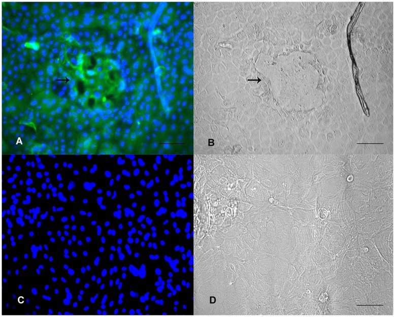
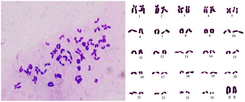
Principal findings: The established buffalo mammary epithelial cell line (BuMEC) exhibited epithelial cell characteristics by immunostaining positively with cytokeratin 18 and negatively with vimentin. The BuMEC maintained the characteristics of its functional differentiation by expression of β-casein, κ-casein, butyrophilin and lactoferrin. BuMEC had normal growth properties and maintained diploid chromosome number (2n = 50) before and after cryopreservation. A spontaneously immortalized buffalo mammary epithelial cell line was established after 20 passages and was continuously subcultured for more than 60 passages without senescence.

Conclusions: We have established a buffalo mammary epithelial cell line that can be used as a model system for studying mammary gland functions.


Conflict of interest statement

Competing Interests: The authors have declared that no competing interests exist.

Figures

Figure 1
Figure 1. Photomicrographs of isolation and culture of Buffalo Mammary Epithelial cells (BuMECs).
A: Mixed population of epithelial and fibroblast cells (×100); B: Purified BuMEC seeded at low density forming islands (×100); C: Confluent mono layer of BuMECs showing cobble stone morphology (×100); D: Post confluent stage BuMECs forming dome structure (×100); E: Phase contrast image of dome structure with focus on the monolayer (×100); F: Phase contrast image of dome structure with focus set at the top of dome (×100). The dome structure represents a raised layer of cells above the plastic substratum.
Figure 2
Figure 2. Organization of dome structure on plastic substratum.
A: Development of inter-connecting structure (arrow head) between domes (arrows) in BuMECs monolayer attached to plastic substratum (×40); B: Magnified view of dome and inter-connecting structure shown in bracket in Fig. A (×100); C: Phase contrast microscopic image of an intermediate stage in the process of development of interconnecting structure (arrow head) between domes (arrow) (×100); D: Branching pattern of interconnecting structure between domes (×100); E: Magnified view of inter-connecting structure with focus on the monolayer (×400); F: Magnified view of interconnecting structure with focus on the top of the structure showing cells above the substratum (×400). This is a unique observation in BuMECs. These inter-connecting structures may represent contact mediated differentiation of BuMECs on plastic substratum.
Figure 3
Figure 3. Development of papillate structures in BuMECs on the plastic substratum.
A: BuMECs from passage 6 forming papillate (arrow) structure after 15 days of growth on plastic substratum (×100); B: BuMECs from passage 8 forming papillate (arrow) structures after 15 days of growth on plastic substratum (×100). Papillate structures represent a small nipple-like projection above the plastic substratum indicating differentiation characteristics of BuMECs.
Figure 4
Figure 4. Insulin and Hydrocortisone increases dome formation in BuMECs.
A: BuMECs grown in the absence of insulin and hydrocortisone (×200); B: BuMECs grown in the presence of insulin and hydrocortisone showing increased formation of domes (arrow) (×200). Results represent three independent experiments.
Figure 5
Figure 5. Growth curves for BuMECs at early passage (10), late passage (60) and after revival from cryopreservation (passage 25).
Results are means ±SD of three independent experiments.
Figure 6
Figure 6. Analysis of cell Senescence–associated β-galactosidase (SA-β-gal) activity in BuMECs.
A: SA- β-gal staining in early passage (10) BuMECs (×400); B: SA β-gal staining in late passage (60) BuMECs; Staining is evident from BuMEC with normal morphology and cells with vacuoles (arrow) (×400); Results represents images from three independent experiments.
Figure 7
Figure 7. Morphological differentiation of BuMECs cultured in Type I collagen matrix.
A: Phase contrast microscopic image showing development of cellular aggregates (thin arrow) and ‘duct–like’ (bold arrow) structures in BuMECs grown in attached collagen Type I matrix for four days; B: Phase contrast microscopic image of BuMECs grown on plastic substratum for four days shows no such morphological changes; C: Phase contrast microscopic image (low magnification) showing development of cellular aggregates (thin arrow) and ‘duct–like’ (bold arrow) structures in BuMECs grown in attached collagen Type I matrix for four days; D: Fluorescent image of acini-like cellular aggregate and duct- like structure in monolayer of BuMECs grown on attached collagen Type I matrix which is evident from the propidium iodide stained nucleus of the cells forming the structures (insert image show the phase contrast image of the field). Acini-like structure (arrow) constitutes a large aggregate of PI stained nuclei and duct-like structure (arrow head) showing a clear arrangement of PI stained nuclei along the sides of duct-like structure suggesting the formation of wall and lumen. Bars, A and B 100 µm, C and D 500 µm Results represent images from two independent experiments.
Figure 8
Figure 8. Immunostaining for cytoskeletal markers in BuMECs.
A: Fluorescent image of BuMECs stained for Cytokeratin 18 showing intermediate filaments; B: Fluorescent image of BuMECs stained for Vimentin; C: Negative control with primary antibody replaced with a normal mouse IgG (Isotype control). The secondary antibodies were goat anti-mouse FITC conjugated antibody. Propidium iodide was used as a nuclear counter stain. Bars 100 µm. Results represent images from three independent experiments.
Figure 9
Figure 9. RT-PCR analysis for β-casein (I), κ-casein (II), Butyrophilin (III) and Lactoferrin (IV) in BuMECs.
L: 100 bp ladder; A: BuMECs; B: Mammary Tissue (Positive control) and C: Skin fibroblasts (Negative control); Loading control represents the house keeping gene Glyceraldehyde 3–phosphate dehydrogenase (GAPDH). Results representative of minimum three independent experiments.
Figure 10
Figure 10. Analysis of casein expression in BuMECs.
A:Western blot analysis for casein in BuMEC lysates, M-Protein MW standard, A-Buffalo milk, B-Mammary tissue lysate (Positive control), C-BuMEC lysate, D-Skin fibroblasts lysate (negative control) ACTIN- Loading control. B: Western Blot analysis for casein in BuMEC conditioned media, M–Protein MW standard, A-Buffalo milk, B-Concentrated growth medium (negative control), C-BuMEC conditioned medium, D-Conditioned media from Skin fibroblasts a; α-casein, b; β-casein, c; κ-casein in A and B.
Figure 11
Figure 11. Immunostaining for casein in BuMECs
A: Fluorecent image of casein producing BuMECs with relatively stronger signal from cells associated with domes (arrow); B: Light microscope image of same field as in A; C: Negative control experiments with rabbit IgG (Isotype control); D: Light microscope image of same field as in C. The secondary antibodies were rabbit anti-mouse FITC conjugated antibody. DAPI was used as nuclear counter stain. Bars 100 µm. Results represent minimum of three independent experiments.
Figure 12
Figure 12. Chromosomal analysis of Buffalo Mammary Epithelial Cells (BuMEC).
Representative metaphase spread and karyotype of BuMECs showing 25 pairs of chromosomes specific to buffalo (2n = 50).

Similar articles

Cited by

References

    1. “Water Buffalo An asset undervalued”. United Nations Food and Agriculture Organization. Retrieved on 2011–09–15. 2000. FAO website: http://www.aphca.org/publications/files/w_buffalo.pdf.
    1. Martinet J, Houdebine LM, Head HH. Biology of Lactation. Paris: INRA publication. 670 p. 1999.
    1. Larson BL. Larson BL, Anderson, RR, editors. Biosynthesis and cellular secretion of milk. 1985. pp. 129–163. Lactation. Ames: The lowa State University Press.
    1. Rosen JM, Jones WK, Campbell SM, Bisbee CA, Yu-Lee LY. Czech MP, Kahn, CR, editors. Structure and regulation of peptide hormone responsive genes. Vol 23. 1985. New York: A. R. Liss. Proceedings UCLA Symposium on Membrane Receptors and Cellular Regulation. (385–396).
    1. Levine JF, Stockdale FE. 3T3-L1 adipocytes promote the growth of mammary epithelium. Exp Cell Res. 1984;151:112–122. - PubMed

Publication types

MeSH terms