. 2012;7(7):e40470.
doi: 10.1371/journal.pone.0040470. Epub 2012 Jul 6.

SCaMC-1Like a member of the mitochondrial carrier (MC) family preferentially expressed in testis and localized in mitochondria and chromatoid body

Affiliations

SCaMC-1Like a member of the mitochondrial carrier (MC) family preferentially expressed in testis and localized in mitochondria and chromatoid body

Ignacio Amigo et al. PLoS One. 2012.

Abstract

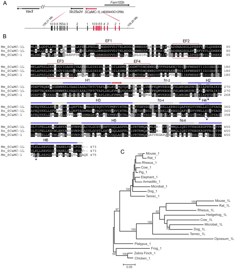
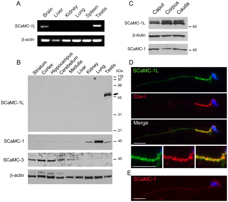
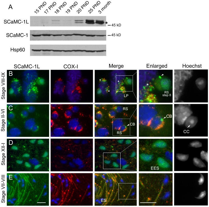
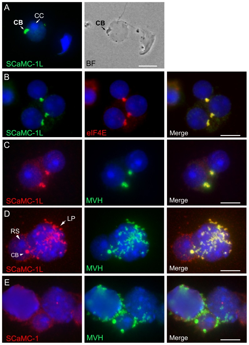
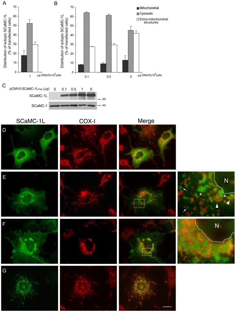
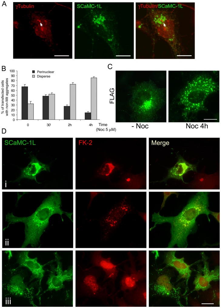
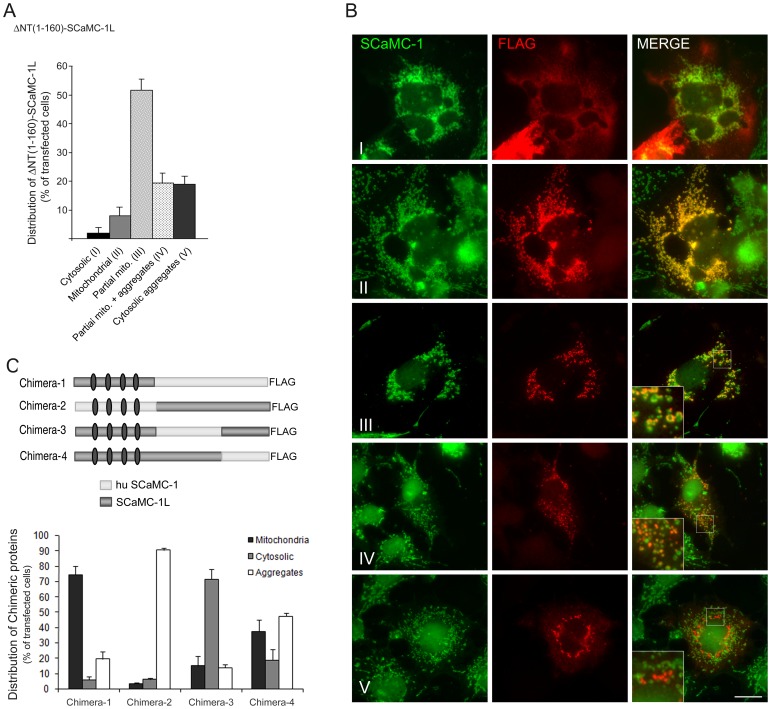
Mitochondrial carriers (MC) form a highly conserved family involved in solute transport across the inner mitochondrial membrane in eukaryotes. In mammals, ATP-Mg/Pi carriers, SCaMCs, form the most complex subgroup with four paralogs, SCaMC-1, -2, -3 and -3L, and several splicing variants. Here, we report the tissue distribution and subcellular localization of a mammalian-specific SCaMC paralog, 4930443G12Rik/SCaMC-1Like (SCaMC-1L), which displays unanticipated new features. SCaMC-1L proteins show higher amino acid substitution rates than its closest paralog SCaMC-1. In mouse, SCaMC-1L expression is restricted to male germ cells and regulated during spermatogenesis but unexpectedly its localization is not limited to mitochondrial structures. In mature spermatids SCaMC-1L is detected in the mitochondrial sheath but in previous differentiation stages appears associated to cytosolic granules which colocalize with specific markers of the chromatoid body (CB) in post-meiotic round spermatids and inter-mitochondrial cement (IMC) in spermatocytes. The origin of this atypical distribution was further investigated by transient expression in cell lines. Similarly to male germ cells, in addition to mitochondrial and cytosolic distribution, a fraction of SCaMC-1L-expressing COS-7 cells display cytosolic SCaMC-1L-aggregates which exhibit aggresomal-like features as the CB. Our results indicate that different regions of SCaMC-1L hinder its import into mitochondria and this apparently favours the formation of cytosolic aggregates in COS-7 cells. This mechanism could be also operational in male germ cells and explain the incorporation of SCaMC-1L into germinal granules.


Conflict of interest statement

Competing Interests: The authors have declared that no competing interests exist.

Figures

Figure 1
Figure 1. SCaMC-1Like, SCaMC-1L, a new SCaMC paralog emerged by a tandem duplication in mammals.
(A) Scheme of the head-to-tail tandem array of mouse SCaMC-1 (slc25a24) and 4930443G12Rik/SCaMC-1L genes. SCaMC-1, SCaMC-1L and flanking loci are represented by arrows indicating transcription orientation. SCaMC-1/SCaMC-1L intron-exon organization is also shown. Exons are indicated by filled boxes (not to scale) and numbered. (B) Alignment of predicted mouse and rat SCaMC-1L protein sequences (Mm_SCaMC-1L and Rn_SCaMC-1L) with that of mouse SCaMC-1 (slc25a24, Mm_SCaMC-1). Alignment was performed with ClustalW and colored with BOXSHADE 3.21 software. Predicted EF-hand calcium-binding motifs are indicated by red boxes. Secondary structure prediction for the region homologous to mitochondrial carriers, amino acids 181 to end, of Mm_SCaMC-1L was obtained using Jpred3 server . The predicted transmembrane helices are indicated (H1–H6), matrix loops are marked in lower case letter and the β-strand region by an arrow. The residues proposed as participants in substrate interactions in H2, H4 and H6 are included in boxes and marked with asterisks. (C) Phylogenetic relationships among SCaMC-1 and SCaMC-1L paralogs. The phylogenetic tree was constructed using amino acid sequences derived from exons 2 to 7 with the neighbor-joining method (MEGA 4.0, [70]) and PAM distances. Non-mammalian vertebrate SCaMCs were used as outgroups. The scale of branch lengths is indicated (number of substitutions per site). Percentage bootstrap values are shown in each node (500 replicates, only bootstrap values of 60% or more are shown). The accession numbers of annotated SCaMC-1 and SCaMC-1L proteins as well as the amino-acid sequences of manually assembled orthologs are compiled in Supplementary Tables S1 and S2. See alignment in Figure S6.
Figure 2
Figure 2. SCaMC-1L is expressed in testis and male germ cells.
(A) RT-PCR analysis of SCaMC-1L expression in mouse tissues. Equivalent aliquots of cDNAs derived from the indicated tissues were used as templates. Amplification of β-actin was used as an internal control. The results obtained indicate that SCaMC-1L is expressed preferentially in testis and, at lower levels, in brain. (B) Expression of SCaMC-1L protein in adult mouse tissues. 10 µg of total protein extracts from the indicated tissues were analyzed by western blot using a specific anti-SCaMC-1L antibody. A single band of the expected size, around 50 kDa, was exclusively detected in testis, marked with an arrow. β-actin levels are shown as loading control. A parallel blot was incubated with antibodies against SCaMC-1 which detected a single band of 45 kDa, and then with antibodies against SCaMC-3 which detected a band of of about 48 kDa. The specific distribution patterns of the labelled bands in mouse tissues rule out any significant crossreactivity among paralogs. (C) 5 µg of total proteins from cauda, corpus and caput spermatids were analyzed by western blot with anti-SCaMC-1L antibody. Membranes were re-probed with β-actin antibody as loading control and anti-SCaMC-1. Equivalent SCaMC-1L levels are found in spermatids from different regions of epididymis. (D) SCaMC-1L-staining is detected in the midpiece of epididymal spermatids. SCaMC-1L was detected using an affinity-purified SCaMC-1L antibody and visualized with a FITC-conjugated secondary antibody, mitochondria were stained with an anti-COX-I monoclonal antibody and visualized with a Cy3-conjugated secondary antibody, nuclei were stained with Hoechst and triple merged panel is also shown. Merge panel shows the co-localization of SCaMC-1L and COX-I staining in the mitochondrial sheath of midpiece. Enlarged images of this region are shown at bottom. (E) SCaMC-1 is not detected in mature spermatids. SCaMC-1 staining was performed using anti-SCaMC-1 antibody at dilution 1∶200 and visualized Alexa Fluor 555 anti-rabbit as secondary antibody, nuclei were stained with Hoechst. Scale bars; 10 µm.
Figure 3
Figure 3. Immunohistochemical localization of SCaMC-1L protein in mouse testis.
(A) Analysis of SCaMC-1L expression during spermatogenesis. SCaMC-1L levels were determined in mitochondrial-enriched extracts prepared from mouse testis at different post-natal days (PND), between 15–25 days and 3-month-old, by western blot. The levels of the mitochondrial proteins SCaMC-1 and hsp60 were used as loading controls. (B–E) Detection of SCaMC-1L in squash preparations of seminiferous tubules by immunofluorescence assays. The stages of seminiferous segments are indicated. SCaMC-1L and COX-I detection were performed as described in Figure 2. SCaMC-1L is detected in granules close to mitochondria in late pachytene spermatocytes (LP) (B), and round spermatids (RS) (B, C), identified by the heterochromatic chromocenter (CC) at the nucleus (C, indicated by an arrow). In elongating spermatids (EES) a diffuse SCaMC-1L staining is detected in the nucleus (D), in elongated spermatid (ES) SCaMC-1L is found at the midpiece matching with COX-I signals (E). Enlarged images of marked insets for merged panels and corresponding Hoechst staining are shown. Scale bars; 10 µm.
Figure 4
Figure 4. Intracellular distribution of SCaMC-1L in drying-down preparations of mouse seminiferous tubules.
Immunofluorescence staining of SCaMC-1L in drying-down slides was performed as in Figure 2. Representative images of SCaMC-1L (green) and the mitochondrial marker COX-I (red) staining in late pachytene spermatocytes (A), haploid round spermatids (B), elongating spermatids at different steps of differentiation (C, D, E) and elongated spermatids (F) are shown. Scale bars; 10 µM.
Figure 5
Figure 5. Localization of SCaMC-1L in chomatoid body.
Drying-down slides were labeled with anti-SCaMC-1L antibody and its localization in chromatoid body (CB) of round spermatids (RS) was confirmed by parallel phase contrast microscopy (A) and by co-staining with the specific markers for CB, eIF4E (B) and MHV (C, D). CBs, identified as eIF4E and MVH protein-positive structures, appear strongly stained for SCaMC-1L. In late pachytene spermatocytes (LP) SCaMC-1L signals co-localizes entirely with cytosolic granules MVH-positive (D). RS were identified by the heterochromatic chromocenter (CC) at the nucleus (indicated by arrow in A). (E) Absence of co-localization of SCaMC-1 and MVH signals in drying-down slides. Scale bars; A, 5 µM; B–E; 10 µM.
Figure 6
Figure 6. Expressed SCaMC-1L shows different intracellular patterns in COS-7 cells.
Quantitative assessment of SCaMC-1L distribution in COS-7 cells. The percentage of SCaMC-1L-expressing cells showing cytosolic, mitochondrial or extra-mitochondrial patterns were analyzed using one µg of DNA (A) or with different DNA concentrations (B). Intracellular pattern was determined in parallel transfections by co-staining with anti-SCaMC-1L and anti-COX-I, as mitochondrial control, and the distribution of SCaMC-1L-signals were examined by visual inspection under a fluorescent microscope. Two hundred cells were analyzed for each transfection assay. Data are the mean ± SEM of three independent experiments. (C) The expression levels of SCaMC-1L at the different plasmid concentrations used were determined by western blot. Five µg of total protein was loaded in each lane. As loading control, SCaMC-1 levels was analyzed (D–F) Representative images of SCaMC-1L-expressing COS-7 cells showing the different intracellular patterns observed; cytosolic (D) mitochondrial (G) and disperse (E) or perinuclear SCaMC-1L extra-mitochondrial aggregates (F). Mitochondrial co-localization was determined by double staining with anti-SCaMC-1L and anti-COX-I antibodies as described in Figure 2, corresponding merge panels are also shown. Scale bar, 20 µm. Insets show higher magnification (600×) of the indicated areas in the merge panels, in both the nuclear area (N) is delimited.
Figure 7
Figure 7. Perinuclear SCaMC-1L-positive aggregates show aggresomal features.
(A) Perinuclear SCaMC-1L aggregates (green) colocalize with γ-Tubulin signals (red) at MTOC (indicated by an arrow). (B) Perinuclear SCaMC-1L aggregates formation depends on microtubule network integrity. COS-7 cells were transfected with SCaMC-1L expression vector and 24 h later incubated with 5 µM nocodazole (Noc) during the indicated time. Cells were then fixed and processed to detect SCaMC-1L distribution by immunofluorescence with anti-FLAG antibody. Only cells showing extra-mitochondrial structures were taken into account. The percentage of SCaMC-1L expressing-cells showing perinuclear and disperse aggregates at different times of Noc incubation was determined. The distribution pattern was scored in at least two hundred cells at each time point. Results are the mean ± SEM of three independent experiments. (C) Representative images of cells showing perinuclear (without Noc, -Noc) and dispersed (after incubation with Noc for 4 h, Noc 4 h) SCaMC-1L aggregates. Incubation with Noc only causes the dispersion of perinuclear aggregates, scattered SCaMC-1L-positive structures are still detected after the treatment. (D) SCaMC-1L aggregates are stained with anti-ubiquitin (FK-2) antibodies. Images of SCaMC-1L transfected cells with anti-FLAG antibody (green) showing perinuclear (i) and disperse aggregates (ii), and mitochondrial distribution (iii) and their corresponding co-staining with FK-2 (red). Merged panels are also shown. Scale bars, 20 µm.
Figure 8
Figure 8. Both N-extension and C-half MC regions of SCaMC-1L are involved in its intracellular distribution.
(A) The N-terminal extension of SCaMC-1L hampers its import into mitochondria. COS-7 cells were transfected with a FLAG-tagged amino truncated SCaMC-1L protein, ΔNT(1–160)-SCaMC-1LFLAG, and 24–30 h later were fixed and co-stained with anti-FLAG (red) and anti-SCaMC-1 (green) antibodies. FLAG and SCaMC-1 images were taken under identical conditions and co-localization was evaluated in merged compositions. Five patterns were clearly identified (I–V). Most of ΔNT(1–160)-SCaMC-1L-positive cells show, at distinct degree, mitochondrial localization. These cells were sub-classified according to their co-localization degree with mitochondria (II–III) and the additional presence of SCaMC-1L aggregates (IV). Results are the mean ± SEM of three independent experiments. At least 50 cells were analyzed for each transfection assay. (B) Representative images of FLAG/SCaMC-1 double stained cells showing ΔNT(1–160)-SCaMC-1L-patterns I, II, III, IV and V merged panels are also shown. Insets magnification 300×; scale bar, 20 µM. (C) Scheme of the chimeric SCaMC-1L/SCaMC-1 proteins used. Relative positions of the EF-hand calcium-binding domains are marked by gray ovals. Quantification of the intracellular patterns observed for each protein (bottom) was performed as described in Figure 6. One hundred cells were counted for each transfection assay. Results are the mean ± SEM of three independent experiments.

Similar articles

Cited by

References

    1. Palmieri F. The mitochondrial transporter family (SLC25): physiological and pathological implications. Pflugers Arch. 2004;447:689–709. - PubMed
    1. Kunji ER. The role and structure of mitochondrial carriers. FEBS Lett. 2004;564:239–44. - PubMed
    1. del Arco A, Satrústegui J. New mitochondrial carriers: an overview. Cell Mol Life Sci. 2005;62:2204–27. - PMC - PubMed
    1. Brower JV, Rodic N, Seki T, Jorgensen M, Fliess N, et al. Evolutionarily conserved mammalian adenine nucleotide translocase 4 is essential for spermatogenesis. J Biol Chem. 2007;282:29658–66. - PubMed
    1. Szklarczyk R, Huynen MA. Expansion of the human mitochondrial proteome by intra- and inter-compartmental protein duplication. Genome Biol. 2009;10:R135. - PMC - PubMed

Publication types

MeSH terms

Substances