. 2012;7(7):e40489.
doi: 10.1371/journal.pone.0040489. Epub 2012 Jul 11.

Novel scabies mite serpins inhibit the three pathways of the human complement system

Affiliations

Novel scabies mite serpins inhibit the three pathways of the human complement system

Angela Mika et al. PLoS One. 2012.

Abstract

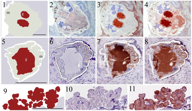
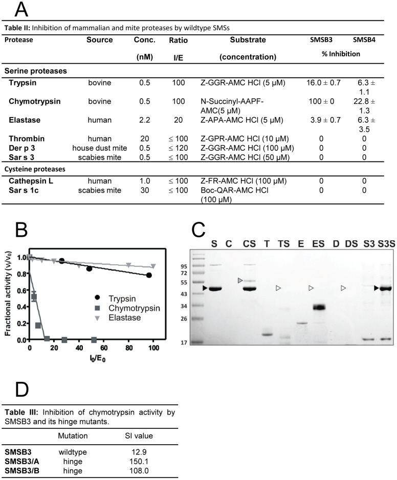
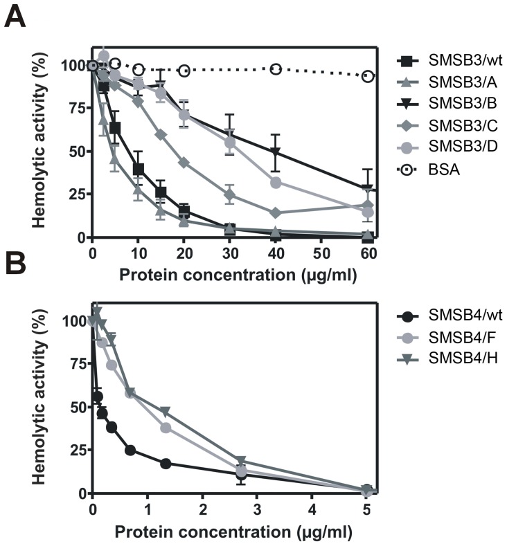
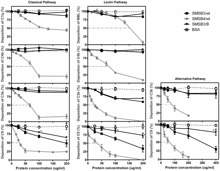
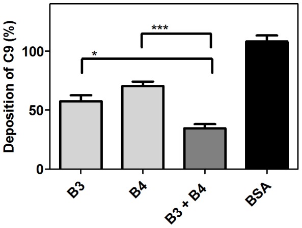
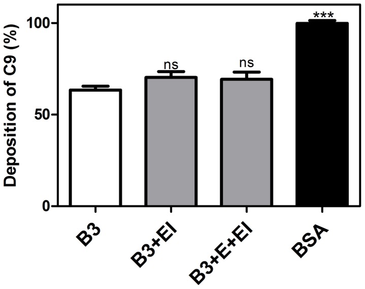
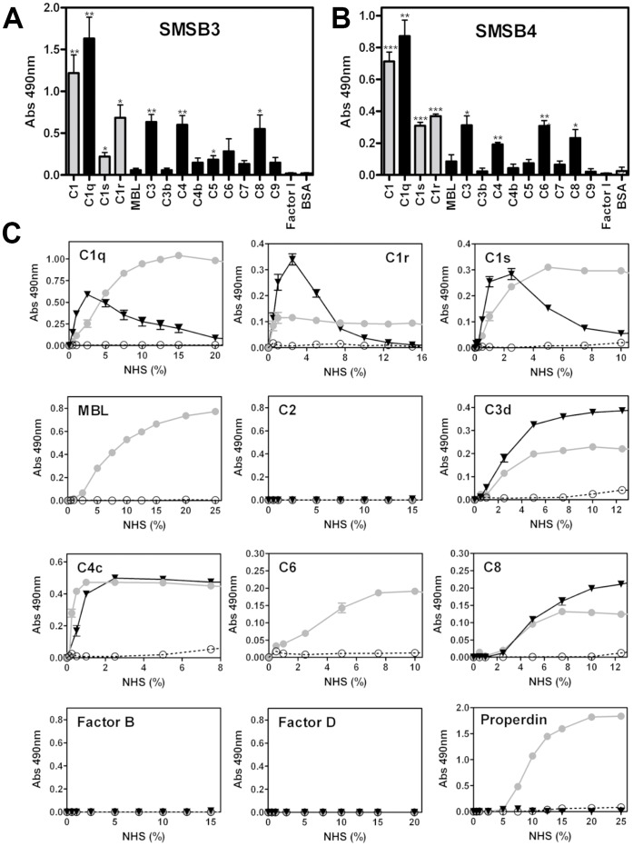
Scabies is a parasitic infestation of the skin by the mite Sarcoptes scabiei that causes significant morbidity worldwide, in particular within socially disadvantaged populations. In order to identify mechanisms that enable the scabies mite to evade human immune defenses, we have studied molecules associated with proteolytic systems in the mite, including two novel scabies mite serine protease inhibitors (SMSs) of the serpin superfamily. Immunohistochemical studies revealed that within mite-infected human skin SMSB4 (54 kDa) and SMSB3 (47 kDa) were both localized in the mite gut and feces. Recombinant purified SMSB3 and SMSB4 did not inhibit mite serine and cysteine proteases, but did inhibit mammalian serine proteases, such as chymotrypsin, albeit inefficiently. Detailed functional analysis revealed that both serpins interfered with all three pathways of the human complement system at different stages of their activation. SMSB4 inhibited mostly the initial and progressing steps of the cascades, while SMSB3 showed the strongest effects at the C9 level in the terminal pathway. Additive effects of both serpins were shown at the C9 level in the lectin pathway. Both SMSs were able to interfere with complement factors without protease function. A range of binding assays showed direct binding between SMSB4 and seven complement proteins (C1, properdin, MBL, C4, C3, C6 and C8), while significant binding of SMSB3 occurred exclusively to complement factors without protease function (C4, C3, C8). Direct binding was observed between SMSB4 and the complement proteases C1s and C1r. However no complex formation was observed between either mite serpin and the complement serine proteases C1r, C1s, MASP-1, MASP-2 and MASP-3. No catalytic inhibition by either serpin was observed for any of these enzymes. In summary, the SMSs were acting at several levels mediating overall inhibition of the complement system and thus we propose that they may protect scabies mites from complement-mediated gut damage.


Conflict of interest statement

Competing Interests: The authors have declared that no competing interests exist.

Figures

Figure 1
Figure 1. Sequence details and recombinant expression of SMSs and mutants. (A)
Shown are the sequence changes in the SMS mutants compared to wild type sequences. Numbering scheme according to . Point mutations were introduced in the two segments of the RCL domain: i) the distal (P9-P5’) variable region that resembles the protease substrate and is attacked by a target protease, and ii) the proximal (P15-P9) hinge region that is well conserved and inserts into β-sheet A during inactivation of a protease in inhibitory serpins. (B) Purification and refolding of SMSB3 and SMSB4. Shown are immobilized nickel affinity chromatography fractions after separation by SDS-PAGE and Coomassie staining. Both SMSs revealed the expected bands of 47 kDa and 54 kDa, respectively. 1, loaded sample; 2, wash; 3, elution with 250 mM imidazole, 4, active serpin after refolding.
Figure 2
Figure 2. Specificity of mouse antisera raised against recombinant SMSB3 and SMSB4.
The purified SMS proteins and BSA (used as a non-related control) were electrophoresed on SDS-PAGE and stained with Coomassie blue R-250 (A) and then immunoblotted with the anti-sera raised against SMSB3 (B) and SMSB4 (C), confirming the specificity of the antibodies and lack of cross-reaction with BSA.
Figure 3
Figure 3. Immunolocalization of SMSs and IgG in scabies mite-infested human skin.
Schematic diagrams 1 and 5 outline the features visible in serial histological sections through a scabies mite in infested human skin. Red staining indicates binding of antibody to protein. Section 2 and 6 probed with pre-immune mouse serum as a negative control remained unstained, while equivalent regions were detected in section 4 and 8 when probed with anti-human IgG, a marker for mite gut , and in section 3 and 7 when probed with antibodies against SMSB4 and SMSB3, respectively. Schematic diagrams outline the features visible in serial histological sections through human epidermis containing scabies mite feces 9. Section 10 probed with pre-immune mouse serum as a negative control remained unstained, while equivalent regions were detected in section 11 when probed with anti-SMSB3 and in section 12 with anti-IgG. Staining of feces was also seen in equivalent experiments performed with antibodies against SMSB4 (not shown). Immunohistological co-localization of SMSs and host IgG indicated that both serpins are localized in the mite gut and mite feces within the burrows. b, burrow; f, feces; g, gut; m, mite. Scale bars (100 µM) indicate the magnification level.
Figure 4
Figure 4. SMSs inhibit mammalian proteases and form serpin/protease complexes.
(A) Inhibitory profile of mammalian and mite proteases by wild type SMSs (overview). Proteases were pre-incubated with different concentrations of SMSB3 or SMSB4 at 37°C for 10 min in the appropriate reaction buffer. The reaction was started by addition of the corresponding fluorescent aminomethyl-coumarin substrate and enzyme inhibition was measured as change in fluorescence in comparison with controls. Shown are means ± SD (n = 3). (B) Stoichiometry of inhibition for SMSB3. The inhibitory activity was assessed by measuring residual enzyme activities of trypsin, chymotrypsin and leucocyte elastase. Enzymes were incubated with a 2-120:1 molar excess of SMSB3 at 37°C for 10 min in the appropriate reaction buffer. Residual enzyme activity was measured in triplicate (SD ≤5%) by adding the appropriate aminomethyl-coumarin peptide substrate and determining the reaction velocity as change in fluorescence. The fractional activity was the ratio of the velocity of inhibited enzyme (vi) over the velocity of uninhibited control (vo). The initial inhibitor/enzyme ratios ([I0]/[E0]) represent the molar excess of serpin over enzyme. (C) Complex formation between SMSB3 and serine proteases. Serpin/protease complexes were analyzed by SDS-PAGE under non-reducing conditions after pre-incubation of SMSB3 (black arrow) with each protease at 37°C for 15 min at serpin/protease ratios of 4∶1. Covalent complex formation was found with chymotrypsin (grey arrow), while SMSB3 was degraded by trypsin, elastase and Der p 3 (white arrows). C, chymotrypsin; D, house dust mite Der p 3; E, neutrophil elastase; S, SMSB3; S3, scabies mite Sar s 3; T, trypsin. (D) Effects of SMSB3 and two mutants on chymotrypsin activity.
Figure 5
Figure 5. SMSs inhibit the hemolytic activity of human serum.
Sheep erythrocytes opsonized with antibodies were incubated with 0.125% NHS to visualize the activity of the classical pathway of the human complement system. Serum was pre-incubated for 10 min at 37°C with various concentrations (2.5–60 µg/ml, i.e. 54–1300 nM) of SMSB3 and its four mutants (A), SMSB4 and its two mutants (0.1–5 µg/ml, i.e. 2–93 nM) (B) and BSA as a negative control. After 1 h of incubation of NHS with erythrocytes at 37°C, the degree of complement-mediated lysis was estimated by measurement of released hemoglobin. The lysis obtained in the absence of SMSs was defined as 100% hemolytic activity. An average of three independent experiments performed in duplicate is presented with bars indicating SEM.
Figure 6
Figure 6. SMSs inhibit the classical, lectin and alternative pathway.
Complement deposition assays were performed on microtiter plates coated with aggregated IgG (classical pathway), mannan (lectin pathway) and zymosan (alternative pathway) in presence or absence of scabies mite serpins, respectively. Shown are means ± SEM of n = 4 independent experiments, each performed in duplicate. The three pathways were measured using 1% (CP), 2% (LP) and 4% (AP) NHS. Assays were performed at inhibitor protein concentrations of 25–400 µg/ml (0.5–8.6 µM) for SMSB3/wt and SMSB3/B and 10–200 µg/ml (0.2–3.7 µM) for SMSB4/wt. Significant differences between wild type SMSB3 and the hinge mutant SMSB3/B (*; p<0.05).
Figure 7
Figure 7. Additive effects of SMS B3 and SMSB4.
Mannan was immobilized on microtiter plates and allowed to activate 4% NHS containing 5 µg/ml of either serpin (110 nM SMSB3 or 93 nM SMSB4) alone or mixed together. As a negative control BSA was used at a concentration of 10 µg/ml. After 20 min of incubation the plates were washed and the deposited C9 was detected with specific antibodies. An average of three independent experiments is presented with bars indicating means ± SEM. *p<0.05, ***p<0.001 by t test (GraphPad Prism).
Figure 8
Figure 8. Digestion of SMSB3 by human neutrophil elastase has no effect on C9 deposition.
SMSB3 was cleaved with human leukocyte elastase, following which the enzyme was inhibited by addition of N-methoxysuccinyl-Ala-Ala-Pro-Val-chloromethyl ketone. Control reactions without addition of elastase (E) and elastase inhibitor N-methoxysuccinyl-Ala-Ala-Pro-Val-chloromethyl ketone were preformed under the same conditions. Mannan was immobilized on microtiter plates and allowed to activate 4% NHS containing 50 µg/ml SMSB3. As a negative control, BSA was used at a concentration of 10 µg/ml. After a 20 min incubation the plates were washed and the deposited C9 was detected with specific antibodies. An average of three independent experiments is presented with bars indicating means ± SEM. *p<0.05, ***p<0.001 by t test (GraphPad Prism).
Figure 9
Figure 9. Direct binding of SMSs to various complement proteins.
Microtiter plates were coated with various purified human complement proteins or BSA as a negative control and incubated with 20 µg/ml SMSB3 (A) or SMSB4 (B). Bound serpins were detected using specific polyclonal antibodies against SMSB3 and SMSB4. Shown are means ± SEM of n = 3 independent experiments. The statistical significance of differences between BSA and the rest of the data groups was estimated using one-way ANOVA. *, p<0.05; **, p<0.01; ***, p<0.001. Grey, serine proteases; black, other complement factors. C Direct binding of complement factors from NHS to SMSs. Increasing concentrations of NHS were added to wells coated with SMSB3 (▾, black), SMSB4 (•, grey) or BSA as a negative control (○, white) and bound complement factors were detected by specific antibodies. Shown are means ± SEM of n = 3 independent experiments measured in duplicates. NHS was tested from 0–100%. Positive controls were used for complement proteins, where no binding to the SMSs was detectable confirming strong immunodetection of the complement factor on 1% NHS coating.

Similar articles

Cited by

References

    1. Hengge UR, Currie BJ, Jäger G, Lupi O, Schwartz RA. Scabies: a ubiquitous neglected skin disease. The Lancet Infectious Diseases. 2006;6:769–779. - PubMed
    1. Walton SF, Currie BJ. Problems in diagnosing scabies, a global disease in human and animal populations. Clin Microbiol Rev. 2007;20:268–279. - PMC - PubMed
    1. Clucas DB, Carville KS, Connors C, Currie BJ, Carapetis JR, et al. Disease burden and health-care clinic attendances for young children in remote aboriginal communities of northern Australia. Bull World Health Organ. 2008;86:275–281. - PMC - PubMed
    1. Brook I. Microbiology of secondary bacterial infection in scabies lesions. J Clin Microbiol. 1995;33:2139–2140. - PMC - PubMed
    1. McDonald M, Currie BJ, Carapetis JR. Acute rheumatic fever: a chink in the chain that links the heart to the throat? Lancet Infect Dis. 2004;4:240–245. - PubMed

Publication types

MeSH terms