. 2012;7(7):e40547.
doi: 10.1371/journal.pone.0040547. Epub 2012 Jul 6.

A new Cre driver mouse line, Tcf21/Pod1-Cre, targets metanephric mesenchyme

Affiliations

A new Cre driver mouse line, Tcf21/Pod1-Cre, targets metanephric mesenchyme

Yoshiro Maezawa et al. PLoS One. 2012.

Abstract

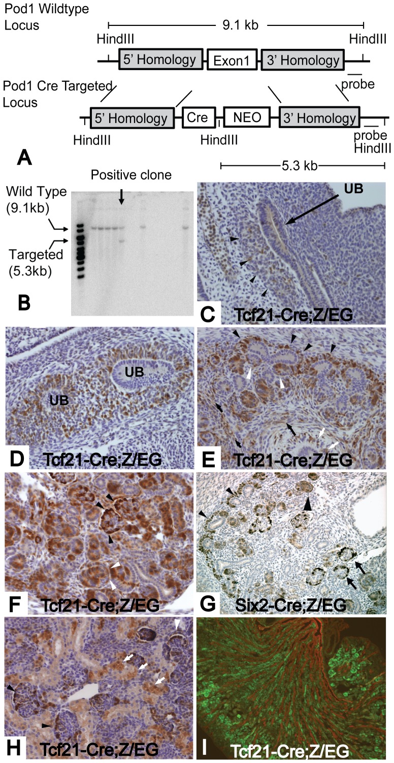
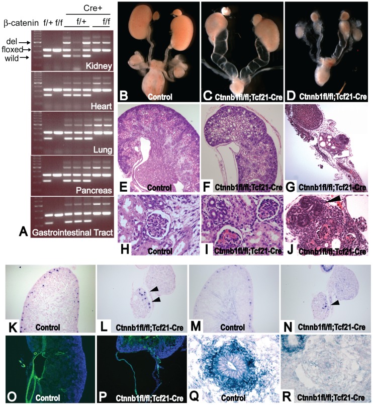
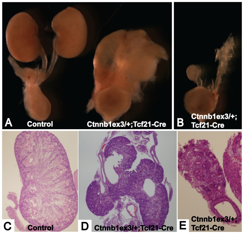
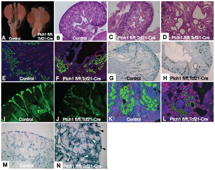
Conditional gene targeting in mice has provided great insight into the role of gene function in kidney development and disease. Although a number of Cre-driver mouse strains already exist for the kidney, development of additional strains with unique expression patterns is needed. Here we report the generation and validation of a Tcf21/Pod1-Cre driver strain that expresses Cre recombinase throughout the condensing and stromal mesenchyme of developing kidneys and in their derivatives including epithelial components of the nephron and interstitial cells. To test the efficiency of this line, we crossed it to mice transgenic for either loss or gain of function β-catenin conditional alleles. Mice with deletion of β-catenin from Tcf21-expressing cells are born with hypoplastic kidneys, hydroureters and hydronephrosis. By contrast, Tcf21-Cre driven gain of function for β-catenin in mice results in fused midline kidneys and hypoplastic kidneys. Finally, we report the first renal mesenchymal deletion of Patched1 (Ptch1), the receptor for sonic hedgehog (Shh), which results in renal cysts demonstrating a functional role of Shh signaling pathway in renal cystogensis. In summary, we report the generation and validation of a new Cre driver strain that provides robust excision in metanephric mesenchyme.


Conflict of interest statement

Competing Interests: The authors have declared that no competing interests exist.

Figures

Figure 1
Figure 1. Tcf21-Cre mouse delete genes in metanephric mesenchyme and its derivatives.
(A) Scheme of Tcf21-Cre targeted allele: Exon1 of the Tcf21 gene was replaced by a Cre transgene and neomycin cassette. (B) Genomic DNA from ES cell clones was isolated and digested with HindIII. The 500 bps probe outside the region of 3′ homology arm recognized a 5.3 kb and a 9.1 kb fragment for the mutant and wild-type alleles, respectively. Tcf21-Cre (C–F and H, I) and Six2-Cre (G) mice were bred to the Z/EG reporter mousline and Cre dependent gene deletion was examined by immunostaining for GFP. (C) At E10.5, thin and mosaic expression of GFP is present in metanephric mesenchyme (black arrowhead). (D) E11.5 embryo shows distinct expression of GFP in metanephric mesenchyme. Ureteric bud is undergoing primary branching. (E) GFP expression is observed in condensing mesenchyme (black arrowhead), pretubular aggregates (white arrowhead), interstitial stromal cells (black arrow), mesenchyme surrounding the ureteric bud (white arrow) at E13.5 embryo. (F) At E16.5, GFP is expressed in developing nephrons and stromal cells. Staining in S shaped body (white arrowhead) and presumptive podocytes (black arrowhead) can be seen in this picture. (G) Six2-Cre gene deletion is restricted to condensing mesenchyme (black arrowhead), developing nephrons (large black arrowhead) and podocytes (black arrow) at E16.5. (H) At postnatal day 0, gene deletion in podocytes (black arrowhead) and Bowman's capsule (white arrowhead) is evident, but tubular staining was mosaic (white arrows). (I) P0 kidney was stained with GFP (green) and pancytokeratin (red). There was no overlap between the two stainings. Magnification: C–H 200×, I 40×.
Figure 2
Figure 2. Tcf21- Cre mouse deletes genes in the mesenchyme of multiple organs.
(A) Dissecting microscope photograph of E10.5 Tcf21-Cre;Z/EG embryo. (B) GFP expression of E10.5 Tcf21-Cre;Z/EG embryo was visualized by fluorescent microscopy. (C–Q) Tcf21-Cre mice were bred to a Z/EG reporter mouse line and Cre dependent gene deletion was examined by immunostaining for GFP. (C) E10.5 embryo shows GFP staining in heart and metanephric mesenchyme. (D) E13.5 and (E) E16.5 embryos show gene deletion in multiple organs. At E 16.5, GFP expression was observed in (F) some part of the epicardium and endocardium, (G) lung mesenchyme, (H) kidney mesenchyme, (I) proper muscle and lamina propria of esophagus, (J) proper muscle and lamina propria of intestine, (K) some part of pancreas, (L) adrenal gland, (M) stroma of gonad, (N) proximal part of aorta, (O) facial muscle, (P) lingual and sublingual muscle, (Q) choroid plexus of ventricles. Magnification: C 40×, F, H, O, P 100×, I, L, N 200×, G, J, K, M, Q 400×. Many 40× pictures were taken for D and E, and stitched together to construct the image of the whole embryo.
Figure 3
Figure 3. Ctnnb1fl/fl;Tcf21-Cre mice shows hydroureter or rudimentary kidneys.
(A) DNA extracted from whole organs was subjected to PCR that detects wild type, floxed allele, and deleted allele of β-catenin. Deleted alleles were detected in kidney, heart, lung, pancreas and gastrointestinal tract. (B, E, H) Control kidney, (C, F, I) Hydroureter and relatively normal kidney of Ctnnb1fl/fl;Tcf21-Cre mice, (D, G, J) Rudimentary kidney and hypoplastic kidney with hydroureter of Ctnnb1fl/fl;Tcf21-Cre mouse. (F) Hydroureter kidneys had relatively normal appearance, but show dilation of tubules, (I) dilation of glomerular capillaries. Some of the glomeruli appear to be normal. (G) Rudimentary kidney was cystic, (J) had random nephrogenic area (black arrowhead) with a few disorganized glomeruli. (K) Fgf8 expression in control kidney. (L) Fgf8 is observed only in the disorganized nephrogenesis area (black arrowhead). (M) Wnt4 expression in control kidney. (N) Wnt4 is observed only in the disorganized nephrogenesis area (Black arrowhead). (O) Expression of α-Sma (green) in control mouse. Blue shows DAPI staining. (P) In hydroureter mutant, α-Sma (green) is present in the ureter wall, but its layer is extremely thin. (Q) Bmp4 expression surrounding the ureteric bud in E13.5 control. (R) Expression of Bmp4 surrounding the ureteric bud is lost in E13.5 Ctnnb1fl/fl;Tcf21-Cre. However, Bmp4 expression is restored in developing nephrons. Experiments were performed on E18.5 embryos if not indicated. B–D are taken at the same magnification. Mice that lack at least one of the transgenes were used as controls. Magnification: E–G, K–P 100×, H–J 200×, Q, R 400×.
Figure 4
Figure 4. Ctnnb1ex3/+; Tcf21-Cre mice show fusion kidney and rudimentary kidney.
(A) Control kidney (left) and fusion kidney of Ctnnb1ex3/+;Tcf21-Cre mouse (right). (B) Severely hypoplastic/dysplastic rudimentary kidney of Ctnnb1ex3/+;Tcf21-Cre mouse. (C) Normal histology of control kidney. (D) Histology of the fusion kidney. (E) Histology of the rudimentary kidney. Magnification: C 40×, E 100×. Many 40× pictures were taken for D, and stitched together to construct the image of the whole fusion kidney.
Figure 5
Figure 5. Ptch1 fl/fl;Tcf21-Cre kidneys develop multiple renal cysts.
(A) Control kidney (left) and Ptch1 fl/fl;Tcf21-Cre kidney (right). (B) Histology of control kidney. (C) Multiple cysts of Ptch1 fl/fl;Tcf21-Cre kidney. (D) Glomerular cysts as well as tubular cysts were observed in the mutant kidney. (E, F) Kidneys were stained with LTL-FITC (green) and THP (red) that mark proximal tubules or loop of Henle, respectively. (E) Control kidney shows beautiful organized structure of proximal tubules and loops of Henle. (F) Disorganized structure of proximal tubule and loop of Henle inPtch1 fl/fl;Tcf21-Cre kidney. Loops of Henle are very short. Some cysts were stained with either LTL or THP. (G–J) Sections were stained with DBA, which marks collecting ducts. (G, I) Control kidney show organized structure of collecting duct. Note the straight configuration of cortical collecting duct. (H, J) Structure of collecting duct system is severely disorganized in Ptch1 fl/fl;Tcf21-Cre kidney. The path of collecting duct is random and winding. (K, L) Sections were stained with LTL (green) and Ki67 for proliferation. (K) normal kidney (L) Ptch1 fl/fl;Tcf21-Cre kidney. Note that Ki67 is frequently positive in the cystic wall. (M and N) In Situ hybridization for Gli1, an established downstream target of Shh pathway. (M) Control kidney shows activation of Shh pathway in the interstitial area (N) Mutant kidney shows increased Gli1 expression in condensing mesenchyme (black arrowhead) and stroma, but is negative in pretubular aggregates (black arrow) and nephrogenic vesicles (white arrowhead). Magnification: B, C 40×, D, G, H 100×, E, F, I–N 200×.

Similar articles

Cited by

References

    1. Belteki G, Haigh J, Kabacs N, Haigh K, Sison K, et al. Conditional and inducible transgene expression in mice through the combinatorial use of Cre-mediated recombination and tetracycline induction. Nucleic Acids Res. 2005;33:e51. - PMC - PubMed
    1. Maezawa Y, Kreidberg JA, Quaggin SE. CHAPTER 1- Embryology of the Kidney. Brenner and Rector's the Kidney. Philadelphia, PA: W.B. Saunders Company. 2012.
    1. Dressler GR. Advances in early kidney specification, development and patterning. Development. 2009;136:3863–3874. - PMC - PubMed
    1. Kobayashi A, Valerius MT, Mugford JW, Carroll TJ, Self M, et al. Six2 defines and regulates a multipotent self-renewing nephron progenitor population throughout mammalian kidney development. Cell Stem Cell. 2008;3:169–181. - PMC - PubMed
    1. Srinivas S, Goldberg MR, Watanabe T, D'Agati V, al-Awqati Q, et al. Expression of green fluorescent protein in the ureteric bud of transgenic mice: a new tool for the analysis of ureteric bud morphogenesis. Dev Genet. 1999;24:241–251. - PubMed

Publication types

MeSH terms

LinkOut - more resources