. 2012;7(7):e40555.
doi: 10.1371/journal.pone.0040555. Epub 2012 Jul 5.

Hilar GABAergic interneuron activity controls spatial learning and memory retrieval

Affiliations

Hilar GABAergic interneuron activity controls spatial learning and memory retrieval

Yaisa Andrews-Zwilling et al. PLoS One. 2012.

Abstract

Background: Although extensive research has demonstrated the importance of excitatory granule neurons in the dentate gyrus of the hippocampus in normal learning and memory and in the pathogenesis of amnesia in Alzheimer's disease (AD), the role of hilar GABAergic inhibitory interneurons, which control the granule neuron activity, remains unclear.

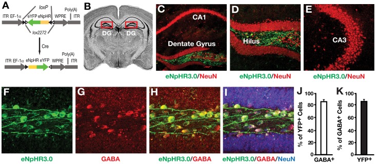
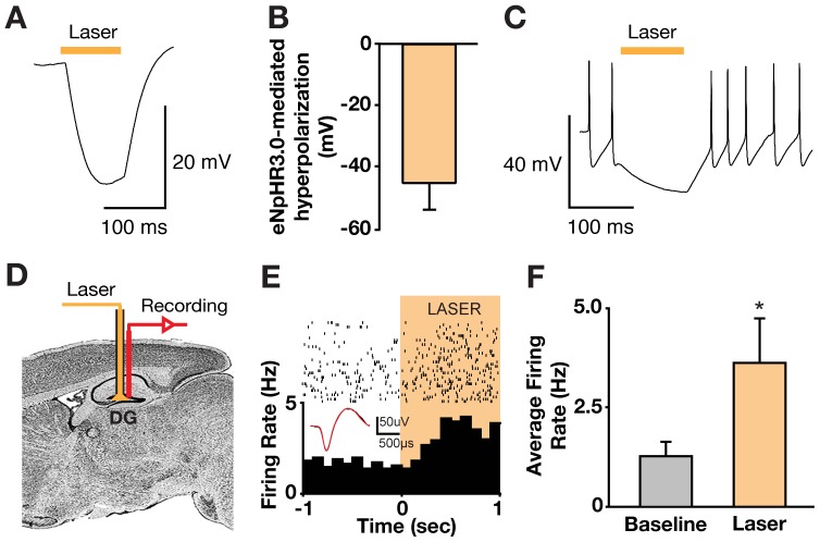
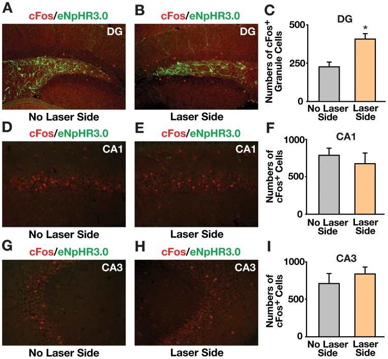
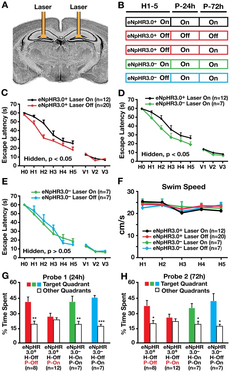
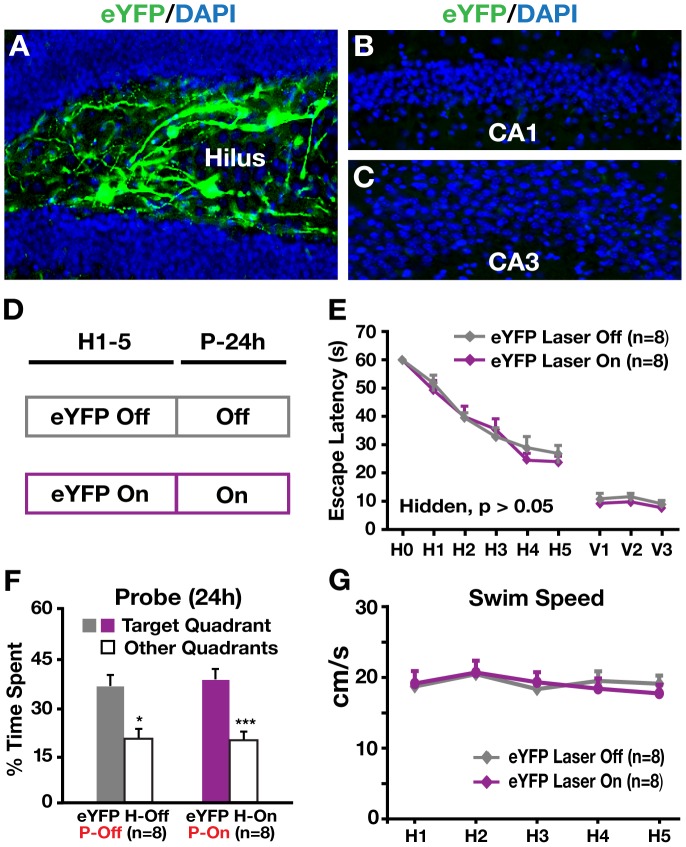
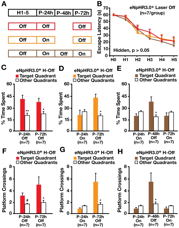
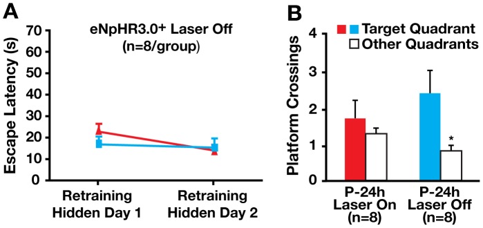
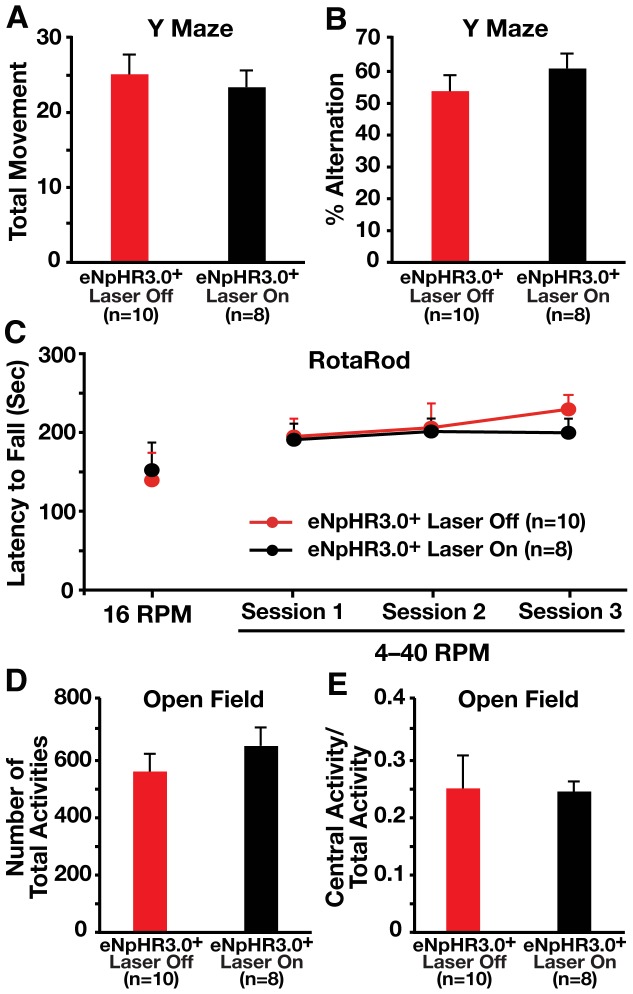
Methodology and principal findings: We explored the function of hilar GABAergic interneurons in spatial learning and memory by inhibiting their activity through Cre-dependent viral expression of enhanced halorhodopsin (eNpHR3.0)--a light-driven chloride pump. Hilar GABAergic interneuron-specific expression of eNpHR3.0 was achieved by bilaterally injecting adeno-associated virus containing a double-floxed inverted open-reading frame encoding eNpHR3.0 into the hilus of the dentate gyrus of mice expressing Cre recombinase under the control of an enhancer specific for GABAergic interneurons. In vitro and in vivo illumination with a yellow laser elicited inhibition of hilar GABAergic interneurons and consequent activation of dentate granule neurons, without affecting pyramidal neurons in the CA3 and CA1 regions of the hippocampus. We found that optogenetic inhibition of hilar GABAergic interneuron activity impaired spatial learning and memory retrieval, without affecting memory retention, as determined in the Morris water maze test. Importantly, optogenetic inhibition of hilar GABAergic interneuron activity did not alter short-term working memory, motor coordination, or exploratory activity.

Conclusions and significance: Our findings establish a critical role for hilar GABAergic interneuron activity in controlling spatial learning and memory retrieval and provide evidence for the potential contribution of GABAergic interneuron impairment to the pathogenesis of amnesia in AD.


Conflict of interest statement

Competing Interests: The authors have declared that no competing interests exist.

Figures

Figure 1
Figure 1. Selective virus-mediated eNpHR3.0 expression in hilar GABAergic interneurons.
(A) Schematic of the double-floxed Cre-dependent AAV1 vector expressing eNpHR3.0-eYFP under control of the EF-1-alpha promoter. eYFP, enhanced yellow fluorescent protein; ITR, inverted terminal repeat; WPRE, woodchuck hepatitis virus posttranscriptional regulatory element. (B) Coronal mouse brain section. Red box indicates the dentate gyrus (DG) of the hippocampus. (C–E) Confocal images of the DG and CA1 (C), the hilus (D), and the CA3 (E) regions of the hippocampus of I12b-Cre mice injected with AAV1-DIO-eNpHR3.0-eYFP virus. Green indicates the expression of eNpHR3.0-eYFP; red indicates neurons stained positive for NeuN. (F–I) Confocal images of hilar cells expressing eNpHR3.0-eYFP (green), GABA (red), or NeuN (blue) in I12b-Cre mice injected with AAV1-DIO-eNpHR3.0-eYFP virus. Yellow indicates the colocalization of eNpHR3.0-eYFP and GABA (H). (J) Percent of eYFP-positive hilar cells also positive for GABA. (K) Percent of GABA-positive hilar cells also positive for eYFP. Values are mean ± SEM (n = 6 mice).
Figure 2
Figure 2. Light-elicited inhibition of hilar GABAergic interneurons and activation of dentate granule neurons.
(A, B) Example trace (A) and summary graph (B, n = 5) of eNpHR3.0-mediated membrane hyperpolarization of hilar GABAergic interneurons in brain slices. In this and subsequent panels, yellow bars indicate illumination time. (C) eNpHR3.0-mediated inhibition of spiking of hilar GABAergic interneurons in brain slices. (D) Schematic of in vivo optical stimulation and recording in the dentate gyrus (DG) of I12b-Cre mice injected with AAV1-DIO-eNpHR3.0-eYFP virus. (E) Example of a granule neuron recorded from the DG of an anaesthetized I12b-Cre mouse injected with AAV1-DIO-eNpHR3.0-eYFP that showed increased firing in response to yellow laser illumination. Inset shows spike waveform with laser illumination. (F) Average change in dentate granule neuron firing rates in response to yellow laser illumination in I12b-Cre mice injected with AAV1-DIO-eNpHR3.0-eYFP virus. Values are mean ± SEM (n = 7, *p<0.05, two-tailed and unpaired t-test).
Figure 3
Figure 3. Light-elicited inhibition of hilar GABAergic interneurons significantly increased cFos-positive neurons in the dentate gyrus.
(A, B) Confocal images of neurons positive for cFos (red) and eNpHR3.0-eYFP (green) on the side of DG without (A) or with (B) laser illumination in I12b-Cre mice injected with AAV1-DIO-eNpHR3.0-eYFP virus. (C) Quantification of cFos-positive dentate granule neurons on the side of DG without or with laser illumination in I12b-Cre mice injected with AAV1-DIO-eNpHR3.0-eYFP virus. Values are mean ± SEM (n = 6, *p<0.05, two tailed and paired t-test). (D, E, G, H) Confocal images of CA1 (D, E) and CA3 (G, H) neurons positive for cFos (red) and eNpHR3.0-eYFP (green) on the side of the hippocampus without (D, G) or with (E, H) laser illumination in the hilus of the dentate gyrus in I12b-Cre mice injected with AAV1-DIO-eNpHR3.0-eYFP virus. (F, I) Quantification of cFos-positive CA1 (F) and CA3 (I) neurons on the side of the hippocampus without or with laser illumination in the hilus of the dentate gyrus in I12b-Cre mice injected with AAV1-DIO-eNpHR3.0-eYFP virus. Values are mean ± SEM (n = 6 mice).
Figure 4
Figure 4. Inhibition of hilar GABAergic interneuron activity impaired spatial learning and memory.
(A) Coronal schematic of cannula placement and bilateral fiber-optic stimulation. (B) Protocols of mice used and laser illumination during hidden platform (H1–5) and probe (P-24 h and P-72 h) trials in the Morris water maze (MWM) test. (C–E) Learning curves of I12b-Cre (eNpHR3.0+) and wildtype (eNpHR3.0) littermates injected with AAV1-DIO-eNpHR3.0-eYFP virus, with or without laser illumination in MWM tests. Points represent averages of daily trials. H, hidden platform sessions (two trials/session, two sessions/day); H0, first trial on H1; V, visible platform sessions (two trials/session, two sessions/day). Y-axis indicates time to reach the target platform (escape latency). Values are mean ± SEM and statistically evaluated by repeated measures ANOVA. (F) Swim speed did not differ significantly among different groups of mice during the MWM test. (G) Percent time spent in the target quadrant versus the other quadrants in the probe trial performed 24 h (probe 1) after the last hidden platform trial. (H) Percent time spent in the target quadrant versus the other quadrants in the probe trial performed 72 h (probe 2) after the last hidden platform trial. Values are mean ± SEM. n = 7–20 mice/group. *p<0.05 **p<0.01, ***p<0.005 (two tailed and unpaired t-test).
Figure 5
Figure 5. Hilar illumination did not alter learning and memory in I12b-Cre mice injected with eYFP virus.
(A–C) Confocal images of the dentate gyrus (A), the CA1 (B), and the CA3 (C) regions of the hippocampus of I12b-Cre mice injected with AAV1-DIO-eYFP virus. Green indicates the expression of eYFP; blue indicates cell nuclei stained positive for DAPI. (D) Protocols of mice used and laser illumination during hidden platform and probe trials in the Morris water maze (MWM) test. (E) Learning curves of AAV1-DIO-eYFP virus-injected I12b-Cre mice with (On) or without (Off) laser illumination did not differ in both hidden and visible platform trials of the MWM test. Points represent averages of daily trials. H, hidden platform sessions (two trials/session, two sessions/day); H0, first trial on H1. Y-axis indicates time to reach the target platform (escape latency). Values are mean ± SEM. (F) Percent time spent in the target quadrant versus the other quadrants in the probe trial performed 24 h after the last hidden platform trial with (P-On) or without (P-Off) laser illumination. Values are mean ± SEM. *p<0.05, ***p<0.005 (two-tailed and unpaired t-test). (G) Swim speed did not differ between the two groups of mice during the MWM test. Values are mean ± SEM. n = 8 mice/group.
Figure 6
Figure 6. Inhibition of hilar GABAergic interneuron activity impaired spatial memory retrieval but not memory retention.
(A) Protocols of mice used and laser illumination during hidden platform (H1–5) and probe (P-24 h, P-48 h, P-72 h) trials in the MWM test. (B) Learning curves of I12b-Cre mice injected with AAV1-DIO-eNpHR3.0-eYFP virus without laser illumination in the hidden platform trials (H-Off) of the MWM test. Points represent averages of daily trials. H, hidden platform sessions (two trials/session, two sessions/day); H0, first trial on H1. Y-axis indicates time to reach the target platform (escape latency). Values are mean ± SEM and statistically evaluated by repeated measures ANOVA. (C–E) Percent time spent in the target quadrant versus the other quadrants in the probe trial performed 24 (P-24 h), 48 (P-48 h), or 72 (P-72 h) hours after the last hidden platform trial with (On) or without (Off) laser illumination. Values are mean ± SEM. *p<0.05, **p<0.01 (two tailed and unpaired t-test). F–H, Platform crossings in the probe trial performed 24 (P-24 h), 48 (P-48 h), or 72 (P-72 h) hours after the last hidden platform trial with (On) or without (Off) laser illumination. Values are mean ± SEM. n = 8 mice/group. *p<0.05, #p = 0.05 (two tailed and unpaired t-test).
Figure 7
Figure 7. Inhibition of hilar GABAergic interneuron activity impaired spatial memory retrieval in retrained mice.
(A) Mice used in Fig. 4 were retrained in the hidden platform trials 2 weeks after the first Morris water maze (MWM) test (see Fig. 4) and showed very good spatial memory. Points represent averages of daily trials. Y-axis indicates time to reach the target platform (escape latency). Values are mean ± SEM. (B) Platform crossings in the probe trial performed with (On) or without (Off) laser illumination 24 h after the last hidden platform trial of the retraining. Values are mean ± SEM. n = 8 mice/group. *p<0.05 (two-tailed and unpaired t-test).
Figure 8
Figure 8. Inhibition of hilar GABAergic interneuron activity did not alter non-hippocampus-dependent behavior.
(A, B) I12b-Cre mice injected with AAV1-DIO-eNpHR3.0-eYFP virus were tested for short-term working memory with (On) or without (Off) laser illumination in a Y maze test. Data were reported as total movement (A) and % alternation (B). Values are mean ± SEM. (C) I12b-Cre mice injected with AAV1-DIO-eNpHR3.0-eYFP virus were tested for motor coordination with (On) or without (Off) laser illumination in a rotarod test. Values are mean ± SEM. (D, E) I12b-Cre mice injected with AAV1-DIO-eNpHR3.0-eYFP virus were tested for exploratory activity with (On) or without (Off) laser illumination in a open field test. Data were reported as the number of total activities (D) and the ratio of central activity to total activity (E). Values are mean ± SEM. n = 8–10 mice/group.

Similar articles

Cited by

References

    1. Squire LR, Wixted JT. The cognitive neuroscience of human memory since H.M. Annu Rev Neurosci. 2011;34:259–288. - PMC - PubMed
    1. Wang SH, Morris RGM. Hippocampal-neocortical interactions in memory formation, consolidation, and reconsolidation. Annu Rev Psychol. 2010;61:49–79. - PubMed
    1. Myers CE, Scharfman HE. A role for hilar cells in pattern separation in the dentate gyrus: a computational approach. Hippocampus. 2009;19:321–337. - PMC - PubMed
    1. Kullmann D. Interneuron networks in the hippocampus. Curr Opin Neurobiol. 2011;21:709–716. - PubMed
    1. Klausberger T, Somogyi P. Neuronal diversity and temporal dynamics: the unity of hippocampal circuit operations. Science. 2008;321:53–57. - PMC - PubMed

Publication types