. 2012;7(7):e40585.
doi: 10.1371/journal.pone.0040585. Epub 2012 Jul 11.

A beta-herpesvirus with fluorescent capsids to study transport in living cells

Affiliations

A beta-herpesvirus with fluorescent capsids to study transport in living cells

Jens B Bosse et al. PLoS One. 2012.

Abstract

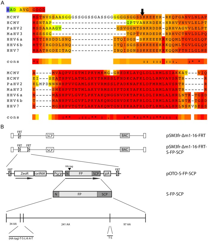
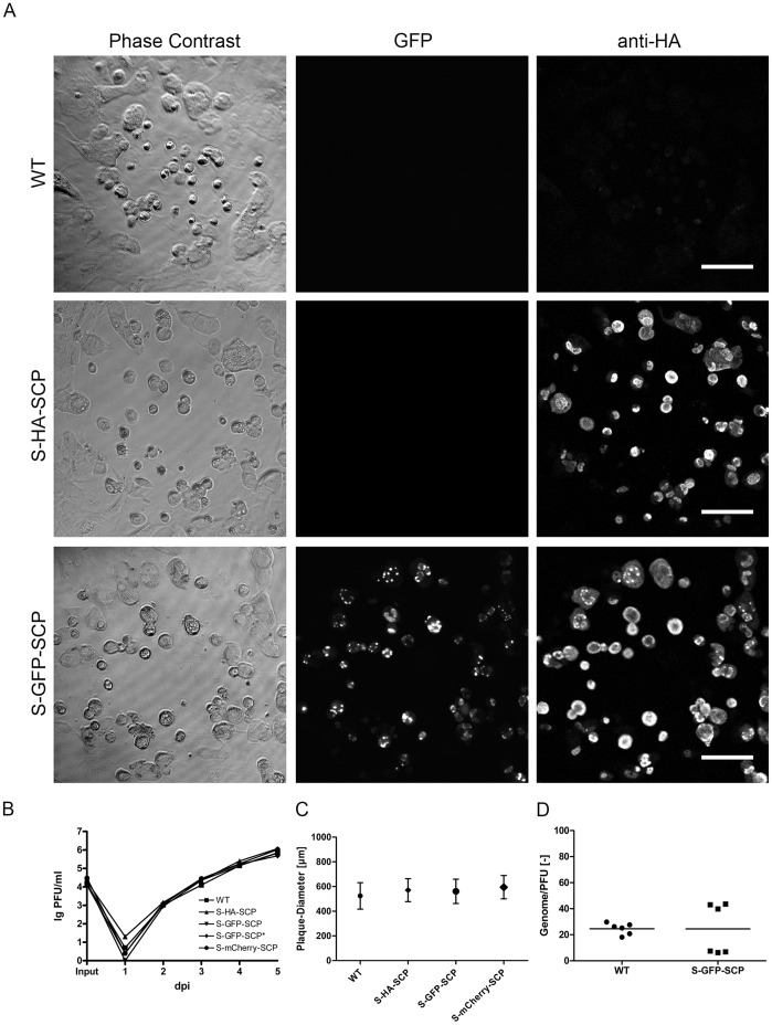
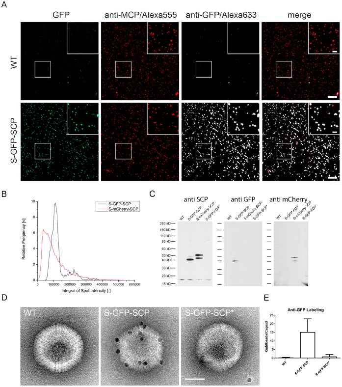
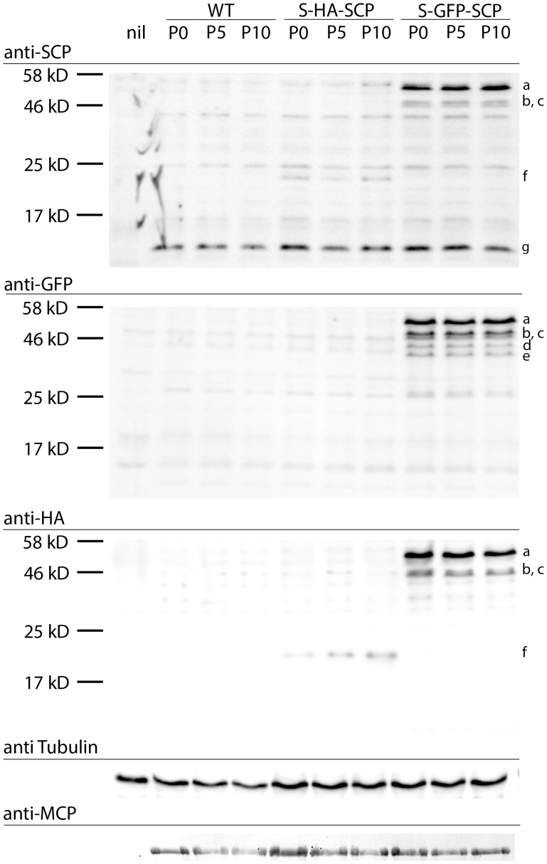
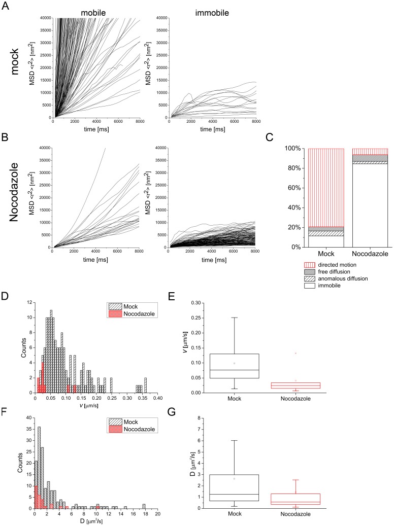
Fluorescent tagging of viral particles by genetic means enables the study of virus dynamics in living cells. However, the study of beta-herpesvirus entry and morphogenesis by this method is currently limited. This is due to the lack of replication competent, capsid-tagged fluorescent viruses. Here, we report on viable recombinant MCMVs carrying ectopic insertions of the small capsid protein (SCP) fused to fluorescent proteins (FPs). The FPs were inserted into an internal position which allowed the production of viable, fluorescently labeled cytomegaloviruses, which replicated with wild type kinetics in cell culture. Fluorescent particles were readily detectable by several methods. Moreover, in a spread assay, labeled capsids accumulated around the nucleus of the newly infected cells without any detectable viral gene expression suggesting normal entry and particle trafficking. These recombinants were used to record particle dynamics by live-cell microscopy during MCMV egress with high spatial as well as temporal resolution. From the resulting tracks we obtained not only mean track velocities but also their mean square displacements and diffusion coefficients. With this key information, we were able to describe particle behavior at high detail and discriminate between particle tracks exhibiting directed movement and tracks in which particles exhibited free or anomalous diffusion.


Conflict of interest statement

Competing Interests: The authors have declared that no competing interests exist.

Figures

Figure 1
Figure 1. Construction of SCP fusion proteins.
(A) Alignment of beta-herpesvirus SCP sequences with T-Coffee . A naturally occurring glycin-serin-linker like sequence separates the N-terminus in MCMV. (B) Detailed representation of the S-GFP-SCP fusion protein as well as the basic genetic layout of the used mutant viruses carrying a fluorescent protein (FP) and a hemagglutinin tag (HA). GFP or mCherry were used as FPs. The FP could be removed by a simple SpeI-mediated digest resulting in a construct encoding for just a HA-tagged SCP. N marks the duplicated N-terminal region of SCP. The plasmids carrying the fusion constructs were inserted into the MCMV BAC by Flp-mediated recombination.
Figure 2
Figure 2. Mutants carrying tagged SCP are viable.
(A) WT (WT) as well as mutant viruses coding for either HA- (S-HA-SCP) or HA-GFP-tagged wt SCP (S-GFP-SCP) were titrated on MEF cells. 4 days post infection (dpi) cells were fixed with PFA and processed for immunofluorescence against the HA-epitope. The scale bars represent 100 µm. (B) Multistep growth curve of mutant viruses used in this study in comparison to WT virus. C) Comparison of plaque diameters of simultaneously titrated WT, HA-, GFP-, and mCherry-tagged virus 4 dpi on MEF cells. (D) Genome to PFU ratio of GFP-tagged and WT virus. The ratio between genome content and titer for two independently prepared and purified virus stocks per virus was determined by titration and quantitative PCR in triplicates.
Figure 3
Figure 3. GFP-tagged SCP is incorporated specifically into virus particles.
(A) Gradient purified virus particles were immobilized on fibronectin-coated cover-slips, fixed and processed for immunofluorescence. A MCP specific polyclonal serum was used as indicator of virus capsids and a GFP specific polyclonal antiserum was used to compare MCP and GFP specific signals. Direct GFP fluorescence was detected by excitation with 488 nm laser light and appropriate emission filters. Inserts depict 2x magnifications. Scale bars represent 10 µm and 2.5 µm in inserts. (B) Quantification of particle fluorescence intensity distributions. Gradient purified S-GFP-SCP or S-mCherry-SCP virus preparations were bound on Poly-Lysin coated glass-bottom dishes and fluorescent spots were recorded in 16 bit mode. Exposure times as well as the EM gain were adjusted to maximize the recorded dynamic range. The graph depicts the integrated fluorescence distributions for S-GFP-SCP (black) and S-mCherry-SCP (red). (C) Immunoblot of gradient-purified virus particles. Approximately 1*105 PFU per lane of wt, (WT), S-GFP-SCP (S-GFP-SCP), S-mCherry-SCP (S-mCherry-SCP) or S-GFP-SCP* (S-GFP-SCP*) were spun down and lysed. Proteins were separated by SDS-PAGE, blotted and immunodetected with polyconal sera against SCP, GFP and mCherry. (D) Immunoelectron microscopy of purified nuclear capsids. Nuclear capsids were purified from wt (WT), S-GFP-SCP (S-GFP-SCP) or S-GFP-SCP* (S-GFP-SCP*) infected cells and immuno gold labeled with an antibody against GFP followed by protein A coupled to 10 nm gold. The scale bar indicates 50 nm. (E) Quantification of gold-labeling intensity. The amount of gold beads per capsid was counted for at least 20 views containing at least 20 capsids per condition as shown in (D). The mean as well as the standard deviation are depicted.
Figure 4
Figure 4. Viruses carrying labeled SCP mutants are genetically stable.
Immunoblot of M2-10B4 cells infected with either wt, S-HA-SCP, or S-GFP-SCP virus derived from passages 0, 5 or 10 after reconstitution. Cells were infected at a MOI of 1, and harvested 48 hpi by lysis in total lysis buffer. Proteins were separated by SDS-PAGE, blotted and immunoprobed for GFP, SCP, HA and MCP as well as beta-Tubulin as loading controls. Lower case letters indicate discussed protein bands.
Figure 5
Figure 5. Ultrastructural assessment of S-GFP-SCP infected cells.
NIH-3T3 and M2-10B4 (upper row) or M2-10B4 cells (lower row) were infected at a MOI of 0.5 and centrifugal enhancement with WT (upper row) or S-GFP-SCP labeled virus (lower row) and incubated for 48 h. Afterwards, cells were high-pressure frozen, freeze-substituted, plastic-embedded and thin-sectioned. Depicted are representative details of two independent experiments showing non-enveloped B- and C-capsids in the nucleus (first column), primary envelopment in the nucleus (2nd column), non-enveloped C-capsids near cellular membranes possibly during secondary envelopment (third column), as well as enveloped capsids in the cytoplasm (right column). Scale bars indicate 200 nm.
Figure 6
Figure 6. Fluorescent virus particles are spread-competent.
(A) Confluent M2-10B4 cells were infected with S-GFP-SCP labeled virus on cover-slips in 24-wells with 100 PFU per well and overlaid with methyl-cellulose. 4 dpi cells were fixed and processed for immunofluorescence. MCP-specific antiserum was used to detect virus producing cells as well as single virus particles while GFP fluorescence was visualized directly. Cell nuclei were counterstained with TO-PRO-3. Inserts depict single virus particles surrounding a cell nucleus (circles) not showing any evidence for being on the late stage of infection and producing infectious particles. Scale bars indicate 20 µm in the upper row and 5 µm in the lower row.
Figure 7
Figure 7. Nocodazole blocks MCMV fluorescent particle mobility.
MEF cells were infected with S-mCherry-SCP for 23 h and treated with 5 µg/ml Noccodazole for 1 h or not. S-mCherry-SCP-emission in live cells recorded under environmentally controlled conditions with 5 frames per second. Fluorescence intensity is coded in false-colors from dark blue to yellow. Three frames from a time-lapse stack each recorded 6 seconds (30 frames) apart are shown for a non-treated (upper row) and Nocodazole treated cell (lower row). Lines indicate the nucleus (Nuc.) as well as the cytoplasm (Cyt.). Circles indicate the position of a fluorescent particle. The right picture depicts all quantified tracks with ellipses marking the tracks from the particles highlighted on the left.
Figure 8
Figure 8. Quantification of particle tracks.
(A) 180 particles from mock treated cells and were tracked and their MSDs plotted against the lag time. Particle tracks were clustered into two classes representing mobile (left diagram) and immobile particles (right diagram) depending on the MSD trend. (B) 163 tracks from Nocodazole treated cells were clustered the same way as in (A). Mobile particles were further clustered depending on which mode of diffusion they exhibited according to their fit to three different diffusion models. (C) Distribution of particle mobility classes for mock- as well as Nocodazole-treated cells depicted in percent. The data is categorized into four classes (immobile, anomalous diffusion, free diffusion, directed motion). (D) Histogram displaying the distribution of mean track velocities of all particles exhibiting directed motion. (E) The same data as in (D) summarized in a box plot with median (line) mean (square), 25th to 75th percentile (box) and whiskers (5th to 95th percentile). (F) Histogram showing the distribution of all measured diffusion coefficients. (G) Box plot of the same data shown in (F).

Similar articles

Cited by

References

    1. McGeoch DJ, Dolan A, Ralph aC. Toward a comprehensive phylogeny for mammalian and avian herpesviruses. Journal of Virology. 2000;74:10401–10406. - PMC - PubMed
    1. Adler H, Messerle M, Wagner M, Koszinowski UH. Cloning and mutagenesis of the murine gammaherpesvirus 68 genome as an infectious bacterial artificial chromosome. Journal of Virology. 2000;74:6964–6974. - PMC - PubMed
    1. Borst EM, Hahn G, Koszinowski UH, Messerle M. Cloning of the human cytomegalovirus (HCMV) genome as an infectious bacterial artificial chromosome in Escherichia coli: a new approach for construction of HCMV mutants. J Virol. 1999;73:8320–8329. - PMC - PubMed
    1. Messerle M, Crnković I, Hammerschmidt W, Ziegler H, Koszinowski UH. Cloning and mutagenesis of a herpesvirus genome as an infectious bacterial artificial chromosome. Proceedings of the National Academy of Sciences of the United States of America. 1997;94:14759–14763. - PMC - PubMed
    1. Nagel C–H, Döhner K, Fathollahy M, Strive T, Borst EM, et al. Nuclear Egress and Envelopment of Herpes Simplex Virus Capsids Analyzed with Dual-Color Fluorescence HSV1 (17+). Journal of Virology. 2008;82:3109–3124. - PMC - PubMed

Publication types

MeSH terms