. 2012;7(7):e40602.
doi: 10.1371/journal.pone.0040602. Epub 2012 Jul 11.

Bone marrow-derived matrix metalloproteinase-9 is associated with fibrous adhesion formation after murine flexor tendon injury

Affiliations

Bone marrow-derived matrix metalloproteinase-9 is associated with fibrous adhesion formation after murine flexor tendon injury

Alayna E Loiselle et al. PLoS One. 2012.

Abstract

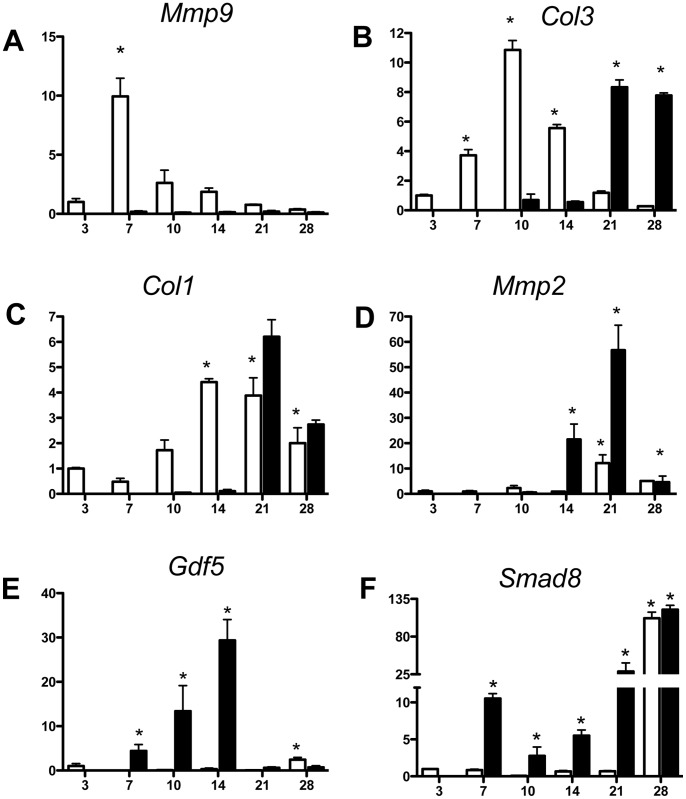
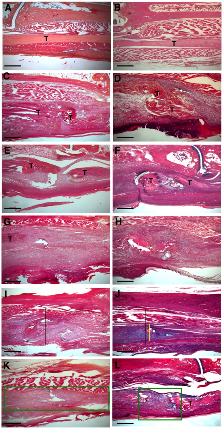
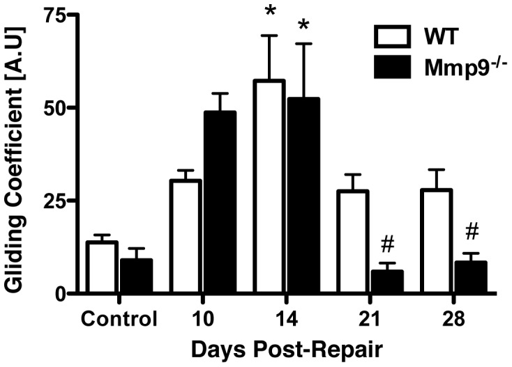
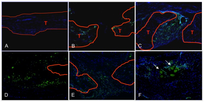
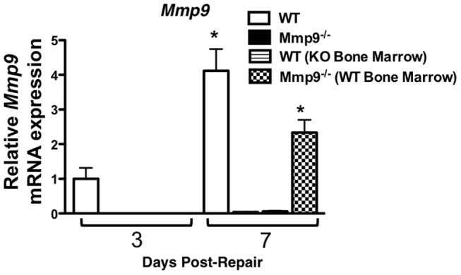
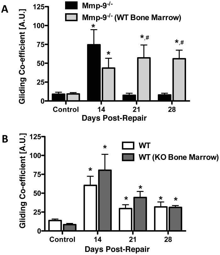
The pathogenesis of adhesions following primary tendon repair is poorly understood, but is thought to involve dysregulation of matrix metalloproteinases (Mmps). We have previously demonstrated that Mmp9 gene expression is increased during the inflammatory phase following murine flexor digitorum (FDL) tendon repair in association with increased adhesions. To further investigate the role of Mmp9, the cellular, molecular, and biomechanical features of healing were examined in WT and Mmp9(-/-) mice using the FDL tendon repair model. Adhesions persisted in WT, but were reduced in Mmp9(-/-) mice by 21 days without any decrease in strength. Deletion of Mmp9 resulted in accelerated expression of neo-tendon associated genes, Gdf5 and Smad8, and delayed expression of collagen I and collagen III. Furthermore, WT bone marrow cells (GFP(+)) migrated specifically to the tendon repair site. Transplanting myeloablated Mmp9(-/-) mice with WT marrow cells resulted in greater adhesions than observed in Mmp9(-/-) mice and similar to those seen in WT mice. These studies show that Mmp9 is primarily derived from bone marrow cells that migrate to the repair site, and mediates adhesion formation in injured tendons. Mmp9 is a potential target to limit adhesion formation in tendon healing.


Conflict of interest statement

Competing Interests: The authors have declared that no competing interests exist.

Figures

Figure 1
Figure 1. Early expression of neo-tendon associated genes during flexor tendon healing in Mmp9
/ mice. Gene expression of (A) Mmp9 (B) Col3a1, (C) Col1a1, (D) Mmp2, (E) Gdf5, and (F) Smad8 in FDL tendon repair tissue over time up to 28 days post-op. Total RNA was extracted and pooled from five tendon repairs per time-point and processed for real-time RT-PCR. Gene expression was standardized with the internal β-actin control and then normalized by the level of expression in day three WT FDL tendon repairs. Data presented as the mean fold induction (over WT day three repairs) ± SEM. * p<0.05 vs. WT day three tendon repair. White bars represent WT mice. Black bars represent Mmp9−/− mice.
Figure 2
Figure 2. Decreased area of remodeling during tendon healing in Mmp9
/ mice. Representative histological sections of sham control WT (A) and Mmp9−/− (B) FDL Tendons. Repaired WT and Mmp9−/− FDL Tendons at days 7 (C, D), 10 (E, F), 14 (G, H) 21 (I, K) and 28 (K, L). Sections were stained with Alcian Blue/Hematoxylin and Orange G. Of note is the fibroblastic granulation tissue that fills in the repair site between tendon ends (marked as T) and is progressively remodeled (green box) with collagen becoming oriented in a parallel fashion with the long axis of the tendon. Black bar represents width of WT tendon; yellow bar represents width of Mmp9−/− tendon at Day 21. Scale bars represent 200 microns. 5X magnification.
Figure 3
Figure 3. Earlier resolution of fibrous adhesions in Mmp9
/ tendon repairs. Gliding coefficient of WT and Mmp9−/− mouse FDL tendon repairs over time up to 28 days post-repair. Repair data is compared to sham control tendons (mean ± SEM). (*) Indicates significant difference versus respective control, while (#) indicates significant difference versus Day 14 Mmp9−/− repair.
Figure 4
Figure 4. Bone marrow cells migrate specifically to the FDL repair site in vivo.
Representative sections of repaired flexor tendons from C57Bl6/J mice that were myeloablated, and reconstituted with bone marrow from GFP transgenic mice. Tendons were repaired after bone marrow cells had engrafted, and tissues were harvested between three and 28 days post-repair. Sections were counterstained with the nuclear dye DAPI (blue) and bone marrow derived cells were identified based on the expression of GFP. Contralateral sham controls (A) did not have any GFP expressing cells indicating a lack of bone marrow cells in un-injured tendon, while bone marrow derived cells are present at the FDL repair site at (B) three, (C) seven, (D) 14, (E) 21, and (F) 28 days post-repair. Tendon tissue is outlined in orange and marked as ‘T’. All images are 10x magnification. Scale bars represent 200 microns.
Figure 5
Figure 5. Mmp9 is not expressed in healing tendons from WT mice with Mmp9
/ bone marrow. Real-time RT-PCR for Mmp9 expression in WT, Mmp9−/− (KO), WT mice transplanted with bone marrow from an Mmp9−/− donor (WT [KO Bone Marrow]), Mmp9−/− mice transplanted with bone marrow from a WT donor (KO [WT Bone Marrow]) in repaired flexor tendons at three and seven days post-repair. Expression is normalized to β-actin, and compared day three expression in WT mice. Data are presented as mean ± SEM. (*) Indicates a significant difference compared to WT day three expression.
Figure 6
Figure 6. WT bone marrow in Mmp9
/ mice results in prolonged presence of adhesions during tendon healing. Gliding coefficient based on range of applied load of FDL tendon repairs from [A] Mmp9−/− mice, and Mmp9−/− mice with WT bone marrow; [B] WT, and WT with Mmp9−/− bone marrow between 14 and 28 days post-repair. (*) Indicates p<0.05 compared to respective control, # indicates p<0.05 compared to Mmp9−/− repair at the same time-point. Data are presented as mean ± SEM.

Similar articles

Cited by

References

    1. Beredjiklian PK. Biologic aspects of flexor tendon laceration and repair. J Bone Joint Surg Am. 2003;85-A:539–550. - PubMed
    1. Lin T. Biomechanics of tendon inury and repair. Journal of Biomechanics. 2004;37:865–877. - PubMed
    1. Gelberman RH, Botte MJ, Spiegelman JJ, Akeson WH. The excursion and deformation of repaired flexor tendons treated with protected early motion. J Hand Surg [Am] 1986;11:106–110. - PubMed
    1. Gelberman RH, Woo SL, Lothringer K, Akeson WH, Amiel D. Effects of early intermittent passive mobilization on healing canine flexor tendons. J Hand Surg [Am] 1982;7:170–175. - PubMed
    1. Sharma P, Maffulli N. Basic biology of tendon injury and healing. Surgeon. 2005;3:309–316. - PubMed

Publication types

Substances