. 2012;7(7):e40620.
doi: 10.1371/journal.pone.0040620. Epub 2012 Jul 9.

IL-15 expression on RA synovial fibroblasts promotes B cell survival

Affiliations

IL-15 expression on RA synovial fibroblasts promotes B cell survival

Marta Benito-Miguel et al. PLoS One. 2012.

Abstract

Introduction: The purpose of this study was to examine the role of RA Synovial Fibroblast (RASFib) IL-15 expression on B cell survival.

Methods: Magnetically sorted peripheral blood memory B cells from 15 healthy subjects were cocultured with RASFib.

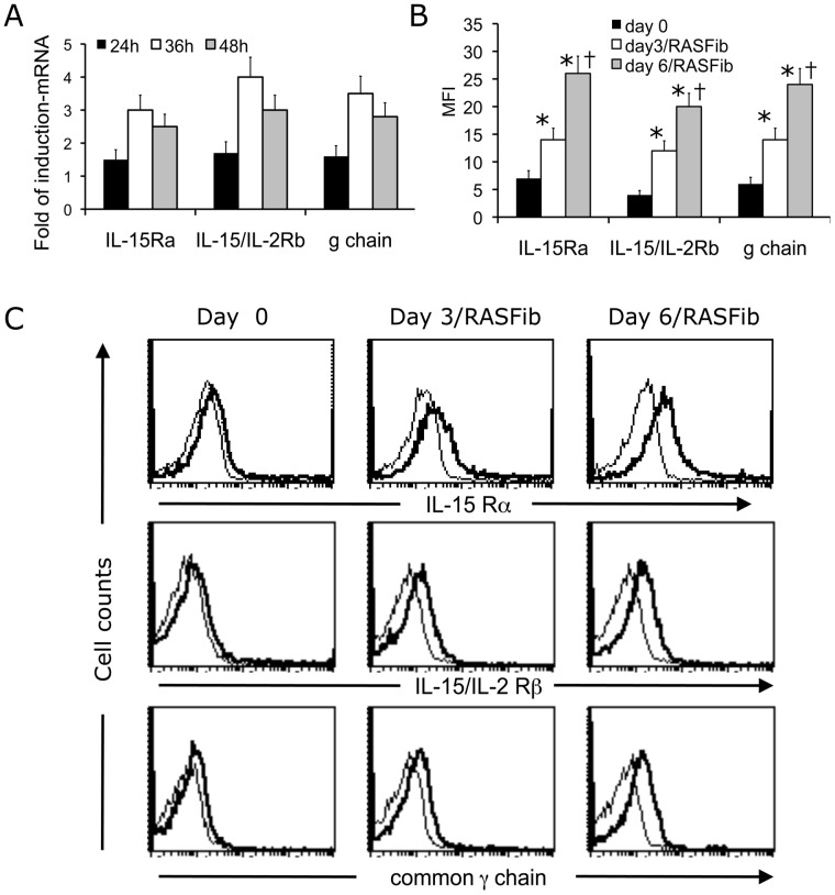
Results: RASFib constitutively expressed membrane IL-15. Survival of isolated B cells cultured for 6 days, below 5%, was extended in coculture with RASFib to 52+/-8% (p<0.001). IL-15 neutralizing agents but not isotype controls, reduced this rate to 31+/-6% (p<0.05). Interestingly, rhIL-15 had no effect on isolated B cells but significantly increased their survival in coculture with RASFib. In parallel, B cell IL-15R chains were upregulated in cocultures. BAFF and VCAM-1, that are expressed on RASFib, were tested as potential candidates involved in upregulating B cell IL-15R. Culture of B cells in the presence of rhBAFF or rhVCAM-1 resulted in significantly increased survival, together with upregulation of all three IL-15R chains; in parallel, rhIL-15 potentiated the anti-apoptotic effect of BAFF and VCAM-1. Both BAFF and VCAM-1 neutralizing agents downmodulated the effect of RASFib on B cell survival and IL-15R expression. In parallel, rhIL-15 had a lower effect on the survival of B cells cocultured with RASFib in the presence of BAFF or VCAM-1 neutralizing agents. Peripheral blood B cells from 15 early RA patients demonstrated an upregulated IL-15R and increased survival in cocultures.

Conclusion: IL-15 expression on RASFib significantly contributes to the anti-apoptotic effect of RASFib on B cells. IL-15 action is facilitated by BAFF and VCAM-1 expressed on RASFib, through an upregulation of IL-15R chains.


Conflict of interest statement

Competing Interests: The work reported in this manuscript has been supported by an unrestricted research grant from Roche Pharmaceuticals. This does not alter the authors’ adherence to all the PLoS ONE policies on sharing data and materials.

Figures

Figure 1
Figure 1. Characterization of cultured RASFib, OASFib and dermal fibroblasts (Third passage) and of freshly isolated memory B cells.
A. Representative flow cytometry histograms show staining of cultured RASFib (Third passage) with anti-CD90, anti-CD1, anti-CD3, anti-CD19, anti-CD14, anti-HLA-DR, anti-CD80 and anti-CD86 (Thick lines) or appropriate isotype controls (Thin lines). B. Bar histogram showing the MFI of RASFib (n = 10 lines), OASfib (n = 10 lines) or dermal fibroblasts (n = 5 lines) on the thrid passage, after staining with the antibodies mentioned in A. C. Representative flow cytometry dot plots after triple staining of isolated memory B cells with anti-CD20 FITC, anti-CD19 APC and anti-CD27PE or isotype control (gamma1) PE. Shown are two-dimension dot plots with different combinations of the above mentioned fluorochrome-labeled antibodies.
Figure 2
Figure 2. Effect of RASFib on B cell survival.
Coculture with RASFib significantly increases B cell survival in a cell-contact dependent manner. A. Representative flow cytometry dot-plots of B cells stained with JC-1 or with 7AAD/Annexin V. When compared with freshly isolated B cells, viability of B cells cultured in plain medium for 6 days is dramatically decreased. Coculture with RASFib for 6 days significantly improves B cell survival. B. Percentage of viable B cells at different time points (0 to 6 days) (JC-1 staining). B cells were cultured in plain medium or cocultured with untreated RASFib, with RASFib and transwell inserts, or cultured with supernatants from 6-day RASFib/Bcell cocultures. Each point represents the mean and SD of 15 subjects. * p<0.05 vs B cells cultured in plain medium. C. RASFib are significantly more effective than OASFib or dermal fibroblasts at promoting B cell suvival. Shown is the percentage of viable cells present at 6 days cocultures of B cells with RASFib versus cocultures with OA or dermal fibroblasts, versus B cells cultured alone (JC-1 staining). Each bar represents the mean and SD of 15 subjects per group. *p<0.05 vs B cells cultured alone in plain medium; †p<0.05 vs B cells cocultured with RASFib. D. Variation of the effect of different RA, OA or dermal fibroblast cell lines on B cell survival was small. B cells from a single donor were cocultured for 6 days with 10 different RA synovial fibroblast lines (RA 1-10), 10 different OA synovial fibroblast lines (OA 1-10) or 5 different dermal fibroblast lines (Der 1–5). Shown is the percentage of viable B cells on the 6th day of coculture with each of the tested lines (JC-1 staining).
Figure 3
Figure 3. Effect of IL-15 neutralizing agents and of exogenous rhIL-15 on the survival of B cells cocultured with RASFib.
The effect of RASFib on B cell survival is modified by IL-15 neutralizing agents and by exogenous rh IL-15. A. Flow cytometry of non-permeabilized cells demonstrates surface IL-15 expression on RASFib, OASFib and dermal fibroblasts. Top panels: representative flow cytometry histograms. Lower panel: bar histogram showing the mean and SD of the MFI (mean fluorescence intensity) of all tested RASFib cell lines (n = 10), OASFib lines (n = 10) and dermal fibroblast lines (n = 5). B. Effect of rhIL-15 (1–400 ng/ml) on isolated B cells cultured alone for 6 days. Each point represents the mean and SD of 15 subjects. C. Effect of IL-15 neutralizing agents on the survival of B cells cocultured with RASFib, OASFib or dermal fibroblasts (single time point, 6th day of coculture). Each point represents the mean and SD of 15 subjects. * p<0.05 vs same condition in the absence of neutralizing agents. †p<0.05 vs B cells cocultured with RASFib in the absence of IL-15 neutralizing agents. D. Effect of IL-15 neutralizing agents on the survival of B cells cocultured with RASFib. Time-course (0–6 day coculture). Each point represents the mean and SD of 15 subjects. * p<0.05 vs conditions in the absence of neutralizing agents. E. rhIL15 has a minimal effect on the survival of B cells cultured alone but significantly upregulates the survival of B cells cocultured with RASFib. Each point represents the mean and SD of 15 subjects. *p<0.05 vs B cells cultured in plain medium. † p<0.05 vs B cells cultured with RASFib in the absence of rhIL-15.
Figure 4
Figure 4. Effect of RASFib on the expression of IL15R α, β and γ chains on cocultured B cells.
B cells cocultured with RASFib upregulate IL-15 α, β and γ chain expression. A. Real-time quantitative analysis of IL-15R mRNA expression in B cells cocultured with RASFib for 24, 36 or 48 h. Shown is fold of induction referred to expression of IL-15R α, β and γ chain in freshly isolated B cells. Each bar represents the mean and SD of 15 subjects. B. Expression of IL-15R α, β and γ chain on the surface of non-permeabilized B cells as determined by flow cytometry. Shown is expression on freshly isolated B cells, on B cells cocultured with RASFib for 3 days and on B cells cocultured with RASFib for 6 days. Each bar represents the mean and SD of 15 subjects. *p<0.05 vs freshly isolated B cells (day 0); † p<0.05 vs B cells cocultured for 3 dys with RASFib (day 3). C. Representative flow cytometry histograms showing the expression of IL-15R α, β and γ chains on freshly isolated B cells and on B cells cocultured with RASFib for 3 or 6 days.
Figure 5
Figure 5. VCAM-1 and BAFF facilitate the effect of IL-15 on B cell survival and upregulate B cell IL-15R expression.
A. Surface expression of VCAM-1 and BAFF on non-permeabilized RASFib, OASFib or dermal fibroblasts. Top panels: representative flow cytometry histograms. Lower panels: bar histograms showing the mean and SD of the MFI (mean fluorescence intensity) of all tested RASFib cell lines (n = 10), OASFIb lines (n = 10) and dermal fibroblast lines (n = 5) B. Effect of rhBAFF (1–400 ng/ml) on isolated B cells cultured alone for 6 days. Each point represents the mean and SD of 15 subjects. C. Percentage of viable B cells cultured for 6 days in plain medium, medium with rhIL-15 or rhBAFF, VCAM-1-coated plates, rhIL-15 plus BAFF or rhIL-15 plus VCAM-1. *p<0.05 vs medium; †p<0.05 vs same condition without rhIL-15. D. Fold of induction of IL-15Rα, β and γ on B cells cultured for 6 days with rhBAFF or rhVCAM-1, as determined by cytometry and referred to freshly isolated cells. E. Percentage of viable B cells cultured for 6 days in medium, or cocultured with RASFib in the absence or presence of anti-VCAM-1 or anti-BAFF antibodies, in combination or not with an anti-IL15 antibody, with or without rhIL-15. *p<0.05 vs B cells cultured in medium; †p<0.05 vs same condition without rh IL-15; ¥p<0.05 vs same condition without anti-VCAM-1 or anti-BAFF antibodies. § p<0.05 vs same condition without anti-IL15. F. Fold of induction of IL-15R α, β and γ on B cells cocultured for 6 days with RASFib, with or without anti-VCAM-1 or anti-BAFF antibodies, as determined by cytometry and referred to freshly isolated B cells. *p<0.05 vs B cells cocultured with RASFib without antibodies. Each point represents the mean/SD of 15 subjects. G. Effect of VCAM-1 or BAFF neutralizing agents on the survival of B cells cocultured with OASFib or dermal fibroblasts. Each point represents the mean and SD of 15 subjects. * p<0.05 vs B cells cultured alone in plain medium.
Figure 6
Figure 6. Memory B cells from the peripheral blood of early RA patients demonstrate and upregulated IL-15R expression together with superior survival rates in response to rhIL-15 and in coculture with RASFib.
A. Surface expression of IL-15 receptor α, β and γ chains on memory B cells from healthy controls (n = 15) (black bars), from early RA patients (n = 15) (open bars), and from 8 early RA patients who donated blood for a second time after achieving remission (grey bars), as determined by flow cytometry of non-permeabilized cells, and expressed as mean fluorescence intensity (MFI). Each bar represents the mean and SD of 15 or 8 subjects. *p<0.05 vs healthy controls. B. Percentage of viable B cells cultured for 6 days in plain medium, or in medium supplemented with rhIL-15, or cocultured with RASFib in plain medium, or cocultured with RASFib in the presence of a neutralizing anti-IL-15 mAb. Black bars represent B cells from healthy controls (n = 15), open bars represent B cells from early RA patients (n = 15) and grey bars represent B cells from 8 early RA patients who donated blood for a second time after achieving remission. Each bar represents the mean and SD of 15 or of 8 subjects. *p<0.05 vs B cells cultured in plain medium. †p<0.05 vs B cells from healthy controls; ¥p<0.05 vs B cells cocultured with RASFib in the absence of an anti-IL-15 neutralizing antibody.

Similar articles

Cited by

References

    1. Zvaifler NJ, Boyle D, Firestein GS. Early synovitis–synoviocytes and mononuclear cells. Semin Arthritis Rheum. 1994;23(6:11–16. - PubMed
    1. Weyand CM, Kang YM, Kurtin PJ, Goronzy JJ. The power of the third dimension: tissue architecture and autoimmunity in rheumatoid arthritis. Curr Opin Rheumatol. 2003;15:259–266. - PubMed
    1. Cantaert T, Kolln J, Timmer T, van der Pouw Kraan TC, Vandooren B, et al. B lymphocyte autoimmunity in rheumatoid synovitis is independent of ectopic lymphoid neogenesis. J Immunol. 2008;181:785–794. - PubMed
    1. Zvaifler NJ. Rheumatoid synovitis. An extravascular immune complex disease. Arthritis Rheum. 1974;17:297–305. - PubMed
    1. Dörner T, Burmester GR. The role of B cells in rheumatoid arthritis: mechanisms and therapeutic targets. Curr Opin Rheumatol. 2003;15:246–252. - PubMed

Publication types

MeSH terms

Substances