. 2012;7(7):e40730.
doi: 10.1371/journal.pone.0040730. Epub 2012 Jul 9.

Yersinia enterocolitica YopT and Clostridium difficile toxin B induce expression of GILZ in epithelial cells

Affiliations

Yersinia enterocolitica YopT and Clostridium difficile toxin B induce expression of GILZ in epithelial cells

Martin Köberle et al. PLoS One. 2012.

Abstract

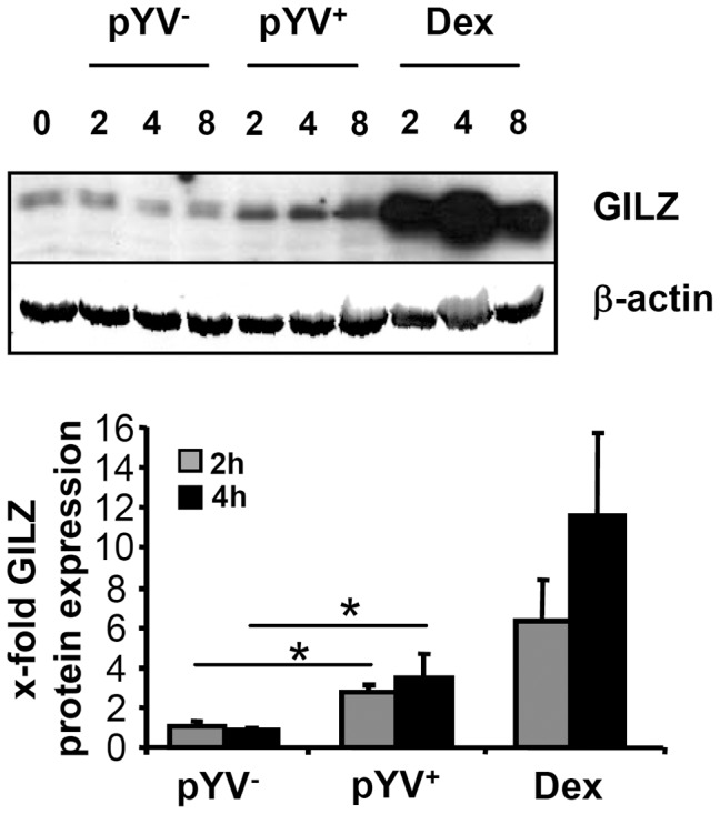
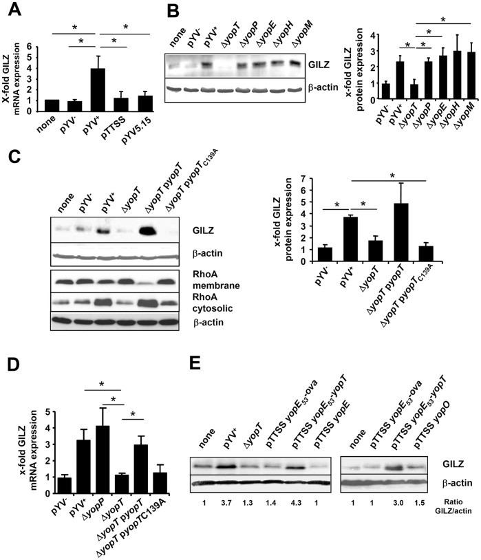
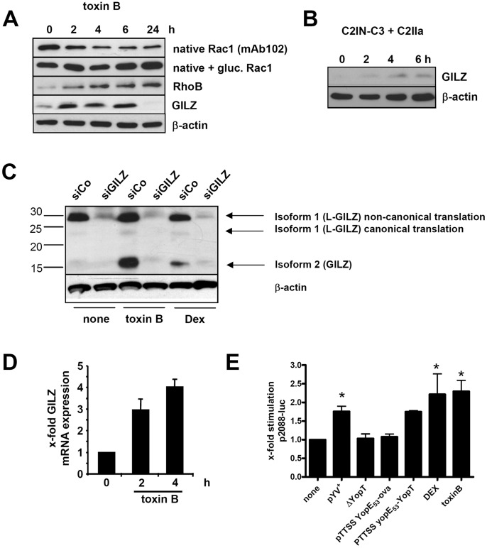
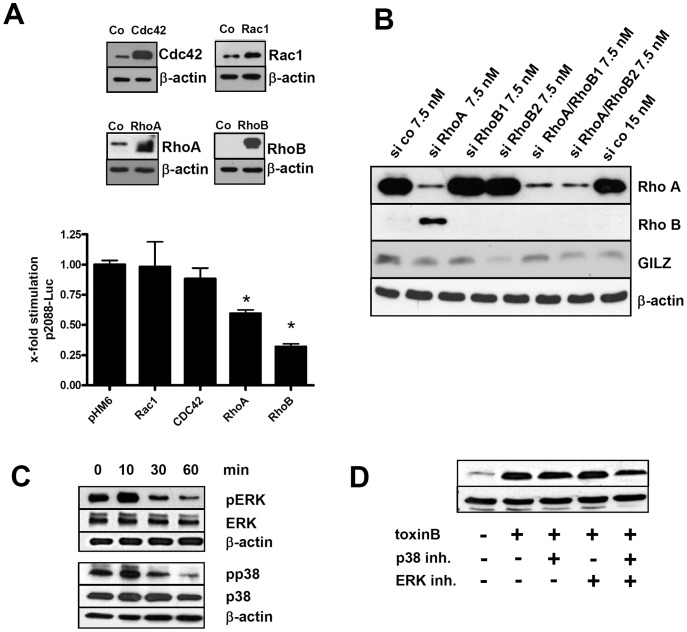
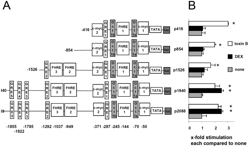
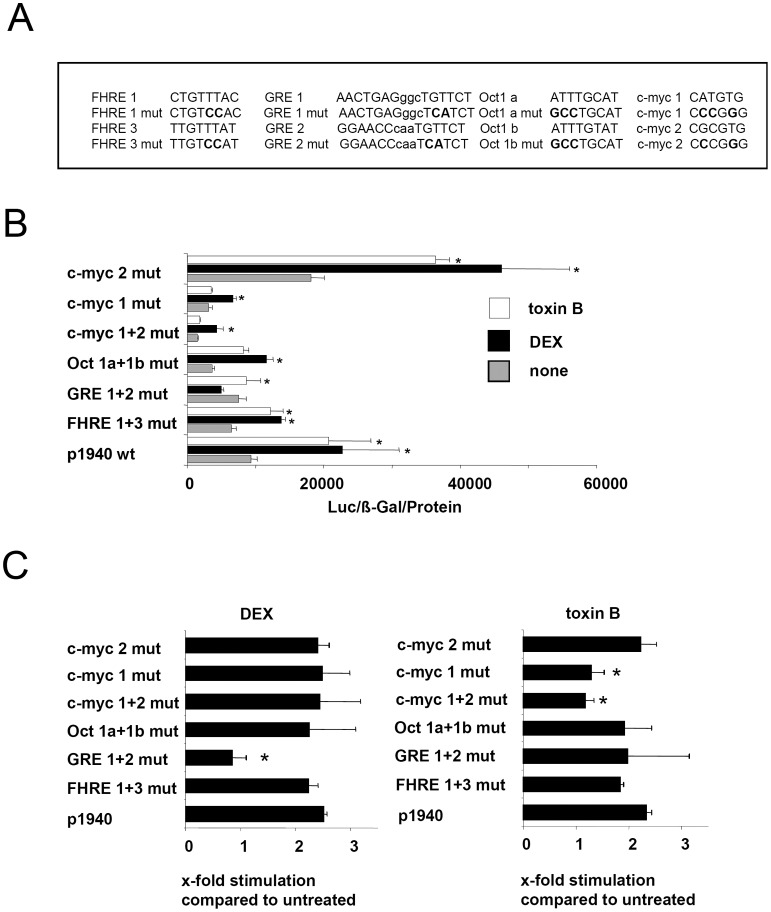
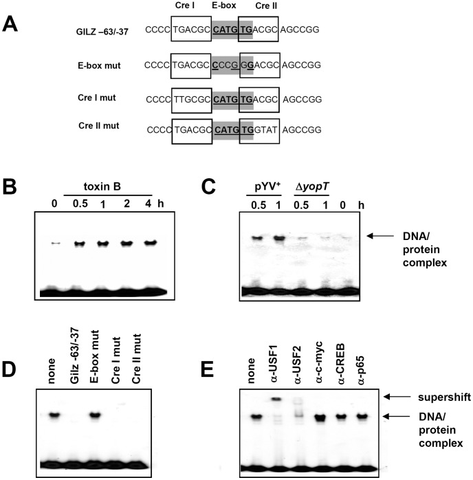
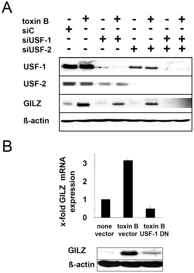
Glucocorticoid induced-leucine zipper (GILZ) has been shown to be induced in cells by different stimuli such as glucocorticoids, IL-10 or deprivation of IL-2. GILZ has anti-inflammatory properties and may be involved in signalling modulating apoptosis. Herein we demonstrate that wildtype Yersinia enterocolitica which carry the pYV plasmid upregulated GILZ mRNA levels and protein expression in epithelial cells. Infection of HeLa cells with different Yersinia mutant strains revealed that the protease activity of YopT, which cleaves the membrane-bound form of Rho GTPases was sufficient to induce GILZ expression. Similarly, Clostridium difficile toxin B, another bacterial inhibitor of Rho GTPases induced GILZ expression. YopT and toxin B both increased transcriptional activity of the GILZ promoter in HeLa cells. GILZ expression could not be linked to the inactivation of an individual Rho GTPase by these toxins. However, forced expression of RhoA and RhoB decreased basal GILZ promoter activity. Furthermore, MAPK activation proved necessary for profound GILZ induction by toxin B. Promoter studies and gel shift analyses defined binding of upstream stimulatory factor (USF) 1 and 2 to a canonical c-Myc binding site (E-box) in the GILZ promoter as a crucial step of its trans-activation. In addition we could show that USF-1 and USF-2 are essential for basal as well as toxin B induced GILZ expression. These findings define a novel way of GILZ promoter trans-activation mediated by bacterial toxins and differentiate it from those mediated by dexamethasone or deprivation of IL-2.


Conflict of interest statement

Competing Interests: The authors have declared that no competing interests exist.

Figures

Figure 1
Figure 1. Yersinia enterocolitica induces GILZ expression.
HeLa cells were infected with a strain harboring the Yersinia virulence plasmid (pYV+) or plasmid cured strain (pYV) with MOI 20 or stimulated with 100 µM DEX for indicated time intervals. The amount of GILZ in cytosolic proteins lysates of HeLa cells was detected by immunoblot at different time points. Actin was used as an internal standard. A representative experiment and quantification means + SEM normalized to untreated of at least 3 experiments are shown.
Figure 2
Figure 2. Yersinia induced GILZ expression depends on YopT protease activity.
HeLa cells were infected with (A) strains with (pYV+) or without (pYV) pathogenicity plasmid or with a pathogenicity plasmid derivate coding for a functional translocation apparatus but not for effector Yops (pTTSS) or with a pathogenicity plasmid coding for a defective translocation apparatus (pYV5.15) with MOI 20 for 2 or 4 h to determine GILZ mRNA expression by real-time RT-PCR. Means + SD of 2 independent experiments. Significant differences compared to pYV+ are indicated by asterisks (p<0.05). (B) Immunoblot of GILZ protein expression induced by strains with single yop deletions. Representative immunoblot and quantification means + SEM of 4 independent experiments normalized to untreated. Significant differences compared to ΔyopT are indicated by asterisks (p<0.05). (C) Infection with a yopT deletion strain and derivative strains complemented with an additional plasmid encoding wildtype (pyopT) or protease deficient (pyopTC139A) YopT. Representative immunoblot analysis of GILZ protein expression in the cytosolic fraction and of RhoA in cytosolic and membrane fraction (left) and quantification means + SD of GILZ expression of two representative experiments normalized to untreated (right). Significant differences compared to pYV+ are indicated by asterisks (p<0.05). (D) Experimental design as in C but analysis of GILZ mRNA expression. Significant differences compared to ΔYopT are indicated by asterisks (p<0.05). Mean + SD of 2 independent experiments normalized to untreated. (E) GILZ protein expression induced by strains translocating only one Yop affecting Rho GTPases. Cells were infected with Yersinia translocating YopE, YopE53-YopT or YopO. Infections with pYV+, ΔyopT and a strain translocating only enzymatically inactive YopE53-Ova247–355 were used as controls.
Figure 3
Figure 3. GILZ is expressed upon stimulation with C3 exotoxin or toxin B.
(A) HeLa cells were incubated with toxin B (50ng/ml) for indicated the time spans and immunoblots were performed from whole cell lysates using anti-Rac1 mAb102 recognizing only non-glucosylated Rac-1, and an anti-Rac1 mAb antibody recognizing total Rac1 as well as antibodies recognizing RhoB, actin or GILZ. (B) Immunoblot of GILZ and actin expression upon stimulation of HeLa cells with C2IN-C3lim (100 ng/mL) + C2IIa (200 ng/mL) for indicated time spans. (C) To explore the expression of additional GILZ isoforms, HeLa cells were transfected with 7.5 nM siRNA specific for GILZ or control siRNA for 48 h and subsequently either left untreated or stimulated with C. difficile toxin B (50 ng/ml) or 100 µM DEX for 4 h. Arrows mark three GILZ isoforms which were inhibited by the used GILZ siRNA. Note that only isoform 1 was induced by the used stimuli. (D) Cells were stimulated with toxin B for 2 or 4 h to determine GILZ mRNA expression by real-time RT-PCR. Mean + SD of 2 independent experiments normalized to untreated. (E) To assay transcriptional activity of the GILZ promoter cells were transfected with a luciferase reporter under control of a 2088 bp GILZ promoter and co-transfected with pCMV-ß-gal (for standardization) 24 h before infection with a Y. enterocolitica pYV+ and various mutant strains or treatment with DEX or toxin B. Means + SD of 4 independent experiments normalized to untreated. Significant differences compared to untreated are indicated by asterisks (p<0.05).
Figure 4
Figure 4. Role of Rho GTPases and MAP kinases for GILZ expression.
(A) Overexpression of RhoA or RhoB lowers basal GILZ levels. HeLa cells were co-transfected with the p2088 GILZ promoter luciferase reporter and pHM6 based plasmid for overexpression of the indicated Rho GTPases. Means + SD of 3 independent experiments normalized to untreated. Significant differences compared to control vector transfection are indicated by asterisks (p<0.05). In a control experiment, HeLa cells were transfected in the same setting and cell lysates were used for immunoblots to detect RhoA, RhoB, Cdc42 and Rac1 expression. (B) HeLa cells were transfected for 48 h with indicated concentrations of siRNA. Immunoblots were performed from cell lysates for RhoA and RhoB and from cytosolic extracts for GILZ. (C) Toxin B treatment leads to fast and transient MAPK phosphorylation. After treatment of HeLa cells with toxin B for the indicated time spans, levels of phosphorylated as well as total ERK and p38 were assayed by immunoblot. (C) Toxin B induced GILZ expression is mediated by both ERK and p38 MAPK. Cells were pretreated with MAPK phosphorylation inhibitors SB 202190 (p38) or PD 98059 (ERK) 2 h prior to toxin B stimulation and GILZ protein was detected by immunoblot analysis at 6 h or 24 h after stimulation.
Figure 5
Figure 5. Differential activation of truncated GILZ promoter elements by toxin B or DEX.
HeLa cells were transiently transfected with pCMV-ß-gal for 24 h and co-transfected (A) with p2088-Luc or luciferase reporters fused to shortened promoter regions, containing the indicated TF binding sites. GRE: glucocorticoid responsive element, FHRE: forkhead responsive elements, c-myc: c-myc binding site (E-box), Oct: octamer binding site (B) Transfected cells were treated with 100 µM DEX or 50 ng/ml toxin B for 6 h. Subsequently luciferase assays were performed. Results are expressed as fold induction compared to unstimulated (none) cells and represent the mean + SEM of three experiments performed in triplicates (p<0.05).
Figure 6
Figure 6. Importance of specific cis-elements for toxin B induced GILZ promoter trans-activation.
(A) Recognition sequences of cis-elements which were mutated. Mutated base pairs are highlighted using bold letters. (B) HeLa cells were transfected with p1940-Luc and different mutated derivatives for 24 h and subsequently stimulated with DEX or toxin B for 6 h. Data are shown as relative light units (RLU) standardized to β-Gal activity and protein concentration or (C) as fold induction after DEX or toxin B stimulation compared to untreated conditions of each individual expression vector. Means + SEM of four independent experiments are shown. Asterisks indicate significant differences between DEX or toxin B stimulation compared to uninfected (p<0.05).
Figure 7
Figure 7. Role of myc-1 E-box in TF binding.
Electromobility shift analyses were performed using a double-stranded oligonucleotide probe representing the GILZ promoter sequence −63 to −37 and for some experiments probes with mutations of the E-box cis-elements and the flanking cAMP response (Cre) elements as depicted in (A). HeLa cells were stimulated/infected for 0.5 h or indicated time intervals with toxin B (B, D, E) or Y. enterocolitica (C) and nuclear extracts of these cells were incubated with P32-labeled GILZ−63/−37 probe. Subsequently band shift analyses were performed. (D) Nuclear extracts were pretreated with a 100-fold excess of indicated cold probes. (E) Nuclear extracts were pretreated with indicated antibodies. Anti-p65 antibody was used as a negative control.
Figure 8
Figure 8. Role of USF-1 and USF-2 for toxin B induced GILZ expression.
(A) HeLa cells were transfected with siRNA silencing USF-1 or USF-2 or with control siRNA (siC) 48 h prior to toxin B treatment and USF-1/2 as wells as GILZ and actin expression was determined by immunoblot. (B) HeLa cells were transfected with empty vector or a dominant negative (DN) USF-1 mutant and subsequently GILZ mRNA or GILZ protein expression was determined by real time RT-PCR (one representative experiment performed in quadruplicates, means + SEM) or immunoblot.

Similar articles

Cited by

References

    1. Smego RA, Frean J, Koornhof HJ. Yersiniosis I: microbiological and clinicoepidemiological aspects of plague and non-plague Yersinia infections. Eur J Clin Microbiol Infect Dis. 1999;18:1–15. - PubMed
    1. Koornhof HJ, Smego RA, Jr, Nicol M. Yersiniosis. II: The pathogenesis of Yersinia infections. Eur J Clin Microbiol Infect Dis. 1999;18:87–112. - PubMed
    1. Schulte R, Wattiau P, Hartland EL, Robins-Browne RM, Cornelis GR. Differential secretion of interleukin-8 by human epithelial cell lines upon entry of virulent or nonvirulent Yersinia enterocolitica. Infect Immun. 1996;64:2106–2113. - PMC - PubMed
    1. Schmid Y, Grassl GA, Buhler OT, Skurnik M, Autenrieth IB, et al. Yersinia enterocolitica adhesin A induces production of interleukin-8 in epithelial cells. Infect Immun. 2004;72:6780–6789. - PMC - PubMed
    1. Auerbuch V, Golenbock DT, Isberg RR. Innate immune recognition of Yersinia pseudotuberculosis type III secretion. PLoS Pathog. 2009;5:e1000686. - PMC - PubMed

Publication types

MeSH terms