Involvement of the penta-EF-hand protein Pef1p in the Ca2+-dependent regulation of COPII subunit assembly in Saccharomyces cerevisiae
- PMID: 22792405
- PMCID: PMC3394733
- DOI: 10.1371/journal.pone.0040765
Involvement of the penta-EF-hand protein Pef1p in the Ca2+-dependent regulation of COPII subunit assembly in Saccharomyces cerevisiae
Abstract
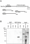
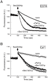
Although it is well established that the coat protein complex II (COPII) mediates the transport of proteins and lipids from the endoplasmic reticulum (ER) to the Golgi apparatus, the regulation of the vesicular transport event and the mechanisms that act to counterbalance the vesicle flow between the ER and Golgi are poorly understood. In this study, we present data indicating that the penta-EF-hand Ca(2+)-binding protein Pef1p directly interacts with the COPII coat subunit Sec31p and regulates COPII assembly in Saccharomyces cerevisiae. ALG-2, a mammalian homolog of Pef1p, has been shown to interact with Sec31A in a Ca(2+)-dependent manner and to have a role in stabilizing the association of the Sec13/31 complex with the membrane. However, Pef1p displayed reversed Ca(2+) dependence for Sec13/31p association; only the Ca(2+)-free form of Pef1p bound to the Sec13/31p complex. In addition, the influence on COPII coat assembly also appeared to be reversed; Pef1p binding acted as a kinetic inhibitor to delay Sec13/31p recruitment. Our results provide further evidence for a linkage between Ca(2+)-dependent signaling and ER-to-Golgi trafficking, but its mechanism of action in yeast seems to be different from the mechanism reported for its mammalian homolog ALG-2.
Conflict of interest statement
Figures


Similar articles
-
ALG-2 attenuates COPII budding in vitro and stabilizes the Sec23/Sec31A complex.PLoS One. 2013 Sep 19;8(9):e75309. doi: 10.1371/journal.pone.0075309. eCollection 2013. PLoS One. 2013. PMID: 24069399 Free PMC article.
-
TRAPPI tethers COPII vesicles by binding the coat subunit Sec23.Nature. 2007 Feb 22;445(7130):941-4. doi: 10.1038/nature05527. Epub 2007 Feb 7. Nature. 2007. PMID: 17287728
-
Human Sec31B: a family of new mammalian orthologues of yeast Sec31p that associate with the COPII coat.J Cell Sci. 2006 Mar 1;119(Pt 5):958-69. doi: 10.1242/jcs.02751. J Cell Sci. 2006. PMID: 16495487
-
Making COPII coats.Cell. 2007 Jun 29;129(7):1251-2. doi: 10.1016/j.cell.2007.06.015. Cell. 2007. PMID: 17604713 Review.
-
Adaptor functions of the Ca2+-binding protein ALG-2 in protein transport from the endoplasmic reticulum.Biosci Biotechnol Biochem. 2019 Jan;83(1):20-32. doi: 10.1080/09168451.2018.1525274. Epub 2018 Sep 27. Biosci Biotechnol Biochem. 2019. PMID: 30259798 Review.
Cited by
-
ALG-2 attenuates COPII budding in vitro and stabilizes the Sec23/Sec31A complex.PLoS One. 2013 Sep 19;8(9):e75309. doi: 10.1371/journal.pone.0075309. eCollection 2013. PLoS One. 2013. PMID: 24069399 Free PMC article.
-
Penta-EF-Hand Protein Peflin Is a Negative Regulator of ER-To-Golgi Transport.PLoS One. 2016 Jun 8;11(6):e0157227. doi: 10.1371/journal.pone.0157227. eCollection 2016. PLoS One. 2016. PMID: 27276012 Free PMC article.
-
Mechanisms for exporting large-sized cargoes from the endoplasmic reticulum.Cell Mol Life Sci. 2015 Oct;72(19):3709-20. doi: 10.1007/s00018-015-1952-9. Epub 2015 Jun 17. Cell Mol Life Sci. 2015. PMID: 26082182 Free PMC article. Review.
-
COPII-dependent ER export in animal cells: adaptation and control for diverse cargo.Histochem Cell Biol. 2018 Aug;150(2):119-131. doi: 10.1007/s00418-018-1689-2. Epub 2018 Jun 18. Histochem Cell Biol. 2018. PMID: 29916038 Free PMC article. Review.
-
Modulation of the secretory pathway rescues zebrafish polycystic kidney disease pathology.J Am Soc Nephrol. 2014 Aug;25(8):1749-59. doi: 10.1681/ASN.2013101060. Epub 2014 Mar 13. J Am Soc Nephrol. 2014. PMID: 24627348 Free PMC article.
References
-
- Dancourt J, Barlowe C. Protein sorting receptors in the early secretory pathway. Annu Rev Biochem. 2010;79:777–802. - PubMed
-
- Zanetti G, Pahuja KB, Studer S, Shim S, Schekman R. COPII and the regulation of protein sorting in mammals. Nat Cell Biol. 2012;14:20–28. - PubMed
-
- Barlowe C, Schekman R. SEC12 encodes a guanine-nucleotide-exchange factor essential for transport vesicle budding from the ER. Nature. 1993;365:347–349. - PubMed
-
- Bi X, Corpina RA, Goldberg J. Structure of the Sec23/24-Sar1 pre-budding complex of the COPII vesicle coat. Nature. 2002;419:271–277. - PubMed
Publication types
MeSH terms
Substances
LinkOut - more resources
Full Text Sources
Molecular Biology Databases
Miscellaneous

