

Primary cardiac tumours: a single-center 41-year experience
- PMID: 22792486
- PMCID: PMC3391967
- DOI: 10.5402/2012/906109
Primary cardiac tumours: a single-center 41-year experience
Abstract
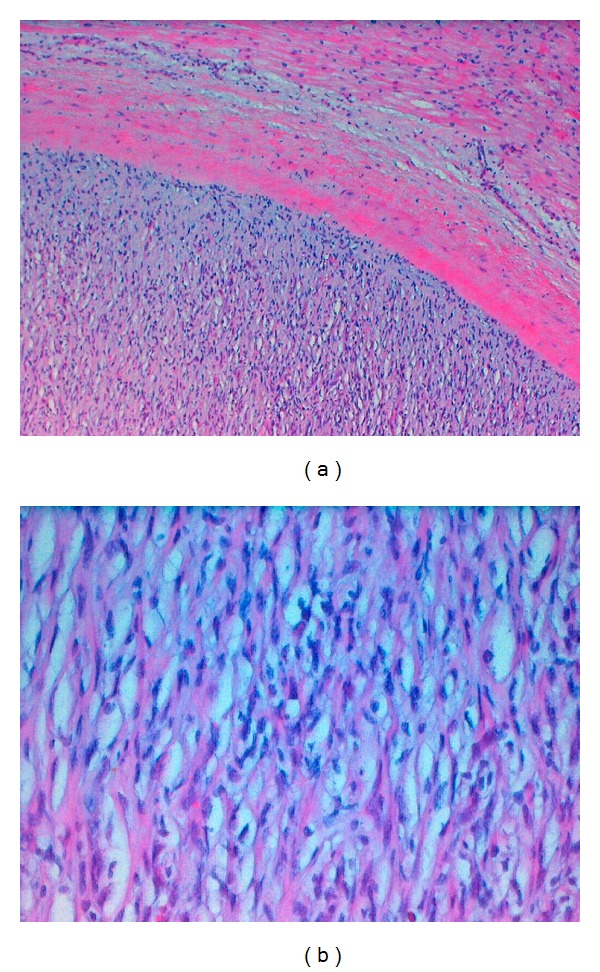
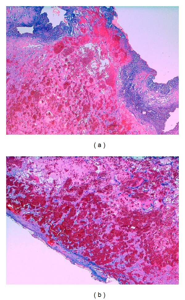
Primary cardiac tumours are extremely rare with the most commonest being left atrial myxomas. In general, surgical resection is indicated, whenever the tumour formation is mobile and embolization can be suspected. Within 17280 patients receiving heart surgery at the Innsbruck Medical University, 78 patients (0.45%) underwent tumourectomy of primary cardiac tumours. The majority of patients (63) suffered from a left or right atrial myxoma, 12 showed a papillary fibroelastoma of the valves at echocardiographical or histological examination, 1 suffered from a hemangioma, 1 from a chemodectoma, and another one from a rhabdomyosarcoma. The mean age of cardiac tumour patients was 54.29 ± 13.28 years (ranging from 18 to 83 years). 67.95% of the patients were female and 32.05% were male. The majority of tumours were found incidentally; 97.44% of the patients showed no tumour recurrence.
Figures


References
-
- Burke A, Virmani R. Atlas of Tumor Pathology: Tumors of the Cardiovascular System. Washington, DC, USA: Armed Forces Institute of Pathology Press; 1996. Classification and incidence of cardiac tumors.
-
- Travis WD, Brambilla E, Müller-Hermelink HK, Harris CC. Pathology & Genetics of Tumours of the Lung, Pleura, Thymus and Heart. Lyon, France: IARC Press; 2004. World Health Organization classification of tumours; p. p. 250.
-
- Burke AP, Tazelaar H, Gomes-Roman JJ, et al. Benign tumors of pluripotent mesenchym. In: Travis W, editor. Pathology & Genetics of Tumours of the Lung, Pleura, Thymus and Heart. Lyon, France: IARC Press; 2004. pp. 260–265.
-
- Hi D, Yoon A, Roberts WC. Sex distribution in cardiac myxomas. American Journal of Cardiology. 2002;90(5):563–565. - PubMed
-
- Burke AP, Virmani R. Cardiac myxoma: a clinicopathologic study. American Journal of Clinical Pathology. 1993;100(6):671–680. - PubMed
LinkOut - more resources
Full Text Sources
