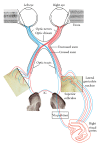
Adaptive neuroplastic responses in early and late hemispherectomized monkeys
- PMID: 22792495
- PMCID: PMC3391903
- DOI: 10.1155/2012/852423
Adaptive neuroplastic responses in early and late hemispherectomized monkeys
Abstract
Behavioural recovery in children who undergo medically required hemispherectomy showcase the remarkable ability of the cerebral cortex to adapt and reorganize following insult early in life. Case study data suggest that lesions sustained early in childhood lead to better recovery compared to those that occur later in life. In these children, it is possible that neural reorganization had begun prior to surgery but was masked by the dysfunctional hemisphere. The degree of neural reorganization has been difficult to study systematically in human infants. Here we present a 20-year culmination of data on our nonhuman primate model (Chlorocebus sabeus) of early-life hemispherectomy in which behavioral recovery is interpreted in light of plastic processes that lead to the anatomical reorganization of the early-damaged brain. The model presented here suggests that significant functional recovery occurs after the removal of one hemisphere in monkeys with no preexisting neurological dysfunctions. Human and primate studies suggest a critical role for subcortical and brainstem structures as well as corticospinal tracts in the neuroanatomical reorganization which result in the remarkable behavioral recovery following hemispherectomy. The non-human primate model presented here offers a unique opportunity for studying the behavioral and functional neuroanatomical reorganization that underlies developmental plasticity.
Figures


Similar articles
-
Functional reorganization after hemispherectomy in humans and animal models: What can we learn about the brain's resilience to extensive unilateral lesions?Brain Res Bull. 2017 May;131:156-167. doi: 10.1016/j.brainresbull.2017.04.005. Epub 2017 Apr 13. Brain Res Bull. 2017. PMID: 28414105 Review.
-
Line bisection following hemispherectomy.Neuropsychologia. 2003;41(11):1523-30. doi: 10.1016/s0028-3932(03)00076-9. Neuropsychologia. 2003. PMID: 12849770 Clinical Trial.
-
Functional reorganization of the human auditory pathways following hemispherectomy: an fMRI demonstration.Neuropsychologia. 2008 Oct;46(12):2936-42. doi: 10.1016/j.neuropsychologia.2008.06.009. Epub 2008 Jun 18. Neuropsychologia. 2008. PMID: 18602934
-
Corollary discharge and spatial updating: when the brain is split, is space still unified?Prog Brain Res. 2005;149:187-205. doi: 10.1016/S0079-6123(05)49014-7. Prog Brain Res. 2005. PMID: 16226585
-
Integrated technology for evaluation of brain function and neural plasticity.Phys Med Rehabil Clin N Am. 2004 Feb;15(1):263-306. doi: 10.1016/s1047-9651(03)00124-4. Phys Med Rehabil Clin N Am. 2004. PMID: 15029909 Review.
Cited by
-
Altered contralateral sensorimotor system organization after experimental hemispherectomy: a structural and functional connectivity study.J Cereb Blood Flow Metab. 2015 Aug;35(8):1358-67. doi: 10.1038/jcbfm.2015.101. Epub 2015 May 13. J Cereb Blood Flow Metab. 2015. PMID: 25966942 Free PMC article.
-
Standardized full-field electroretinography in the Green Monkey (Chlorocebus sabaeus).PLoS One. 2014 Oct 31;9(10):e111569. doi: 10.1371/journal.pone.0111569. eCollection 2014. PLoS One. 2014. PMID: 25360686 Free PMC article.
-
A reproducible and translatable model of focal ischemia in the visual cortex of infant and adult marmoset monkeys.Brain Pathol. 2014 Sep;24(5):459-74. doi: 10.1111/bpa.12129. Epub 2014 Feb 28. Brain Pathol. 2014. PMID: 25469561 Free PMC article.
References
-
- Wilson PJE. Cerebral hemispherectomy for infantile hemiplegia: a REPORT of 50 cases. Brain. 1970;93(1):147–180. - PubMed
-
- French LA, Johnson DR, Brown IA, van Bergen FB. Cerebral hemispherectomy for control of intractable convulsive seizures. Journal of Neurosurgery. 1955;12(2):154–164. - PubMed
-
- Krynauw RW. Infantile hemiplegia treated by removal of one cerebral hemisphere. South African Medical Journal. 1950;24(27):539–546. - PubMed
-
- White HH. Cerebral hemispherectomy in the treatment of infantile hemiplegia; review of the literature and report of two cases. Confinia Neurologica. 1961;21:1–50. - PubMed
-
- van Empelen R, Jennekens-Schinkel A, Buskens E, Helders PJM, van Nieuwenhuizen O. Functional consequences of hemispherectomy. Brain. 2004;127(9):2071–2079. - PubMed
Publication types
MeSH terms
LinkOut - more resources
Full Text Sources

