

Intestinal schistosomiasis as unusual aetiology for acute appendicitis, nowadays a rising disease in Western countries
- PMID: 22792502
- PMCID: PMC3389664
- DOI: 10.1155/2012/896820
Intestinal schistosomiasis as unusual aetiology for acute appendicitis, nowadays a rising disease in Western countries
Abstract
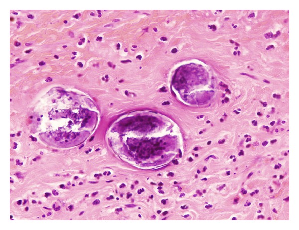
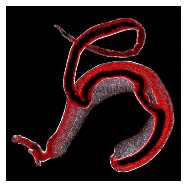
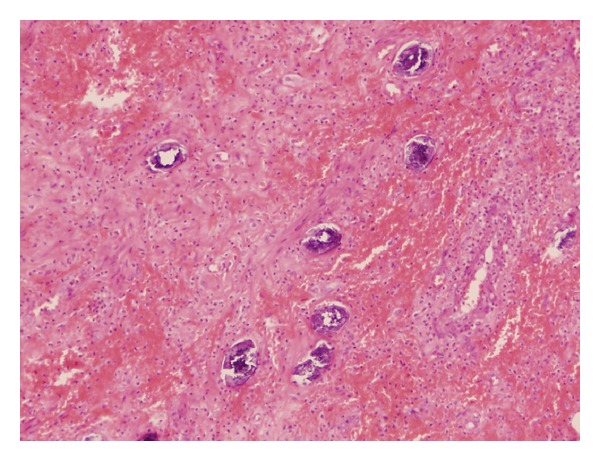
Intestinal schistosomiasis as unusual aetiology for acute appendicitis, nowadays a rising disease in western countries. Recent changes in global migration has led to an immigration growth in our scenario, upsurging people coming from endemic areas of schistosomiasis. Schistosomal appendicitis, seldom reported in developed countries, is now an expected incrising entity in our hospitals during the near future. Due to this circumstances, we believe that schistosomiasis should be consider as a rising source for acute appendicitis in western countries. In order to illustrate this point, we present a case of a 45-years-old black man, from Africa, was admitted via A&E because of acute abdominal pain, located in right lower quadrant. Acute appendicitis was suspected, and he underwent laparotomy and appendectomy. Pathological study by microscope revealed a gangrenous appendix with abscesses and parasitic ova into the submucosal layer of the appendix, suggesting Schistosomiasis.
Figures
References
-
- Hatz CFR. Schistosomiasis: an underestimated problem in industrialized countries? Journal of Travel Medicine. 2005;12(1):1–2. - PubMed
-
- Bierman WFW, Wetsteyn JCFM, van Gool T. Presentation and diagnosis of imported schistosomiasis: relevance of eosinophilia, microscopy for ova, and serology. Journal of Travel Medicine. 2005;12(1):9–13. - PubMed
-
- Grobusch MP, Mühlberger N, Jelinek T, et al. Imported schistosomiasis in Europe: sentinel surveillance data from TropNetEurop. Journal of Travel Medicine. 2003;10(3):164–169. - PubMed
-
- Schwartz E, Kozarsky P, Wilson M, Cetron M. Schistosome infection among river rafters on Omo River, Ethiopia. Journal of Travel Medicine. 2005;12(1):3–8. - PubMed
-
- Helmy AH, Shousha TA, Magdi M, Sabri T. Appendicitis; appendicectomy and the value of endemic parasitic infestation. Egyptian Journal of Surgery. 2000;19(2):87–91.
LinkOut - more resources
Full Text Sources
