Review
. 2012 Jun;4(12):1509-25.
doi: 10.4155/bio.12.133.

Microfluidic 3D cell culture: potential application for tissue-based bioassays

Affiliations
Review

Microfluidic 3D cell culture: potential application for tissue-based bioassays

Xiujun James Li et al. Bioanalysis. 2012 Jun.

Abstract

Current fundamental investigations of human biology and the development of therapeutic drugs commonly rely on 2D monolayer cell culture systems. However, 2D cell culture systems do not accurately recapitulate the structure, function or physiology of living tissues, nor the highly complex and dynamic 3D environments in vivo. Microfluidic technology can provide microscale complex structures and well-controlled parameters to mimic the in vivo environment of cells. The combination of microfluidic technology with 3D cell culture offers great potential for in vivo-like tissue-based applications, such as the emerging organ-on-a-chip system. This article will review recent advances in the microfluidic technology for 3D cell culture and their biological applications.


Figures

Figure 1
Figure 1
A microfluidic perfusion 3D cell culture system using micropillars to separate cells with perfusion medium. Cells are perfused with cell culture medium through gaps between micropillars [14]. Reprinted with permission from the Royal Society of Chemistry.
Figure 2
Figure 2
Schematic of the fabrication of cell-laden agarose-based microfluidic devices without (left) and with (right) embedded cells. Standard soft lithography technique was used to mold molten agarose on a SU-8 patterned silicon wafer. Two agarose substrates were then thermally bonded together [47]. Reprinted with permission from the Royal Society of Chemistry.
Figure 3
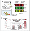
Figure 3
Paper-based 3D cell culture. (A) Schematic of the generation of paper-based 3D cell culture on a single layer of paper. Chromatography or filter paper is permeated with Matrigel or other hydrogel precursor and cell suspension by spotting, producing cell-laden hydrogels of thickness equal to the paper substrate. (B) Fluorescence images of cells cultured in paper. (C) Schematic illustration of stacking and destacking of multiple layers of paper impregnated with cells for the study of gradient-dependent 3D cell culture [40]. Reprinted with permission from National Academy of Sciences.
Figure 4
Figure 4
(a-c) Schematic of microfluidic spheroid formation in a two-layer microfluidic PDMS device. Top layer consisted of dead end channels with 28 side-chambers for cell capture and spheroid culture formation, while the bottom layer allowed the flow of medium through the channel. (d-e) Actual time-lapse images of PC-3DsRed co-culture spheroid formation within microchannels [5]. Reprinted with permission from Biomaterials.
Figure 5
Figure 5
Summarized applications of microfluidic 3D cell culture according to cell type
Figure 6
Figure 6
Perivascular association between HUVEC and MSC in co-culture (b-d) inside a micropillar-bound microchannel (a). Formation of capillary networks inside microfluidic channel was observed by confocal microscope imaging. Green indicates presence of laminin in (b); MSCs stained for α-SMA staining (green) lie in close proximity to the newly deposited basement membrane (laminnin, red) in (c-d), and all nuclei were stained with DAPI, blue [18]. Reprinted with permission from Biotechnology and Bioengineering.
Figure 7
Figure 7
3D microfluidic cell culture system for mimicking four human organs [62]. (a) Microfluidic system set-up. (b) Evaluation of cell viability. Reprinted with permission from the Royal Society of Chemistry.

Similar articles

Cited by

References

    1. Marimuthu M, Kim S. Microfluidic cell coculture methods for understanding cell biology, analyzing bio/pharmaceuticals, and developing tissue constructs. Analytical biochemistry. 2011;413(2):81–89. - PubMed
    1. Elliott NT, Yuan F. A review of three-dimensional in vitro tissue models for drug discovery and transport studies. Journal of pharmaceutical sciences. 2011;100(1):59–74. - PubMed
    1. A fair recent review about tissue bodels, but not specificly related to microfluidics.
    1. Chen SY, Hung PJ, Lee PJ. Microfluidic array for three-dimensional perfusion culture of human mammary epithelial cells. Biomed Microdevices. 2011;13(4):753–758. - PubMed
    1. Agastin S, Giang UB, Geng Y, Delouise LA, King MR. Continuously perfused microbubble array for 3D tumor spheroid model. Biomicrofluidics. 2011;5(2):24110. - PMC - PubMed

Publication types

MeSH terms