. 2013 Jul 10;19(2):89-101.
doi: 10.1089/ars.2012.4586. Epub 2012 Sep 17.

Identification and functional studies of a new Nrf2 partner IQGAP1: a critical role in the stability and transactivation of Nrf2

Affiliations

Identification and functional studies of a new Nrf2 partner IQGAP1: a critical role in the stability and transactivation of Nrf2

Jung-Hwan Kim et al. Antioxid Redox Signal. .

Abstract

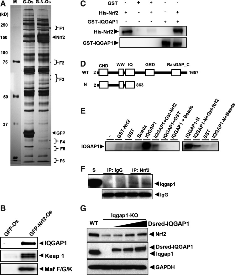
Aims: Nuclear factor-erythroid-related factor 2 (Nrf2) is a critical transcriptional factor that is used in regulating cellular defense against oxidative stress. This study is aimed at investigating new interacting protein partners of Nrf2 using One-strep tag pull-down coupled with LTQ Orbitrap LC/MS/MS, and at examining the impact on Nr2 signaling by the newly identified IQ motif containing GTPase activating protein 1 (IQGAP1).

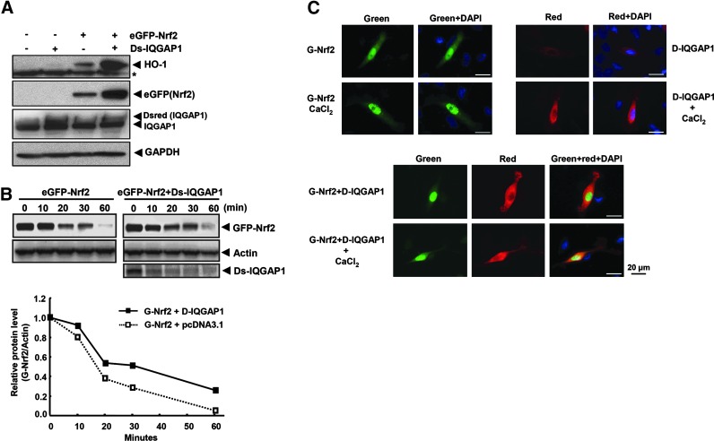
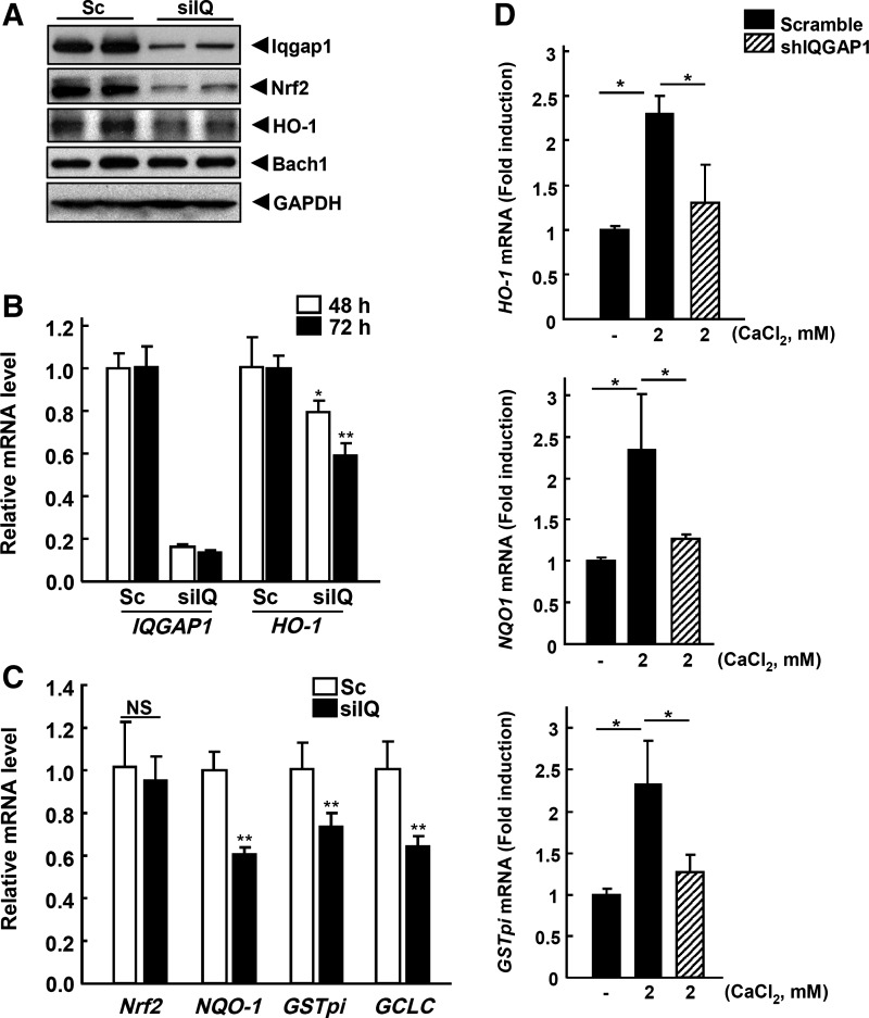
Results: Using the One-strep tag pull-down and LTQ Orbitrap LC/MS/MS, we identified IQGAP1 as a new Nrf2 interacting partner. Direct interactions between IQGAP1 and Nrf2 proteins were verified using in vitro glutathione S-transferase (GST) pull-down, transcription/translation assays, and in vivo utilizing Nrf2 overexpressing cells. Coexpression of Dsredmono-IQGAP1 and eGFP-Nrf2 increased the stability of eGFP-Nrf2 and enhanced the expression of Nrf2-target gene heme oxygenase-1 (HO-1). To confirm the functional role of IQGAP1 on Nrf2, knock-downed IQGAP1 using siIQGAP1 attenuated the expression of endogenous Nrf2, HO-1 proteins, and Nrf2-target genes GSTpi, GCLC, and

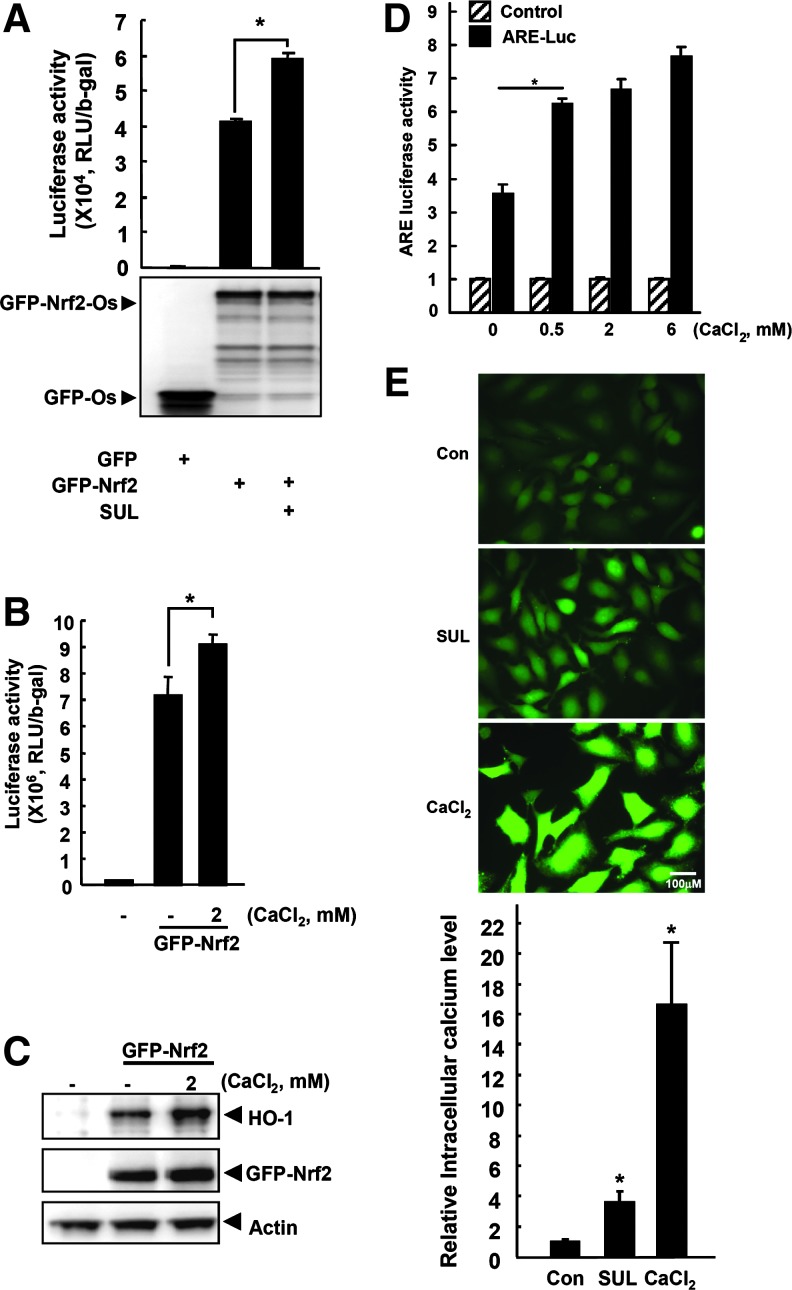
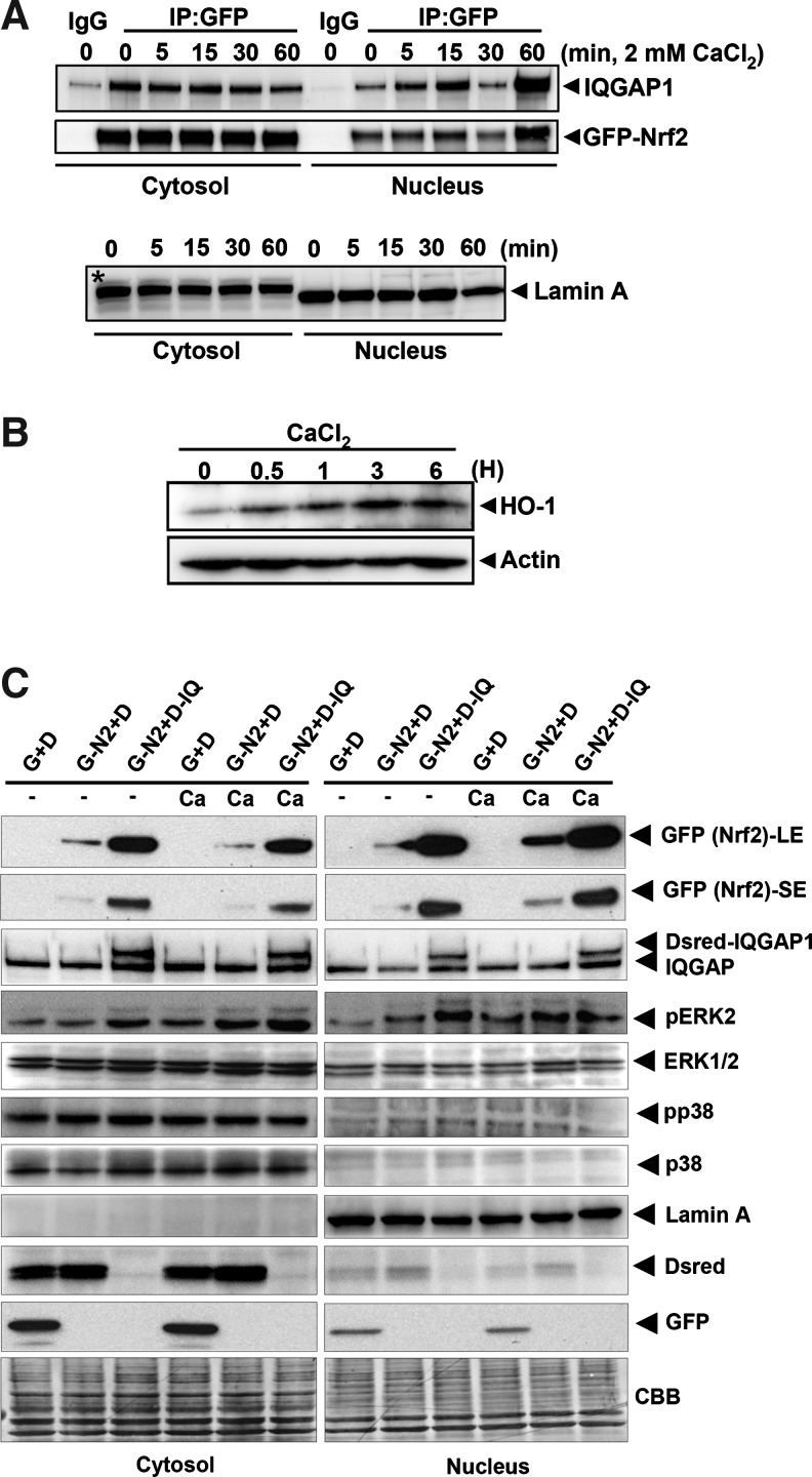
Nad(p)h: quinone oxidoreductase 1 (NQO-1). Furthermore, the stability of Nrf2 was dramatically decreased in IQGAP1-deficient mouse embryonic fibroblast (MEF) cells. Since IQGAP1 signaling could be mediated by calcium, treating the cells with calcium showed the translocation of IQGAP1/Nrf2 complex into the nucleus, suggesting that IQGAP1 may play a critical role in Nrf2 stability. Interestingly, consistent with calcium signaling for IQGAP1, treating the cells with calcium functionally enhanced Nrf2-mediated antioxidant responsive element-transcription activity and enhanced the expression of the endogenous Nrf2-target gene HO-1.

Innovation: In the aggregate, our current study identifies and functionally characterizes a new Nrf2 partner protein IQGAP1, which may contribute to Nrf2's regulation of antioxidant enzymes such as HO-1.

Conclusion: IQGAP1 may play a critical role in the stability and transactivation of Nrf2.


Figures

FIG. 1.
FIG. 1.
Purification and identification of novel Nrf2 partners. (A) Eluted samples from the One-strep pull-down assay were concentrated using Microcon YM-50 (Millipore) and subjected to polyacrylamide gel electrophoresis gel (6%–15% gradient) electrophoresis for silver staining. The representative protein bands in the lane of eGFP-Nrf2-Os (G-N-Os) were excised for LC/MS/MS analysis. To minimize the nonspecific background between eGFP-One-strep (G-Os; control) and eGFP-Nrf2-One-strep (G-N-Os), the same region of G-Os was also cut as a counterpart of G-N-Os. All excised samples were measured by LTQ_Orbitrap LC/MS/MS equipment (http://cabm-ms.cabm.rutgers.edu), and all mass data were analyzed using The Global Proteome Machine Organization Proteomics Database (Open Source Software, www.thegpm.org). (B) Based on the mass identification, same samples from (A) were subjected to Western blot analysis to check and verify the identified proteins. Newly identified IQGAP1 was blotted using anti-IQGAP1antibody, and four known Nrf2 partners, Keap1 and Maf F/G/K, were also blotted using anti-Keap1and anti-Maf F/G/K antibodies, respectively. (C) To verify the binding between IQGAP1 and Nrf2, GST-IQGAP1 protein was incubated with purified His-Nrf2 protein in vitro and followed by GST pull-down assay. Protein-bead complexes were then subjected to Western blot analysis against His-Nrf2 using anti-Nrf2 (C-20). (D) Schematic diagram shows the structure of different segments of IQGAP1 utilized for in vitro transcription/translation-binding assay. (E) IQGAP proteins (full-length or N-terminal fragment) were synthesized by in vitro transcription/translation, as described in Materials and Methods, and were incubated with GST-Nrf2. The protein complex was pulled down by glutathione-sepharose beads and subjected to Western blot analysis using an antibody against IQGAP1. (F) Whole-protein extract from MEF cells were subjected to co-IP to examine the binding affinity between the endogenous mouse Iqgap1 and Nrf2. (G) Western blot analysis showed that suppressed mouse Nrf2 in the Iqgap1-deficient MEF cells was rescued by transfection of the different amounts (2, 3, 5 μg) of Dsred-IQGAP1. CHD, calponin homology domain; WW, WW domain; IQ, IQ motif; GRD, GAP-related domain; RasGAP_C, RasGAP binding C-terminal. S, standard mouse Iqgap1 protein; WT, wild type; KO, knock-out; Dsred-IQGAP1, Dsredmono-IQGAP1; GST, glutathione S-transferase; IQGAP1, IQ motif containing GTPase activating protein 1; Keap1, kelch-like ECH-associated protein 1; MEF, mouse embryonic fibroblast; co-IP, coimmunoprecipitation;.
FIG. 2.
FIG. 2.
IQGAP1 enhances the expression of Nrf2 and induction of HO-1 expression as well as the stability of Nrf2. (A) Based on mass data shown in Table 1, the newly identified IQGAP1 was then tested to see whether it could regulate the expression of HO-1, which is also regulated by Nrf2. HeLa cells were cotransfected with pDsredmono-IQGAP1 and pEGFP-Nrf2 plasmids for 24 h using jetPEI reagent. The cells were then lysed with RIPA lysis buffer, and 20 μg of proteins were subjected to Western blot analysis against HO-1, eGFP-Nrf2, and Dsred-IQGAP1 using anti-HO-1, anti-GFP, and anti-IQGAP1 antibodies, respectively. GAPDH was used for the equal loading control. pCDNA3.1 vector was used for the equal amount of transfection. GFP-Nrf2, pEGFP-Nrf2; Dsred-IQGAP1, pDsredmono-IQGAP1. (B) To measure the stability of Nrf2 by IQGAP1, HeLa cells were transfected with pEGFP-Nrf2 (500 ng) and either empty vector (pcDNA3.1) or pDsredmono-IQGAP1 (500 ng) constructs using jetPEI reagent for 24 h. The cells were then treated with cycloheximide (5 μg/ml) for different time intervals as just indicated. Cell lysates (20 μg) were subsequently subjected to immunoblotting against eGFP-Nrf2 or Dsred-IQGAP1 using anti-Nrf2 or anti-Dsred antibodies, respectively. Actin was used for the equal loading control. GFP-Nrf2, pEGFP-Nrf2; Dsred-IQGAP1, pDsredmono-IQGAP1. (C) HeLa cells were plated in glass-bottom culture dishes and transfected with pEGFP-Nrf2 and pDsredmono-IQGAP1 using jetPEI transfection reagent. After 24 h of transfection, the cells were fixed with 4% paraformaldehyde, and the images were taken by fluorescent microscopy. Green, Red, and DAPI filters were used for GFP, Dsred, and nucleus, respectively. GFP-Nrf2, pEGFP-Nrf2; Dsred-IQGAP1, pDsredmono-IQGAP1. magnification (34×). HO-1, heme oxygenase-1.
FIG. 3.
FIG. 3.
siIQGAP1 decreases the stability of Nrf2 and attenuates HO-1 expression and ARE-luciferase activity. (A) HeLa cells were transfected with siIQGAP1 for 72 h using Lipofectamin 2000 reagent to silence or knockdown the endogenous IQGAP1 gene expression. The expression levels of endogenous IQGAP1, Nrf2, HO-1 and Bach1 were measured by Western blot analysis. (B) IQGAP1 and HO-1 mRNAs were measured by qPCR after transfection with siIQGAP1 for different time periods (48 and 72 h). (C) Nrf2, NQO-1, GSTpi, and GCLC mRNAs were also measured from the siIQGAP1-72 h-transfected samples. (D) HeLa cells were transfected with pGFP-V-RS-Sc plasmid or pGFP-V-RS-IQGAP1-77 for 3 days and treated with 2 mM CaCl2 for 6 h. Isolated mRNA was subjected to real-time qPCR for HO-1, NQO-1, and GSTpi. Beta-Actin was used for the normalization. *p<0.05 **p<0.01. Sc, scramble; siIQ, siIQGAP1; ARE, antioxidant responsive element; NQO-1, NAD(P)H: quinone oxidoreductase 1; NS, not significant.
FIG. 4.
FIG. 4.
Calcium induces the activity of ARE-luciferase and the expression of Nrf2 target proteins. (A) To verify the Nrf2-One-strep tag construct for this system, HeLa cells were transfected with pEGFP-Nrf2-Os and pTI-ARE-Luc plasmids for 24 h, and the Nrf2 transactivation activity was examined by reporter assay after 6 h of treatment with 25 μM SUL, as an Nrf2 inducer. pEGFP-Os was also transfected as a control. (B) To check the involvement of the calcium ion in Nrf2 activity or Nrf2 partner complex, after cotransfection with pEGFP-Nrf2 and pTI-ARE-Luc constructs, HeLa cells were treated for 6 h with various concentrations of CaCl2. ARE-luciferase activity was then measured by reporter assay. (C) The level of endogenous HO-1 was also measured by Western blotting using the same samples from (B). (D) HeLa cells were transfected with pGL3-luc or pTI-ARE constructs for 24 h and treated with different concentrations of CaCl2 for 3 h, and ARE luciferase activity was measured. (E) To measure the changes in the intracellular calcium level, HeLa cells were treated with SUL, an ARE activator and CaCl2. The cells were seeded in 12-well plates and cultured overnight. Intracellular calcium level was measured using FluoForte® Calcium Assay Kit (ENZO Life Sciences) according to the manufacturer's instructions. Briefly, the cultured medium was removed, and the cells were incubated with 1 ml of FluoForte™ Dye-Loading Solution for 40 min at room temperature. Then, 20 μM SUL or 2 mM CaCl2 was added to the incubation solution. After 1 min of incubation, the cells were immediately imaged with a fluorescence microscope (Carl Weiss, Axiovert S100) using the FITC channel (left panel). Right panel shows a densitometry analysis of the pictures, which represent the relative intracellular calcium levels. Con, Control (0.1% DMSO); SUL, sulforaphane; Magnitude,×32. *p<0.001 (vs. control).
FIG. 5.
FIG. 5.
Translocation of Nrf2-IQGAP1 complex into the nucleus by calcium treatment and induction of HO-1 protein. (A) To verify the involvement of calcium in endogenous Nrf2-IQGAP1 complexes, HeLa cells were transfected with pEGFP-Nrf2 (8 μg/100 mm dish) for 24 h and treated with CaCl2 (2 mM) for different times. Then, cytosolic and nuclear proteins were isolated using the M-PER kit (Thermo Scientific). Equal volumes of cytosolic and nuclear fractions were subjected to IP with the GFP antibody using Dynabead G (Invitrogen) beads. The procedures of this experiment are described in Materials and Methods. The IP samples were then subjected to Western blotting against endogenous IQGAP1 and eGFP-Nrf2 using anti-IQGAP1 or GFP antibody. Lamin A was used for nuclear fraction marker protein. *, represents nonspecific protein. (B) To examine the time course of induction of HO-1 by CaCl2, HeLa cells were treated with CaCl2 for different times. Then, cell lysates were subjected to Western blotting against HO-1 using anti-HO-1 (C-20) antibody. Actin was used for the control of the equal loading of protein. (C) To test whether calcium treatment facilitates the Nrf2 translocation into the nucleus, HeLa cells were transfected with pEGFP-Nrf2 (4 μg) or/and pDsredmomo-IQGAP1 (4 μg) constructs on 100 mm dishes for 24 h and treated with 2 mM CaCl2 for 30 min. Cytosolic and nuclear fractions were isolated using the NE-PER kit (Thermo Scientific). Then, fractionated lysates (15 μg) were subjected to Western blot analysis. Lamin A was used as an indicator of a nuclear fraction. Both eGFP and Dsredmono control vectors were used for equal transfection. CBB staining was used as an equal loading control. G, EGFP; D, Dsredmono; G-N2, eGFP-Nrf2; D-IQ, Dsredmono-IQGAP1; CBB, Coomassie brilliant blue staining; LE, longer exposure; SE, shorter exposure; Ca, CaCl2.

Similar articles

Cited by

References

    1. Alam J. Stewart D. Touchard C. Boinapally S. Choi AM. Cook JL. Nrf2, a Cap'n'Collar transcription factor, regulates induction of the heme oxygenase-1 gene. J Biol Chem. 1999;274:26071–26078. - PubMed
    1. Berridge MJ. Lipp P. Bootman MD. The versatility and universality of calcium signalling. Nat Rev Mol Cell Biol. 2000;1:11–21. - PubMed
    1. Blank V. Small Maf proteins in mammalian gene control: mere dimerization partners or dynamic transcriptional regulators? J Mol Biol. 2008;376:913–925. - PubMed
    1. Briggs MW. Sacks DB. IQGAP1 as signal integrator: Ca2+, calmodulin, Cdc42 and the cytoskeleton. FEBS Lett. 2003;542:7–11. - PubMed
    1. Brown MD. Sacks DB. IQGAP1 in cellular signaling: bridging the GAP. Trends Cell Biol. 2006;16:242–249. - PubMed

Publication types

MeSH terms

LinkOut - more resources