Review
. 2012 Jul;96(4):729-43.
doi: 10.1016/j.mcna.2012.05.003.

The role and potential of imaging in COPD

Affiliations
Review

The role and potential of imaging in COPD

George R Washko. Med Clin North Am. 2012 Jul.

Abstract

Chronic obstructive pulmonary disease is a heterogeneous condition of the lungs and body. Techniques in chest imaging and quantitative image analysis provide novel in vivo insight into the disease and potentially examine divergent responses to therapy. This article reviews the strengths and limitations of the leading imaging techniques: computed tomography, magnetic resonance imaging, positron emission tomography, and optical coherence tomography. Following an explanation of the technique, each section details some of the useful information obtained with these examinations. Future clinical care and investigation will likely include some combination of these imaging modalities and more standard assessments of disease severity.


Figures

Figure 1A and B
Figure 1A and B
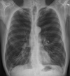
PA and Lateral chest x-ray of a subject with severe COPD and emphysema. Note the lucency of the lung fields, flattening of the diaphragms, narrowed cardiac silhouette, and paucity of peripheral vascular markings.
Figure 1A and B
Figure 1A and B
PA and Lateral chest x-ray of a subject with severe COPD and emphysema. Note the lucency of the lung fields, flattening of the diaphragms, narrowed cardiac silhouette, and paucity of peripheral vascular markings.
Figure 2
Figure 2
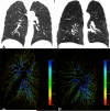
Panels A and C provide a coronal view of the lungs of a smoker with normal lung function (Panel A) and one with moderate emphysema (Panel C). Panels B and D provide volumetric reconstructions of the left lung pulmonary vasculature from a left mid axillary view. Vessels are color coded by diameter. Notice the loss of vasculature and thinning of the vessels in regions most affected by emphysema.
Figure 3
Figure 3
3D coronal maps of a healthy human subject depicting regional differences in oxygen tension using 3He. At right is a color coded bar of oxygen tension in mbar (1mbar=0.75mm Hg). Slide courtesy of Dr. Samuel Patz.
Figure 4
Figure 4
Figure 4 provides a view using OCT. At the center can be seen the fiberoptic probe. Adjacent to the outer surface of the probe are alveolar ducts and alveoli. Slide courtesy of Dr. Anthony Lee of the British Columbia Cancer Research Centre.

Similar articles

Cited by

References

    1. Rabe KF, Hurd S, Anzueto A, Barnes PJ, Buist SA, Calverley P, Fukuchi Y, Jenkins C, Rodriguez-Roisin R, van Weel C, et al. Global strategy for the diagnosis, management, and prevention of chronic obstructive pulmonary disease: Gold executive summary. Am J Respir Crit Care Med. 2007;176:532–555. - PubMed
    1. Barr RG, Bluemke DA, Ahmed FS, Carr JJ, Enright PL, Hoffman EA, Jiang R, Kawut SM, Kronmal RA, Lima JA, et al. Percent emphysema, airflow obstruction, and impaired left ventricular filling. N Engl J Med. 362:217–227. - PMC - PubMed
    1. Bon J, Fuhrman CR, Weissfeld JL, Duncan SR, Branch RA, Chang CC, Zhang Y, Leader JK, Gur D, Greenspan SL, et al. Radiographic emphysema predicts low bone mineral density in a tobacco-exposed cohort. Am J Respir Crit Care Med. 183:885–890. - PMC - PubMed
    1. Snider GL. Emphysema: The first two centuries--and beyond. A historical overview, with suggestions for future research: Part 1. Am Rev Respir Dis. 1992;146:1334–1344. - PubMed
    1. Webb WR. Thin-section ct of the secondary pulmonary lobule: Anatomy and the image--the 2004 fleischner lecture. Radiology. 2006;239:322–338. - PubMed

Publication types

MeSH terms