. 2012 Jul 13:7:34.
doi: 10.1186/1750-1326-7-34.

Bone marrow-derived fibroblast growth factor-2 induces glial cell proliferation in the regenerating peripheral nervous system

Affiliations

Bone marrow-derived fibroblast growth factor-2 induces glial cell proliferation in the regenerating peripheral nervous system

Victor Tulio Ribeiro-Resende et al. Mol Neurodegener. .

Abstract

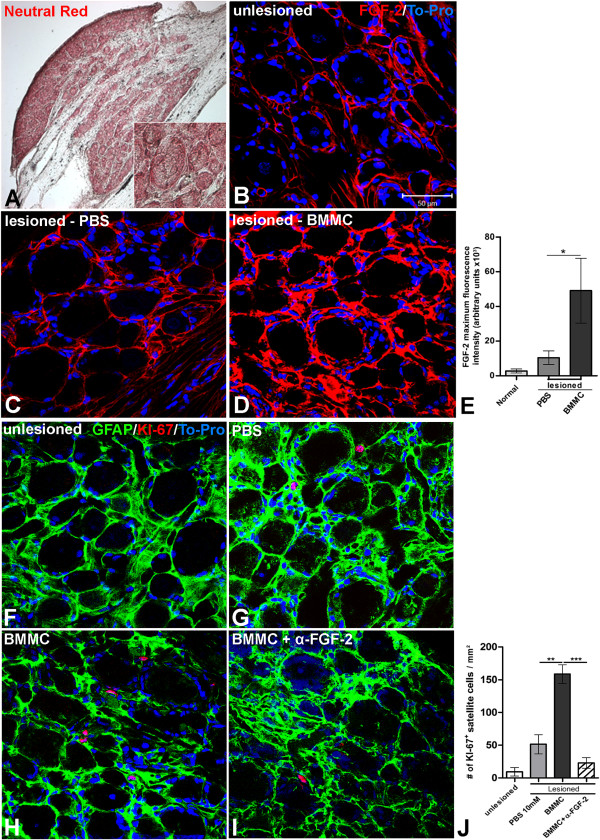
Background: Among the essential biological roles of bone marrow-derived cells, secretion of many soluble factors is included and these small molecules can act upon specific receptors present in many tissues including the nervous system. Some of the released molecules can induce proliferation of Schwann cells (SC), satellite cells and lumbar spinal cord astrocytes during early steps of regeneration in a rat model of sciatic nerve transection. These are the major glial cell types that support neuronal survival and axonal growth following peripheral nerve injury. Fibroblast growth factor-2 (FGF-2) is the main mitogenic factor for SCs and is released in large amounts by bone marrow-derived cells, as well as by growing axons and endoneurial fibroblasts during development and regeneration of the peripheral nervous system (PNS).

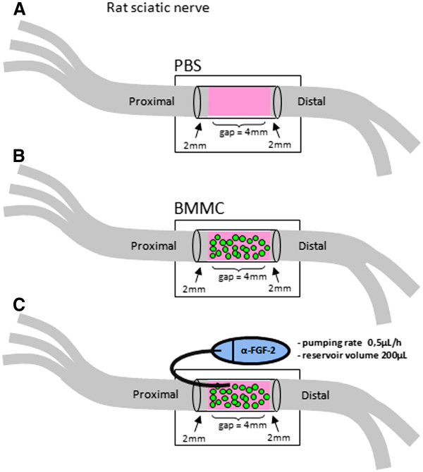
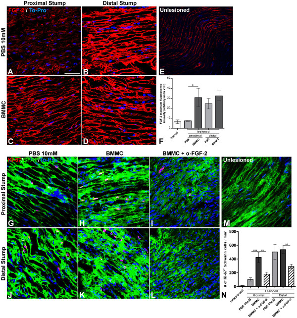
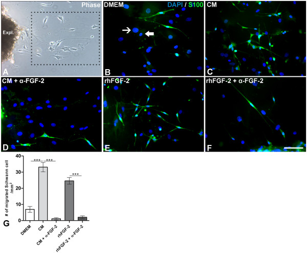
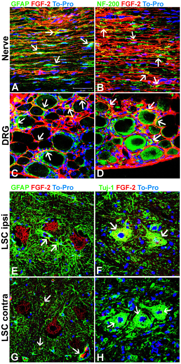
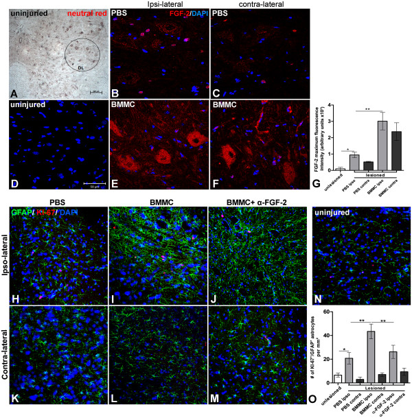
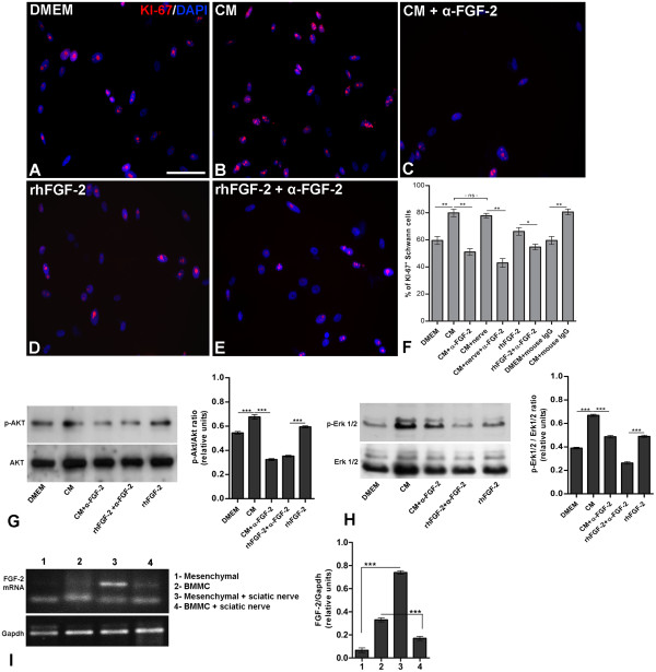
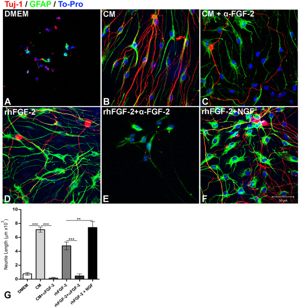
Results: Here we show that bone marrow-derived cell treatment induce an increase in the expression of FGF-2 in the sciatic nerve, dorsal root ganglia and the dorsolateral (DL) region of the lumbar spinal cord (LSC) in a model of sciatic nerve transection and connection into a hollow tube. SCs in culture in the presence of bone marrow derived conditioned media (CM) resulted in increased proliferation and migration. This effect was reduced when FGF-2 was neutralized by pretreating BMMC or CM with a specific antibody. The increased expression of FGF-2 was validated by RT-PCR and immunocytochemistry in co-cultures of bone marrow derived cells with sciatic nerve explants and regenerating nerve tissue respectivelly.

Conclusion: We conclude that FGF-2 secreted by BMMC strongly increases early glial proliferation, which can potentially improve PNS regeneration.


Figures

Figure 1
Figure 1
Illustration of in vivo experimental model of rat sciatic nerve. A-C: Full transection of sciatic nerve followed by connection of proximal and distal stumps inside a hollow tube of silicon and treatment with PBS (A), bone marrow mononuclear cells (BMMC) (B), or BMMC + neutralizing FGF-2 antibody delivered inside the gap by a cannula coupled to an osmotic mini-pump (C).
Figure 2
Figure 2
Increased expression of FGF-2 is time correlated with increased Schwann cell proliferation in the sciatic nerve after lesion and BMMC treatment. A-E: Optical sections of sciatic nerve by confocal microscopy taken 10 days after total transection immunolabeled for FGF-2 and treated with PBS (A, proximal stump and B, distal stump) or with BMMC (C, proximal stump and D, distal stump). An intact nerve is showed in E. Cell nuclei were counterstained with TO-PRO. F: Histogram of FGF-2 expression in sciatic nerve tissue comparing different experimental groups. Statistics: p < 0.0001 ANOVA. G-M: Optical sections of sciatic nerve by confocal microscopy taken 10 days after total transection double immunolabeled for KI-67 (red) and GFAP (green) treated with PBS (G, proximal stump and J, distal stump), with BMMC (H, proximal stump and K, distal stump) or BMMC + neutralizing antibody for FGF-2 (I, proximal stump and L, distal stump) delivered by osmotic mini-pumps. Uninjured nerve is showed in M. N: Histogram of the number of KI-67+ SCs in the nerve tissue under the different experimental conditions. Statistics: p < 0.001 ANOVA. Scale bar: A-E; G-M = 50 μm.
Figure 3
Figure 3
FGF-2 secreted by BMMC increases SC migration from sciatic nerve explants. A-F: Crushed sciatic nerve explants from distal nerve stump kept for ten days in culture under specific experimental conditions. A: Phase contrast image showing one explant with cells migrating from the tissue. B-F: Epifluorescent microscopy of migrating SCs immunolabeled for S-100β (green) and incubated with control medium (A), BMMC-CM (B), BMMC-CM+ neutralizing FGF-2 (C), rhFGF-2 added to the control medium (D) or rhFGF-2 + neutralizing FGF-2 (E). Cell nuclei were counterstained with DAPI. F: Quantitative analysis of the number of S-100β migrated SCs from sciatic nerve explants after incubation under different experimental conditions. Statistics: p<0.0001 ANOVA. Non-Schwann cells are indicated by thick or thin arrows in B. Scale bar: A-F= 50 µm.
Figure 4
Figure 4
Localization of FGF-2 in the regenerating nerve tissue, DRG and lumbar spinal cord. A-H: Optical slices by confocal microscopy of longitudinal sections of sciatic nerve at proximal stump (A and B) or dorsal root ganglia (C and D) and transversal section of lumbar spinal cord (E-H) at the ipsi-lateral (E and F) or contra-lateral region (G and H). All samples were obtained from rats 10 days after treatment with BMMC. Sciatic nerve and DRG sections were double immunolabeled for GFAP/FGF-2 (A and C) and NF-200/FGF-2 (B and D). LSC sections were double immunolabeled for GFAP/FGF-2 and Tuj-1/FGF-2. White arrows indicate yellow areas of colocalisation between glial marker (GFAP) and FGF-2 and neuronal markers (NF-200/Tuj-1) and FGF-2. Scale bar: A-H = 50 βm.
Figure 5
Figure 5
Increased expression of FGF-2 is time correlated with increased satellite cell proliferation in the DRG after nerve lesion and BMMC treatment. A-D; F-I: Longitudinal sections of dorsal root ganglia (DRG) of adult rats. A: Low magnification photomicrograph of a longitudinal section of an uninjured DRG stained with neutral red. Insert show neurons in different cell size (low, medium and big size). B-D: Optical sections of DRGs slices by confocal microscopy taken 10 days after total transection immunolabeled for FGF-2 (red) and counterstained with TO-PRO (blue) treated with PBS (C) or BMMC (D) or uninjured sciatic nerve (B). E: Quantification of the fluorescence intensity compared between the experimental groups cited above. Statistics: p<0,0001 ANOVA. F-I: Optical.
Figure 6
Figure 6
Increased expression of FGF-2 is time correlated with increased astrocyte proliferation in the LSC after nerve lesion and treatment with BMMC. A: Low magnification photomicrogragh of lumbar spinal cord with an uninjured sciatic nerve stained with neutral red. The DL region is circled. B-F: Optical sections by confocal microscopy of LSC at DL region immunolabeled for FGF-2 ten days after sciatic nerve lesion treated with PBS (B, ipsi-lateral and C, contralateral) or BMMC (E, ipsi-lateral and F, contralateral). D: LSC section of an uninjured rat. Cell nuclei were counterstained with TO-PRO G: histogram of FGF-2 fluorescence between experimental groups. Statistics: p<0.001 ANOVA. H-N: Optical slides by confocal microscopy of the DL region of LSC double immunolabeled for KI-67 (red) and GFAP (green) ten days after sciatic nerve lesion treated with PBS (H, ipsi-lateral and K, contralateral), BMMC (I, ipsilateral and L, contralateral) or BMMC+ neutralizing FGF-2 antibody (J, ipsilateral and M, contralateral) delivered by osmotic mini-pumps. N: LSC section of an uninjured rat. O: Histogram of the number of KI-67/GFAP + astrocytes at DL/LSC region per mm2. Statistics: p<0.001 ANOVA. Scale bars: A = 200 µm; B-F, H-M=50 µm.
Figure 7
Figure 7
Neutralizing FGF-2 antibody blocks SC proliferation and survival induced by BMMC-CM in vitro. A-E: ST-8814 SCs cultured for 48 h in low density (2.00 x 10⁴ cells/well) with DMEM F-12 + 10% FBS (A), CM (B), CM+ neutralizing FGF-2 antibody (C), rhFGF-2 (D) or rhFGF-2 + neutralizing FGF-2 antibody (E). Cells were immunolabeled for KI-67 and cell nuclei were counterstained with DAPI. F: Quantification of the percentage of KI-67+ SC per mm2. Statistics: p<0.0001 ANOVA. G and H: Western blotting and quantitative analysis of the active Akt (p-Akt, G) and active ERK 1/2 (p-ERK 1/2, H) from SCs cultured for 48 h with control medium, CM, CM+ neutralizing FGF-2, rhFGF-2 + neutralizing FGF-2 or rhFGF-2. I: Semi quantitative polymerase chain reaction (RT-PCR) for FGF-2 transcripts from samples of BMMC or MSC in the presence or absence of sciatic nerve pieces. Quantitative analysis showing the transcript level following the same experimental conditions. Statistics: p<0.0001 ANOVA. Scale bar: A-E=50 µm.
Figure 8
Figure 8
FGF-2 secreted by BMMC stimulates neuritogenesis of DRG neurons co-cultured with SC. A-F: Optical sections obtained by confocal microscopy of E16 rat embryo DRG sensory neurons (Tuj-1, red) and SCs (GFAP, green) incubated for 48 h with DMEM F-12 (control medium, A), bone marrow conditioned medium (CM, B), CM + neutralizing FGF-2 antibody (C), rhFGF-2 added to the control medium (D), rhFGF-2 + neutralizing FGF-2 antibody (E) or rhFGF-2 + rhNGF (F). Both cell types derived from cultured DRG explants of E16 rat embryos. Cell nuclei were counterstained with TO-PRO. All culture media were serum-free. rh: recombinant human protein. G: Quantification of the average neurite length per neuron under the experimental conditions described above. Statistics: p < 0.001. Scale bars: A-F = 50 μm.

Similar articles

Cited by

References

    1. Chen ZL, Yu WM, Strickland S. Peripheral Regeneration. Annu Rev Neurosci. 2007;30:209–233. doi: 10.1146/annurev.neuro.30.051606.094337. - DOI - PubMed
    1. Jessen KR, Mirsky R. Schwann cells and their precursors emerge as major regulators of nerve development. Trends Neurosci. 1999;22:402–410. doi: 10.1016/S0166-2236(98)01391-5. - DOI - PubMed
    1. Goldberg JL, Barres BA. The relationship between neuronal survival and regeneration. Annu Rev Neurosci. 2000;23:579–612. doi: 10.1146/annurev.neuro.23.1.579. - DOI - PubMed
    1. Vargas ME, Barres BA. Why is Wallerian degeneration in the CNS so slow? Annu Rev Neurosci. 2007;30:153–179. doi: 10.1146/annurev.neuro.30.051606.094354. - DOI - PubMed
    1. Ribeiro-Resende VT, Pimentel-Coelho PM, Mesentier-Louro LA, Mendez RM, Mello-Silva JP, Cabral-da-Silva MC, de Mello FG, de Melo Reis RA, Mendez-Otero R. Trophic activity derived from bone marrow mononuclear cells increases peripheral nerve regeneration by acting on both neuronal and glial cell populations. Neuroscience. 2009;159:540–549. doi: 10.1016/j.neuroscience.2008.12.059. - DOI - PubMed

Publication types

MeSH terms

Substances

LinkOut - more resources