Review
. 2013 Mar 10;18(8):851-73.
doi: 10.1089/ars.2011.4203. Epub 2012 Sep 5.

Biological and therapeutic relevance of nonreplicative DNA polymerases to cancer

Affiliations
Review

Biological and therapeutic relevance of nonreplicative DNA polymerases to cancer

Jason L Parsons et al. Antioxid Redox Signal. .

Abstract

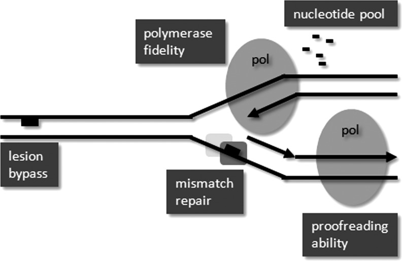
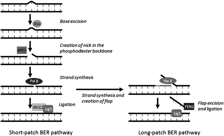
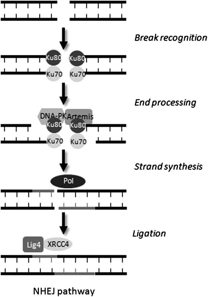
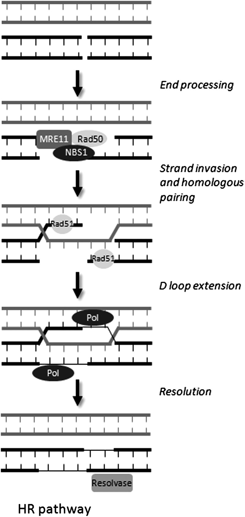
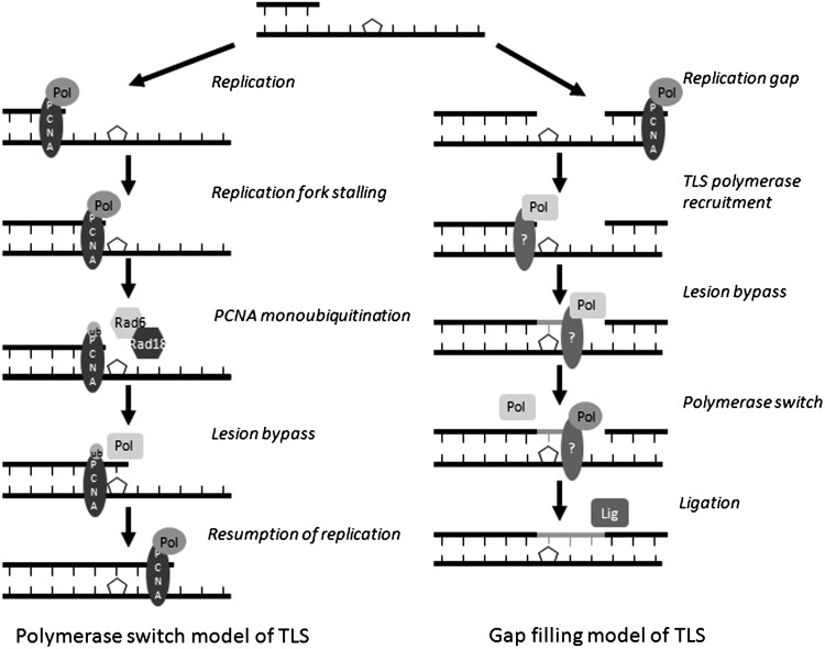
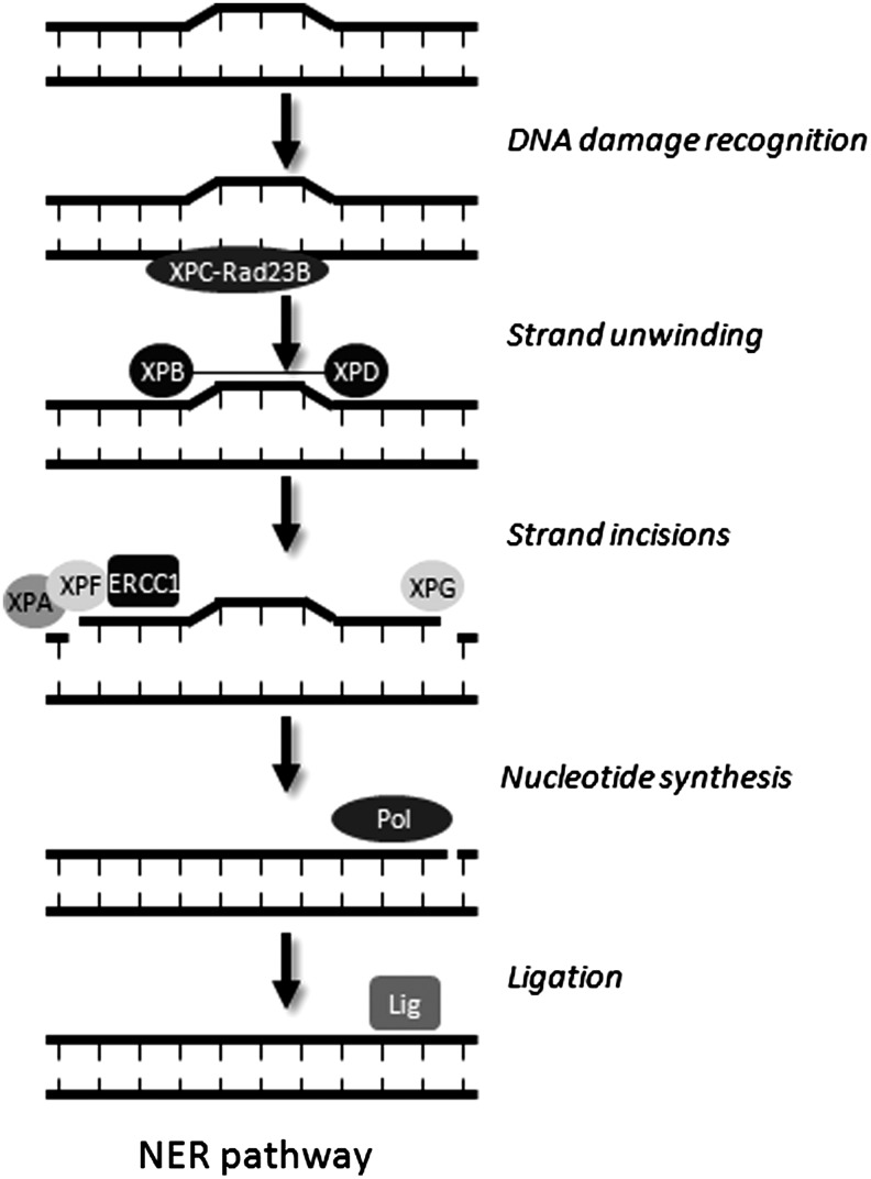
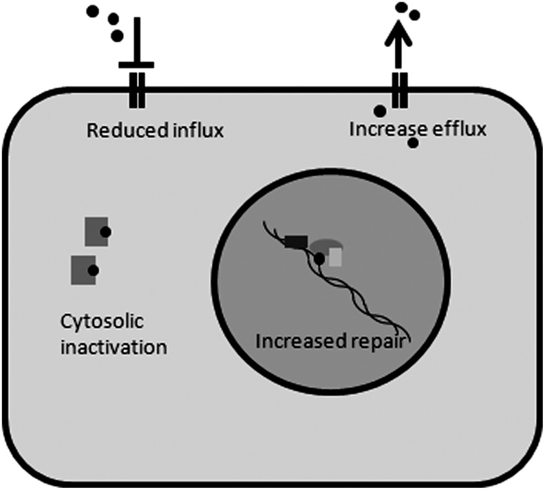
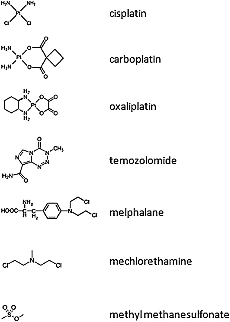
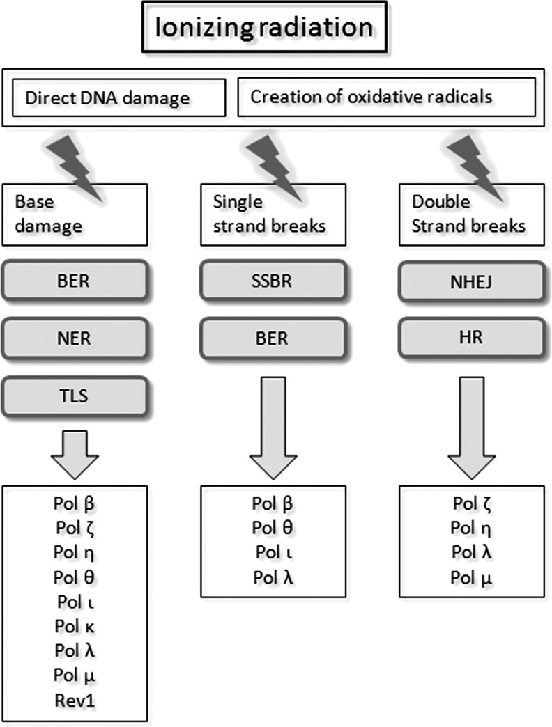
Apart from surgical approaches, the treatment of cancer remains largely underpinned by radiotherapy and pharmacological agents that cause damage to cellular DNA, which ultimately causes cancer cell death. DNA polymerases, which are involved in the repair of cellular DNA damage, are therefore potential targets for inhibitors for improving the efficacy of cancer therapy. They can be divided, according to their main function, into two groups, namely replicative and nonreplicative enzymes. At least 15 different DNA polymerases, including their homologs, have been discovered to date, which vary considerably in processivity and fidelity. Many of the nonreplicative (specialized) DNA polymerases replicate DNA in an error-prone fashion, and they have been shown to participate in multiple DNA damage repair and tolerance pathways, which are often aberrant in cancer cells. Alterations in DNA repair pathways involving DNA polymerases have been linked with cancer survival and with treatment response to radiotherapy or to classes of cytotoxic drugs routinely used for cancer treatment, particularly cisplatin, oxaliplatin, etoposide, and bleomycin. Indeed, there are extensive preclinical data to suggest that DNA polymerase inhibition may prove to be a useful approach for increasing the effectiveness of therapies in patients with cancer. Furthermore, specialized DNA polymerases warrant examination of their potential use as clinical biomarkers to select for particular cancer therapies, to individualize treatment for patients.


Figures

FIG. 1.
FIG. 1.
Determinants of replication fidelity. The major determinants of the fidelity of DNA replication by eukaryotic polymerases are shown in boxes. Polymerase-inherent replication fidelity and the enzymatic 3′–5′ exonuclease activity are the main determinants of replication fidelity. Additional exogenous determinants of replication fidelity are the cellular DNA damage bypass ability and the efficient repair of newly created mismatches. A further factor is the composition of the cellular deoxynucleotide pool.
FIG. 2.
FIG. 2.
Schematic depiction of the short- and long-patch base excision repair (BER) pathways. The excision of a damaged base from a DNA strand is initiated by a damage-specific glycosylase (DSG), and then the remaining abasic site (AP) is incised by AP endonuclease 1 (APE1), creating a 5′-deoxyribose phosphate (dRP) group. This group is removed by pol β, which subsequently fills the one nucleotide gap created by APE1, and the strand is ligated by DNA ligase 3–x-ray repair cross-complementing protein-1 (XRCC1) complex (short-patch BER). Alternatively, following pol β one-nucleotide addition, strand extension from that nucleotide is performed by pol δ or ɛ, thereby creating an oligonucleotide overhang. The overhang is then excised by flap endonuclease 1 (FEN1), and the remaining nick is sealed by DNA ligase 1 (long-patch BER).
FIG. 3.
FIG. 3.
Schematic depiction of the nonhomologous end-joining (NHEJ) pathway. NHEJ is initiated by the Ku70-Ku80 complex (Ku) that recognizes DNA double-strand breaks (DSBs) and recruits DNA-PK. If the DNA ends cannot be ligated directly due to complex DNA damage, end resection is carried out by the Artemis endonuclease. The removed strand segments are resynthesized by a DNA polymerase (pol), before ligation is carried out by the DNA ligase 4–XRCC4 complex.
FIG. 4.
FIG. 4.
Schematic overview of the homologous recombination (HR) pathway. The HR pathway requires a double-stranded homolog to serve as a template for the damaged strand, and therefore only occurs during the late S and G2 phases of the cell cycle. The 3′-end of the damaged strand, following end-processing by the MRE11-Rad50-NBS1 complex, invades and pairs with the homologous sequence of the sister chromatid. Strand invasion leads to the formation of a D-loop structure, from which the lesion-containing strand is resynthesized by a DNA polymerase (pol) and finally a resolvase restores the two sister chromatids.
FIG. 5.
FIG. 5.
Schematic depiction of the translesion synthesis (TLS) polymerase switch and gap-filling models. In the TLS polymerase model, the replication machinery cannot bypass distorting or bulky DNA lesions and therefore stalls. Replication fork stalling leads to the monoubiquitination of proliferating cell nuclear antigen (PCNA) by the Rad18-Rad6 heterodimer, resulting in the removal of the replicative DNA polymerase from the strand and the recruitment of a TLS polymerase that has the ability to carry out translesional bypass. After insertion of nucleotides opposite the lesion, a second polymerase switch takes place, replacing the TLS polymerase with a replicative enzyme. In TLS gap filling, this mechanism occurs independently of replication and deals with single-strand gaps that may have been left behind by the replication machinery. The TLS polymerase is recruited to the gap in the DNA and is able to insert nucleotides opposite the DNA lesion, before a polymerase switch that may lead to a replicative polymerase taking over the strand elongation before the remaining nick is sealed by a DNA ligase.
FIG. 6.
FIG. 6.
Schematic depiction of the nucleotide excision repair (NER) pathway. The NER pathway can carry out repair of most nucleotidic damage and lesions that cause distortion of the DNA double helix. The DNA damage is recognized by the XPC-Rad23B complex, and the helicase activity of the transcription factor IIH (TFIIH), consisting of XPB and XPD, carries out strand unwinding at the damage site. The first incision is carried out by the endonuclease XPG 3′—to the lesion, and the second incision, 5′—to the damage—is performed by the XPF/ERCC1 complex. After removal of the damaged nucleotide sequence, the remaining gap is filled by a DNA polymerase, and the nick is sealed by a DNA ligase.
FIG. 7.
FIG. 7.
Mechanisms for the resistance of cancer cells to cytotoxic drugs. Cancer cells employ several mechanisms that lead to the formation of resistance to chemotherapeutic drugs, such as cisplatin or etoposide. Decreased uptake by copper transporters or increased removal by the multidrug-resistance receptor proteins may lead to a diminished concentration of the drug at the target structure. Binding or inactivation in the cytosol, for example, through glutathione metabolism, has also been shown to contribute to increased resistance. Alterations and increases in several different DNA repair pathways lead to an increased removal of drug-induced DNA lesions and have the potential to reduce the efficacy of cancer treatment.
FIG. 8.
FIG. 8.
Chemical structures of commonly used cytotoxic chemotherapy drugs used to treat cancer.
FIG. 9.
FIG. 9.
Types of DNA damage caused by ionizing radiation. Ionizing radiation causes DNA damage directly, either by depositing its energy onto the DNA strands or by ionizing cellular water, creating reactive oxygen species that lead to secondary DNA lesions. Common types of ionizing radiation-induced DNA damage include base damage, single-strand breaks (SSBs), and DSBs. The repair pathways that deal with these types of DNA damage include the BER, NER, TLS, single-strand break repair (SSBR), nonhomologous end joining (NHEJ), and HR pathways.
FIG. 10.
FIG. 10.
Chemical structures of pharmacological inhibitors of DNA polymerases.

Similar articles

Cited by

References

    1. Abdulovic AL. Jinks-Robertson S. The in vivo characterization of translesion synthesis across UV-induced lesions in Saccharomyces cerevisiae: insights into Pol zeta- and Pol eta-dependent frameshift mutagenesis. Genetics. 2006;172:1487–1498. - PMC - PubMed
    1. Acharya N. Johnson RE. Prakash S. Prakash L. Complex formation with Rev1 enhances the proficiency of Saccharomyces cerevisiae DNA polymerase zeta for mismatch extension and for extension opposite from DNA lesions. Mol Cell Biol. 2006;26:9555–9563. - PMC - PubMed
    1. Acharya N. Yoon JH. Gali H. Unk I. Haracska L. Johnson RE. Hurwitz J. Prakash L. Prakash S. Roles of PCNA-binding and ubiquitin-binding domains in human DNA polymerase eta in translesion DNA synthesis. Proc Natl Acad Sci U S A. 2008;105:17724–17729. - PMC - PubMed
    1. Adachi M. Ijichi K. Hasegawa Y. Ogawa T. Nakamura H. Yasui Y. Fukushima M. Ishizaki K. Hypersensitivity to cisplatin after hRev3 mRNA knockdown in head and neck squamous cell carcinoma cells. Mol Med Report. 2008;1:695–698. - PubMed
    1. Albertella MR. Green CM. Lehmann AR. O'Connor MJ. A role for polymerase eta in the cellular tolerance to cisplatin-induced damage. Cancer Res. 2005;65:9799–9806. - PubMed

Publication types

MeSH terms

Substances