. 2012 Jun;26(2):177-88.
doi: 10.3109/01677063.2012.692412.

A role for the cytoplasmic DEAD box helicase Dbp21E2 in rhodopsin maturation and photoreceptor viability

Affiliations

A role for the cytoplasmic DEAD box helicase Dbp21E2 in rhodopsin maturation and photoreceptor viability

Karen L Hibbard et al. J Neurogenet. 2012 Jun.

Abstract

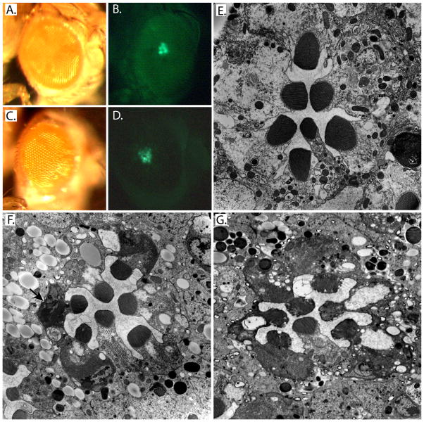
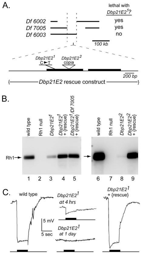
The Dbp21E2 (DEAD box protein 21E2) is a member of a family of DEAD box helicases active in RNA processing and stability. The authors used genetic mosaics to identify mutants in Dbp21E2 that affect rhodopsin biogenesis and the maintenance of photoreceptor structure. Analysis of a green fluorescent protein (GFP)-tagged Rh1 rhodopsin construct placed under control of a heat shock promoter showed that Dbp21E21 fails to efficiently transport Rh1 from the photoreceptor cell body to the rhabdomere. Retinal degeneration is not dependent on the Rh1 transport defects. The authors also showed that GFP- and red fluorescent protein (RFP)-tagged Dbp21E2 proteins are localized to discrete cytoplasmic structures that are not associated with organelles known to be active in rhodopsin transport. The molecular genetic analysis described here reveals an unexpected role for the Dbp21E2 helicase and provides an experimental system to further characterize its function.


Figures

Figure 1
Figure 1
Phenotypic characterization of the Dbp21E2 mutant. (A–D). Images of the compound eye and fluorescent pseudopupil images of the control 2L FRT40A mosaic (A, B) and the Dbp21E21 mutants (C, D) are shown. The pseudopupil of the Dbp21E21 mutant is dimmer than parental strain and has been digitally enhanced in this image to allow visualization of the loss of the distinctive trapezoidal shape of the pseudopupil resulting from the rough surface of compound eye. (E–G). Electron micrographs show cross-sectioned ommatidia at the level of the R1-6 photoreceptor nuclei. The one day old control 2L FRT40A mosaic is shown in (E). The one-day old Dbp21E21 mutant is shown in (F). Some photoreceptors in the Dbp21E21 mutant show apoptotic cell death of the photoreceptor cells as indicated by the electron dense cell body (arrow). The R7 cell is marked with an asterisk. At three days post-eclosion (G), all R1-R6 cells show cytoplasmic condensation and vacuole formation indicative of retinal degeneration. The rate of degeneration in the R7 photoreceptor is slower.
Figure 2
Figure 2
Identification of the Dbp21E2 gene. (A) The Dbp21E21 lethal mutation is cytologically mapped to the 21E2 region. The deficiencies Df(2L)Exel6002 and Df(2L)Exel7005 uncover the Dbp21E21 lethal mutation. Df(2L)Exel6003 complemented Dbp21E21. Sequencing of the EMS derived mutant identified Dbp21E2 (CG3561) as a candidate gene due to the base change of cytosine to thymine at base position 172 within the first exon creating a nonsense codon at this site. Sequencing of a second mutant allele, Dbp21E22, revealed the insertion of a Copia transposon at base position 531 in the first exon. The rescue transgene of Dbp21E2, P{Dbp21E2+} was constructed from genomic DNA, including 0.8 kb upstream and 0.4 kb downstream of the coding sequence. (B) Native Rh1 protein levels were assessed in the Dbp21E21 and Dbp21E22 mutant allele in a non-GFP background. Rh1 antibodies detected low Rh1 protein levels in Dbp21E21 and Dbp21E2 (lanes 3 and 8). Addition of the rescue construct, P{Dbp21E2+}, to Dbp21E21 and Dbp21E22 restored Rh1 protein to comparable wild type levels (lanes 4 and 9). The P{Dbp21E2+} construct also rescued the lethal phenotype of Dbp21E21/Df(2L)Exel7005 and the Rh1 protein was at wild type levels in these flies (lane 5). Wildtype and the Rh1 null mutant were positive and negative controls respectively (lanes 1, 2, 6, and 7). (C) Electroretinogram analysis showed that P{Dbp21E2+} restored the activation of the phototransduction cascade in the Dbp21E21 mutant. The phototransduction cascade was activated with a stimulus of blue light for 5 seconds. Wild type responds as expected to the light stimulus (left trace) but newly eclosed Dbp21E21 show a slight response to the pulse of light (second trace) and by one day, Dbp21E21 no longer responds to the pulse of light (third trace). Addition of the rescue construct, P{Dbp21E2+}, to the genome of the Dbp21E21 mutant restored the light response (right trace).
Figure 3
Figure 3
The trafficking of Rh1GFP is impeded in Dbp21E21 mutants. An Rh1::GFP fusion gene was expressed under the control of a heat shock promoter and Rh1GFP examined in Dbp21E21 mutants and Dbp21E21 heterozygous siblings at specific times after heat shock. (A) Confocal images of single ommatidial bundles at specific time point following induction of Rh1GFP. At four hours after heat shock, Rh1GFP was visible in the cell body in both the Dbp21E21 mutant and the Dbp21E21/+ control. At fourteen hours after heat shock, the Dbp21E21 mutant retained the majority of the Rh1GFP within the cell body while the Dbp21E21/+ controls showed most of the Rh1GFP within the rhabdomeres (R) and little within the cell body (CB). (B) Protein blot analysis of the heat shock-induced Rh1GFP levels in Dbp21E21 mutants and Dbp21E21/+ controls. In the SDS-PAGE experiment, the mutant lanes are loaded with a 5-fold excess of protein extract to facilitate detection of Rh1. At 4 hours after heat shock induction, both dimer form (~134 kDa, black arrowheads) and a glycosylated immature form of Rh1GFP (~67 KDa, white arrowheads) is easily detected in both mutant and controls (lanes 4 and 9). For the Dbp21E21/+ control, the 6, 10, and 14 hour time points show that the immature Rh1GFP forms gradually diminish and the mature non-glycosylated form of Rh1GFP (~61 kDa, asterisk) accumulates. Even though the lanes of the Dbp21E21 mutant contained 5-fold more protein, the mature non-phosphorylated form of Rh1GFP (asterisk) is found at lower levels than seen in the controls. (C) Protein blots analyses of Rh4, RdgB, Trp, and NorpA in Dbp21E21 mutants and control strains. In Dbp21E2 mutants, these 4 eye-specific proteins all show reduced protein levels. The reduction in protein amounts appear unique to each protein, with NorpA and RdgB being more affected than Trp and Rh4. Following detection of Rh4, the same membrane was reprobed for native Rh1 levels. The Rh1 levels in the Dbp21E2 mutant are impacted to a greater extent than Rh4. This blot was reprobed with an actin antibody to confirm similar loading of protein, as were the 3 other protein blots (data not shown).
Figure 4
Figure 4
Dbp21E2 induces retinal degeneration in the absence of Rh1. Dbp21E21 mutants were reared in Rh1+ and a null Rh1 (Rh1) genetic background. Ommatidial cross sections were imaged at 1 (A-C) and 3 (D-F) days of age. There was no observable difference in the rate of retinal degeneration of Dbp21E21 mutants in the presence or absence of Rh1 (A vs. B, D vs. E). (C) The Rh1 null mutant at 1 day has small rhabdomeres (white arrow) and cytoplasmic extension of the rhabdomeric membranes (asterisk). The R7 photoreceptor is marked. (F) By 3 days, the R1-R6 rhabdomeres of the Rh1 null mutant are further decreased in size, but R1-6 cell bodies remain intact (one is marked by a star).
Figure 5
Figure 5
Cytoplasmic localization of GFP and RFP-tagged Dbp21E2 proteins. (A) Image of a cryosectioned retina from flies expressing both Rh1GFP and Dbp21E2RFP under control of the Rh1-GAL4 driver. Rh1 localizes to rhabdomeric membranes and Dbp21E2RFP is found in discrete cytoplasmic regions. (B) Image of a cryosectioned retina from flies expressing both PDIRFP and Dbp21E2GFP. Dbp21E2GFP is also found in a small number of cytoplasmic regions. These do not colocalize with PDIRFP, a protein expected to decorate the endoplasmic reticulum. (C–E.) Images of retina from flies expressing GFP and RFP-tagged Dbp21E2 with the cytoplasmic organelle markers Grasp65GFP, to label the Golgi compartment, Rab7GFP, to label endomembrane organelles, and mitoGFP, to label the mitochondria. For A–E, the GFP and RFP images are shown separately below the composite image.

Similar articles

Cited by

References

    1. Baker EK, Colley NJ, Zuker CS. The cyclophilin homolog NinaA functions as a chaperone, forming a stable complex in vivo with its protein target rhodopsin. EMBO J. 1994;13:4886–4895. - PMC - PubMed
    1. Bier E, Vaessin H, Shepherd S, Lee K, McCall K, Barbel S, Ackerman L, Carretto R, Uemura T, Grell E, et al. Searching for pattern and mutation in the Drosophila genome with a P-lacZ vector. Genes & Development. 1989;3:1273–1287. - PubMed
    1. Boudet N, Aubourg S, Toffano-Nioche C, Kreis M, Lecharny A. Evolution of Intron/Exon Structure of DEAD Helicase Family Genes in Arabidopsis, Caenorhabditis, and Drosophila. Genome Res. 2001;11:2101–2114. - PMC - PubMed
    1. Brand AH, Perrimon N. Targeted gene expression as a means of altering cell fates and generating dominant phenotypes. Development. 1993;118:401–415. - PubMed
    1. Caruthers JM, McKay DB. Helicase structure and mechanism. Curr Opin Struct Biol. 2002;12:123–133. - PubMed

Publication types

MeSH terms

LinkOut - more resources